. 2022 Jun 17:13:909109.
doi: 10.3389/fmicb.2022.909109. eCollection 2022.

Reduction Kinetic of Water Soluble Metal Salts by Geobacter sulfurreducens: Fe2+/Hemes Stabilize and Regulate Electron Flux Rates

Affiliations

Reduction Kinetic of Water Soluble Metal Salts by Geobacter sulfurreducens: Fe2+/Hemes Stabilize and Regulate Electron Flux Rates

Maksym Karamash et al. Front Microbiol. .

Abstract

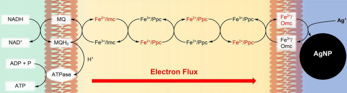
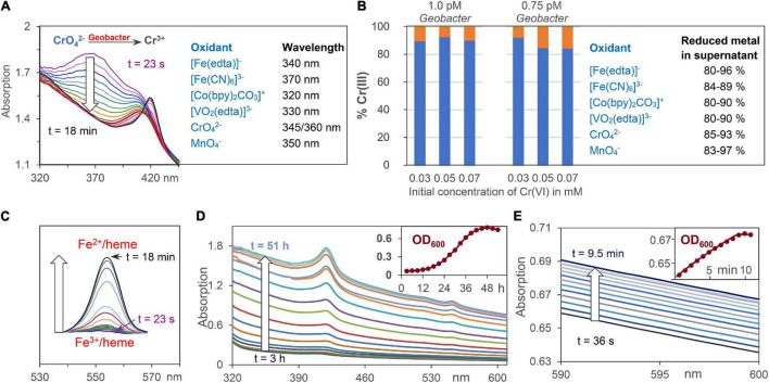
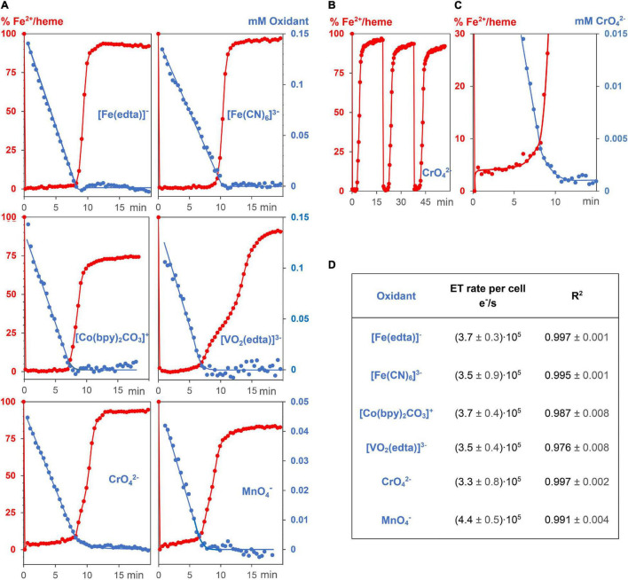
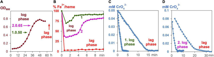
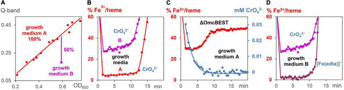
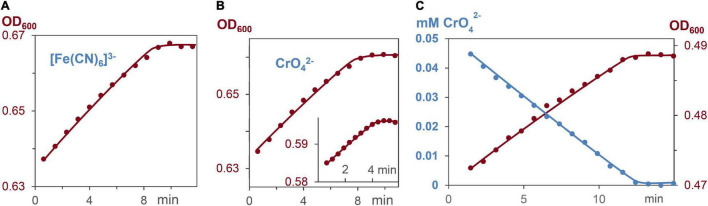
Geobacter sulfurreducens is a widely applied microorganism for the reduction of toxic metal salts, as an electron source for bioelectrochemical devices, and as a reagent for the synthesis of nanoparticles. In order to understand the influence of metal salts, and of electron transporting, multiheme c-cytochromes on the electron flux during respiration of G. sulfurreducens, the reduction kinetic of Fe3+, Co3+, V5+, Cr6+, and Mn7+ containing complexes were measured. Starting from the resting phase, each G. sulfurreducens cell produced an electron flux of 3.7 × 10⁵ electrons per second during the respiration process. Reduction rates were within ± 30% the same for the 6 different metal salts, and reaction kinetics were of zero order. Decrease of c-cytochrome concentrations by downregulation and mutation demonstrated that c-cytochromes stabilized respiration rates by variation of their redox states. Increasing Fe2+/heme levels increased electron flux rates, and induced respiration flexibility. The kinetic effects parallel electrochemical results of G. sulfurreducens biofilms on electrodes, and might help to optimize bioelectrochemical devices.

Keywords: Geobacter sulfurreducens; bioelectrochemistry; c-cytochrome; reaction kinetic; remediation.


Conflict of interest statement

The authors declare that the research was conducted in the absence of any commercial or financial relationships that could be construed as a potential conflict of interest.

Figures

FIGURE 1
FIGURE 1
Synthesis of Ag nanoparticles (AgNPs) during respiration of G. sulfurreducens with water soluble Ag+ ions. The c-type cytochromes at the inner cell membrane (Imc), in the periplasm (Ppc), and at the outer cell membrane (Omc) transport electrons from intracellular electron donors like NADH to Ag+/Omc complexes, which leads to AgNPs, attached to the outer cell membrane (Chabert et al., 2020). In recent cell growth experiments with CrO42– (Gong et al., 2018), Cr3+ reduction products could be detected within the cells, if the bacteria reacted for several hours with the chromium salts. Therefore, our reduction experiments of CrO42– might also occur partly inside of the cells, although the reaction conditions are different.
FIGURE 2
FIGURE 2
Analytical tools for the analysis of extracellular metal ion, Fe2+/heme, and cell concentrations. (A) Reduction of oxidizing metals salts by G. sulfurreducens by time dependent UV/Vis spectroscopy at wavelengths where product absorptions are negligible. CrO42– respiration is shown as an example. (B) ICP analysis of Cr3+ ions outside of the cell (blue) and at the cell membrane (orange) after reduction of CrO42–. Percentage data for reduced metal salts in the supernatant are given. (C) Changes of Fe2+/heme concentration during respiration using the areas of Q-bands. (D) Increase of UV/Vis absorptions during bacteria growth with fumarate as intracellular oxidant. The insert shows scattering increases at 600 nm (OD600) during 2 days of cell growth. (E) UV/Vis absorptions during cell growth using low chromate concentrations for G. sulfurreducens respiration, starting from the resting state.
FIGURE 3
FIGURE 3
Kinetic experiments of G. sulfurreducens with extracellular metal salts. (A) Reduction of 6 different oxidizing metal salt solutions (blue, 0.05–0.15 mM) with G. sulfurreducens solutions in the resting state (8.9 ± 0.3 pM). Their linear time dependences of the metal salt concentrations demonstrate zero kinetic order of the reduction process. At the start of the experiments, all iron ions of the bacterial multiheme cytochromes were in the Fe2+ state (red). They were rapidly oxidized upon addition of extracellular metal salts, their levels remained low during reduction of the metal salts, and increased again after major consumption of the oxidants. (B) Experiments could be repeated several times. Shown are Fe2+/heme levels upon three consecutive additions of CrO42–. (C) A closer look at the Fe2+/heme (red) and CrO42– (blue) concentrations. The Fe2+ /heme levels increased after major part of CrO42– was reduced, which obviously stabilized EET rates at low concentrations of the extracellular metal salts. (D) Zero order reduction rates per cell for the 6 different metal salts at 30°C and pH = 7.4.
FIGURE 4
FIGURE 4
CrO42– induce respiration of G. sulfurreducens, starting from the log phase. (A) Fumarate- respiring growth of G. sulfurreducens. Bacteria after 30 and 36 h growth, respectively, were used for metal induced respiration of G. sulfurreducens in the log phase. (B) Fe2+/heme levels during CrO42– induced respiration of bacteria starting from log phases 1 and 2, as well as the resting (lag) phase. (C) Reduction of CrO42– (0.05 mM) by bacteria (0.63 ± 0.2 pM) in the log phase 1 and the lag phase. (D) Reduction of CrO42– (0.1 mM) by bacteria (0.83 ± 0.2 pM) in the log phase 2 and the lag phase.
FIGURE 5
FIGURE 5
Changes of Fe2+/heme levels during metal induced respiration of G. sulfurreducens. (A) Linear correlation (red line) between Fe2+/heme concentrations (Q-bands) and G. sulfurreducens concentrations (OD600 data). The data (red points) were gained by dilution and different cell growth experiments in medium A. Measurement (magenta point) with G. sulfurreducens, grown in medium B, where c-cytochromes are downregulated. (B) Fe2+/heme levels during CrO42– induced respiration (0.05 mM) of cells grown in media A (0.63 pM) and B (0.73 pM). (C) Fe2+/heme levels (red) and CrO42– concentrations (blue) during CrO42– induced respiration (0.05 mM) of mutants lacking OmcBEST (0.76 pM) grown in medium A. (D) Fe2+/heme levels of experiments with G. sulfurreducens (0.74 pM), grown in medium B, with [Fe(edta)] (0.15 mM) and CrO42– (0.05 mM), respectively.
FIGURE 6
FIGURE 6
Increase of cell concentration and decrease of oxidants [Fe(CN)6]3– and CrO42– during G. sulfurreducens respiration in grow medium A, lacking fumarate, and stating from the lag phase. (A) Linear increase of the bacterial growth with 0.15 mM [Fe(CN)6]3–, R2 = 0.98. (B) Linear increase of the bacterial growth with 0.05 and 0.03 mM CrO42– (insert), R2 = 0.98. (C) Linear CrO42– decrease (blue, R2 = 0.99), and linear bacterial growth (brown, R2 = 0.98) of the same respiration experiment.

Similar articles

Cited by

References

    1. Aklujkar M., Coppi M. V., Leang C., Kim B. C., Chavan M. A., Perpetua L. A., et al. (2013). Proteins involved in electron transfer to Fe(III) and Mn(IV) oxides by Geobacter sulfurreducens and Geobacter uraniireducens. Microbiology 159 515–535. 10.1099/mic.0.064089-0 - DOI - PubMed
    1. Bochdansky A. B., Stouffer A. N., Washington N. N. (2021). Adenosine triphosphate (ATP) as a metric of microbial biomass in aquatic systems: new simplified protocols, laboratory validation, and a reflection on data from the literature. Limnol. Oceanogr. Methods 19 115–131. 10.1002/lom3.10409 - DOI
    1. Bond D. R., Lovley D. R. (2003). Electricity production by geobacter sulfurreducens attached to electrodes. Appl. Environ. Microbiol. 69 1548–1555. 10.1128/AEM.69.3.1548-1555.2003 - DOI - PMC - PubMed
    1. Brown G. C. (1992). Control of respiration and ATP synthesis in mammalian mitochondria and cells. Biochem. J. 284 1–13. 10.1042/bj2840001 - DOI - PMC - PubMed
    1. Bruderer R., Bernhardt O. M., Gandhi T., Miladinović S. M., Cheng L. Y. (2015). Extending the limits of quantitative proteome profiling with data-independent acquisition and application to acetaminophen-treated three-dimensional liver microtissues. Mol. Cell. Proteomics 14 1400–1410. 10.1074/mcp.M114.044305 - DOI - PMC - PubMed

LinkOut - more resources