

Rhino-Orbital-Cerebral Mycosis and Extranodal Natural Killer or/and T-Cell Lymphoma, Nasal Type
- PMID: 35783622
- PMCID: PMC9248758
- DOI: 10.3389/fmed.2022.851208
Rhino-Orbital-Cerebral Mycosis and Extranodal Natural Killer or/and T-Cell Lymphoma, Nasal Type
Abstract
Background: Extranodal natural killer/T-cell lymphoma, nasal type is a syndrome of middle face destruction with an association to Epstein-Barr virus. Fungi have been recovered from the diseased tissue now and then but were often seen as a lymphoma-associated secondary infection. However, there are ENKTL-NT cases with the recoveries of fungi and complete recovery with antifungal therapy, which are quite similar to rhino-orbital-cerebral mycosis (ROCM) that often confuses the physicians.
Methods: We searched Medline for English-language manuscripts limited to "human" and "case reports," "letters," "reviews," and "clinical conferences" from 1966 to 2022. We used MeSH terms "lymphoma, extranodal nk-t-cell" [MeSH Terms] or "lethal midline granuloma" [MeSH Terms], in combination with MeSH terms "microbiology" [subheading] or "microbiology" [all fields] or "fungi" [all fields] or "fungi" [MeSH Terms] for ENKTL-NT with infections. We used MeSH terms "Mycoses" in combination with "Nose" [Mesh] OR "Orbital Diseases" [Mesh] for rhino-orbital-cerebral fungal infections.
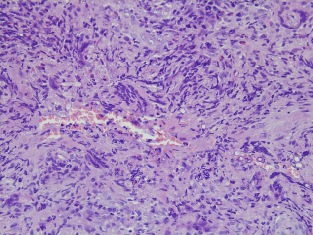
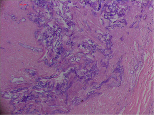
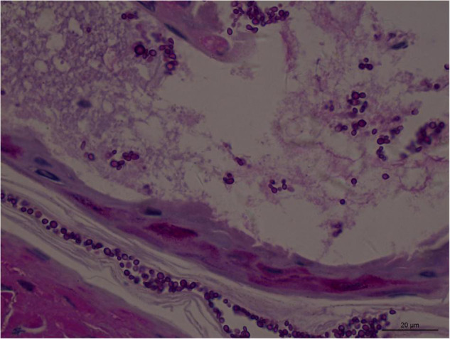
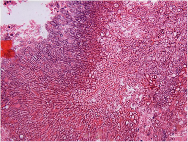
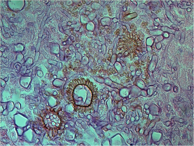
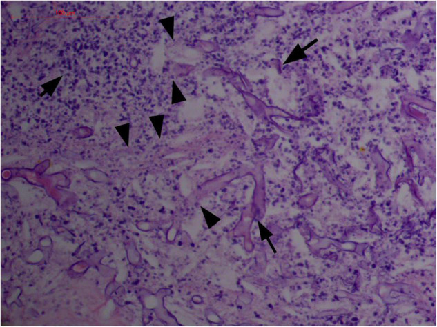
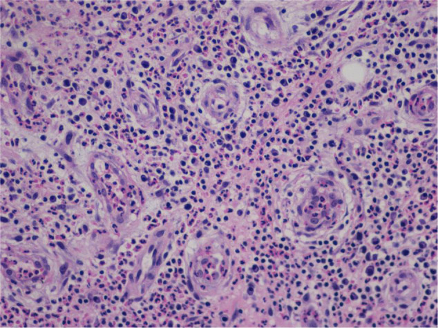
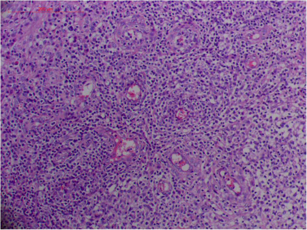
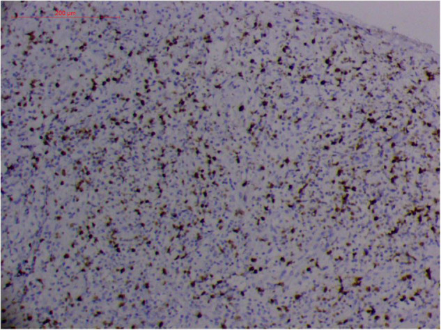
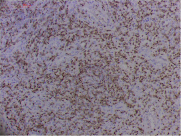
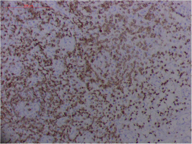
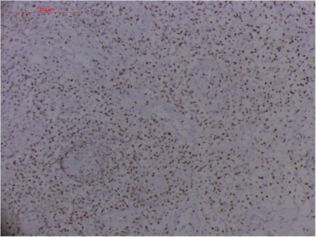
Results: We appraised 149 included articles and extracted references related to ENKTL-NT and/or ROCM. Themes and subcategories were subsequently derived. Our findings revealed that ROCM and ENKTL-NT are characterized by progressive and destructive ulcers in the midline face or rhino-orbital structures. ROCM is mainly caused by fungi in the order of Mucorales, and ENKTL-NT is usually associated with Epstein-Barr virus and sometimes fungi. Radiologically, both are characterized by non-specific features of sinusitis, soft tissue infection, and necrosis. Pathologically, ROCM and ENKTL-NT share the same characteristics of inflammation, necrosis, and granuloma. ROCM is characterized by the detection of fungi in tissue, while ENKTL-NT is typically positive for NK/T-cell markers and cytotoxic granule-associated proteins, proliferation, and vascular damage of angioinvasion, which could be incited by Mucor irregularis and Rhizopus arrhizus in patients and mice.
Conclusion: ENKTL-NT and ROCM share many similarities in clinical presentations, radiology, and histopathology, and might have the same etiology. This may explain why the two diseases are tangled together in the reported cases, and suggests the role that the fungi may play in the development of these ENKTL-NT/ROCM diseases. The reason why ENKTL-NT and ROCM are sometimes confused is that the main pathogens of ROCM, Mucor irregularis and Rhizopus arrhizus, are the fungal causative agents of ENKTL-NT.
Keywords: Rhizopus arrhizus; extranodal nK/T-cell lymphoma; facial destruction; lethal midline granuloma; mucor irregularis; nasal type; rhino-orbital-cerebral mycosis.
Copyright © 2022 Li and Lun.
Conflict of interest statement
The authors declare that the research was conducted in the absence of any commercial or financial relationships that could be construed as a potential conflict of interest.
Figures


Similar articles
-
Case Report: Rhizopus arrhizus Rhino-Orbital-Cerebral Mycosis and Lethal Midline Granuloma: Another Fungal Etiological Agent.Front Med (Lausanne). 2021 Jun 2;8:578684. doi: 10.3389/fmed.2021.578684. eCollection 2021. Front Med (Lausanne). 2021. PMID: 34150783 Free PMC article.
-
Extranodal natural-killer/T-cell lymphoma, nasal type: An immunomorphological study from a regional cancer institute in India.J Cancer Res Ther. 2022 Jul-Sep;18(4):1137-1143. doi: 10.4103/jcrt.JCRT_226_20. J Cancer Res Ther. 2022. PMID: 36149173
-
Extranodal NK/T Cell Lymphoma, Nasal Type (ENKTL-NT): An Update on Epidemiology, Clinical Presentation, and Natural History in North American and European Cases.Curr Hematol Malig Rep. 2016 Dec;11(6):514-527. doi: 10.1007/s11899-016-0355-9. Curr Hematol Malig Rep. 2016. PMID: 27778143 Free PMC article. Review.
-
Two young patients with extranodal natural killer/T-cell non-Hodgkin lymphoma, nasal-type (ENKTL-NT) masquerading inflammatory processes: A case series.Taiwan J Ophthalmol. 2021 Feb 12;12(2):231-236. doi: 10.4103/tjo.tjo_80_20. eCollection 2022 Apr-Jun. Taiwan J Ophthalmol. 2021. PMID: 35813787 Free PMC article.
-
Clinicopathological study of pulmonary extranodal nature killer/T-cell lymphoma, nasal type and literature review.Pathol Res Pract. 2015 Jul;211(7):544-9. doi: 10.1016/j.prp.2015.04.002. Epub 2015 Apr 17. Pathol Res Pract. 2015. PMID: 25953340 Review.
Cited by
-
Nasal NK/T-Cell Lymphoma with Unusual Fonsecaea pedrosoi Infection: A Rare Case Report from Northeast India.Indian J Otolaryngol Head Neck Surg. 2025 May;77(5):2149-2152. doi: 10.1007/s12070-025-05455-y. Epub 2025 Apr 8. Indian J Otolaryngol Head Neck Surg. 2025. PMID: 40321399
References
-
- Haverkos BM, Pan Z, Gru AA, Freud AG, Rabinovitch R, Xu-Welliver M, et al. Extranodal NK/T cell lymphoma, nasal type (ENKTL-NT): an update on epidemiology, clinical presentation, and natural history in north american and european cases. Curr Hematol Malig Rep. (2016) 11:514–27. 10.1007/s11899-016-0355-9 - DOI - PMC - PubMed
LinkOut - more resources
Full Text Sources
Research Materials