

Diagnostic Value of Spectral-Domain Optical Coherence Tomography for Polypoidal Choroidal Vasculopathy: A Systematic Review and Meta-Analysis
- PMID: 35783657
- PMCID: PMC9242399
- DOI: 10.3389/fmed.2022.878946
Diagnostic Value of Spectral-Domain Optical Coherence Tomography for Polypoidal Choroidal Vasculopathy: A Systematic Review and Meta-Analysis
Abstract
Purpose: To evaluate the diagnostic value of spectral-domain optical coherence tomography (SD-OCT) for polypoidal choroidal vasculopathy (PCV).
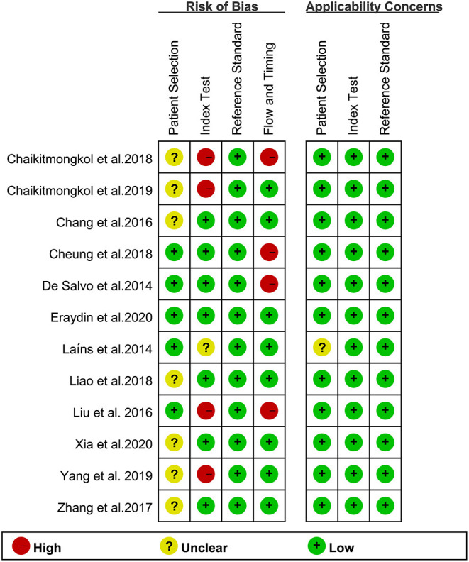
Methods: A search of electronic databases was conducted from 2010 to 2021 to review the relevant literature on SD-OCT to identify PCV and other lesions causing serious or serosanguinous retinal pigment epithelial detachment (PED), specifically neovascular age-related macular degeneration (nvAMD). The QUADAS-2 scale was used to evaluate the quality of the literature. We performed a meta-analysis, including heterogeneity tests, analyze and synthesize the study data, meta-regression analysis, subgroup analysis, Fagan's plot, sensitivity analysis and publication bias tests.
Results: A total of 12 related studies involving 1,348 eyes were included in this study, and the random-effects model was used for meta-analysis. The results showed that the pooled sensitivity of SD-OCT in the diagnosis of PCV was 0.87 (95% CI: 0.84-0.89), the pooled specificity was 0.83 (95% CI: 0.80-0.86), and the pooled positive/negative likelihood ratios were 5.38 (95% CI: 3.28-8.80) and 0.16 (95% CI: 0.10-0.25), respectively. The diagnostic odds ratio (DOR) was 36.07 (95% CI: 15.98-81.40), and the area under the sROC curve (AUC) was 0.9429. When the pre-test probability was set at 20%, the post-test positive and negative probabilities were 58% and 4%, respectively. Meta-regression indicated that race was the primary source of heterogeneity (P <0.05). The Deeks' funnel plot showed no significant publication bias in this study (P>0.05).
Conclusion: SD-OCT has high sensitivity and specificity for the diagnosis of PCV, as well as significant clinical applicability. Since color fundus photography (CFP) is more clinically available and can improve the diagnostic efficacy, we recommend SD-OCT combined with CFP to diagnose PCV, especially without indocyanine green angiography (ICGA).
Systematic review registration: https://inplasy.com/inplasy-2021-12-0048/, identifier: INPLASY2021120048.
Keywords: diagnostic value; meta-analysis; polypoidal choroidal vasculopathy; sensitivity; specificity; spectral-domain optical coherence tomography.
Copyright © 2022 Jiang and Qi.
Conflict of interest statement
The authors declare that the research was conducted in the absence of any commercial or financial relationships that could be construed as a potential conflict of interest.
Figures


Similar articles
-
Sensitivity and Specificity of Potential Diagnostic Features Detected Using Fundus Photography, Optical Coherence Tomography, and Fluorescein Angiography for Polypoidal Choroidal Vasculopathy.JAMA Ophthalmol. 2019 Jun 1;137(6):661-667. doi: 10.1001/jamaophthalmol.2019.0565. JAMA Ophthalmol. 2019. PMID: 30973593 Free PMC article.
-
Optical coherence tomography in diagnosing polypoidal choroidal vasculopathy. Looking into the future: a systematic review and meta-analysis.Int J Retina Vitreous. 2022 Feb 28;8(1):14. doi: 10.1186/s40942-022-00365-5. Int J Retina Vitreous. 2022. PMID: 35227320 Free PMC article. Review.
-
IMPROVED DETECTION AND DIAGNOSIS OF POLYPOIDAL CHOROIDAL VASCULOPATHY USING A COMBINATION OF OPTICAL COHERENCE TOMOGRAPHY AND OPTICAL COHERENCE TOMOGRAPHY ANGIOGRAPHY.Retina. 2019 Sep;39(9):1655-1663. doi: 10.1097/IAE.0000000000002228. Retina. 2019. PMID: 29927796
-
Color Fundus Photography, Optical Coherence Tomography, and Fluorescein Angiography in Diagnosing Polypoidal Choroidal Vasculopathy.Am J Ophthalmol. 2018 Aug;192:77-83. doi: 10.1016/j.ajo.2018.05.005. Epub 2018 May 29. Am J Ophthalmol. 2018. PMID: 29753852
-
Non-ICGA treatment criteria for Suboptimal Anti-VEGF Response for Polypoidal Choroidal Vasculopathy: APOIS PCV Workgroup Report 2.Ophthalmol Retina. 2021 Oct;5(10):945-953. doi: 10.1016/j.oret.2021.04.002. Epub 2021 Apr 16. Ophthalmol Retina. 2021. PMID: 33866022
Cited by
-
Polypoidal choroidal vasculopathy with an exceptionally elevated pigment epithelial detachment.Am J Ophthalmol Case Rep. 2024 Sep 12;36:102171. doi: 10.1016/j.ajoc.2024.102171. eCollection 2024 Dec. Am J Ophthalmol Case Rep. 2024. PMID: 39314252 Free PMC article.
References
-
- Liu R, Ding X. Xirouyang mailuomo xueguang bingbian yu shenchuxing laonian huangbanbianxing de yitong [Similarities and differences between polypoid choroidal vasculopathy and exudative age-related macular degeneration]. Chin J Ocul Fundus Dis. (2012) 9:533–9. 10.3760/cma.j.issn.1005-1015.2012.05.029 - DOI
-
- Zhang S, Zhang J, Xu X, Gao R, Sun X, Huan X. Progress in treatment of submacular hemorrhage due to polypoid choroidal vasculopathy. Int J Ophthalmol. (2019) 19:950–5. 10.3980/j.issn.1672-5123.2019.6.13 - DOI
Publication types
LinkOut - more resources
Full Text Sources
Miscellaneous