

Comparison and optimization of sheep in vivo intervertebral disc injury model
- PMID: 35783908
- PMCID: PMC9238284
- DOI: 10.1002/jsp2.1198
Comparison and optimization of sheep in vivo intervertebral disc injury model
Abstract
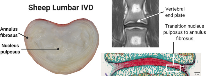
Background: The current standard of care for intervertebral disc (IVD) herniation, surgical discectomy, does not repair annulus fibrosus (AF) defects, which is partly due to the lack of effective methods to do so and is why new repair strategies are widely investigated and tested preclinically. There is a need to develop a standardized IVD injury model in large animals to enable comparison and interpretation across preclinical study results. The purpose of this study was to compare in vivo IVD injury models in sheep to determine which annulus fibrosus (AF) defect type combined with partial nucleus pulposus (NP) removal would better mimic degenerative human spinal pathologies.
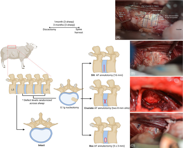
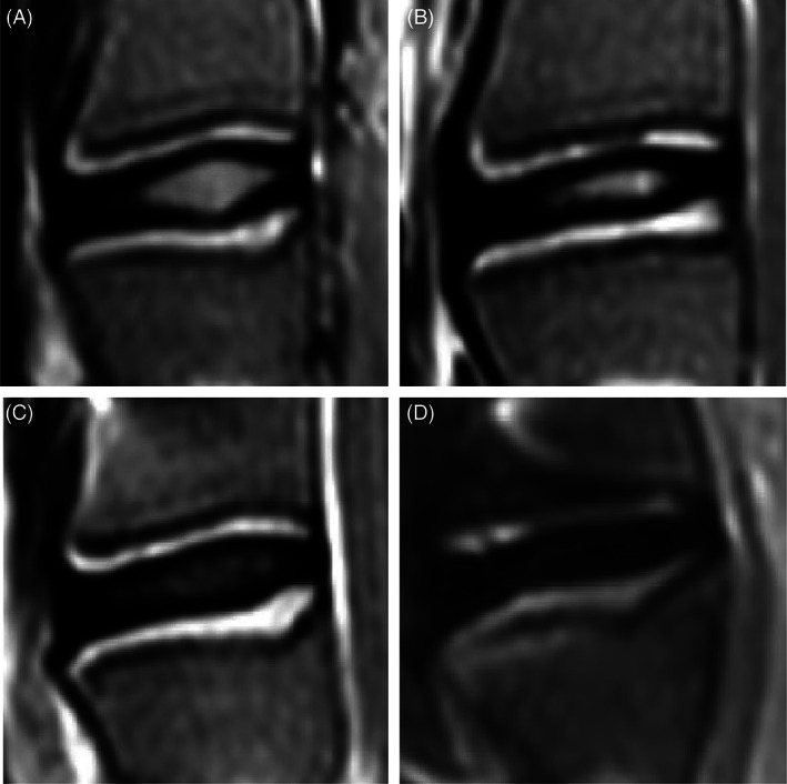
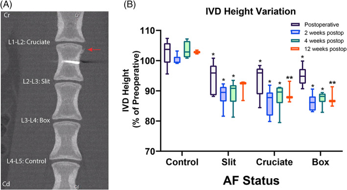
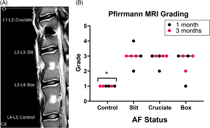
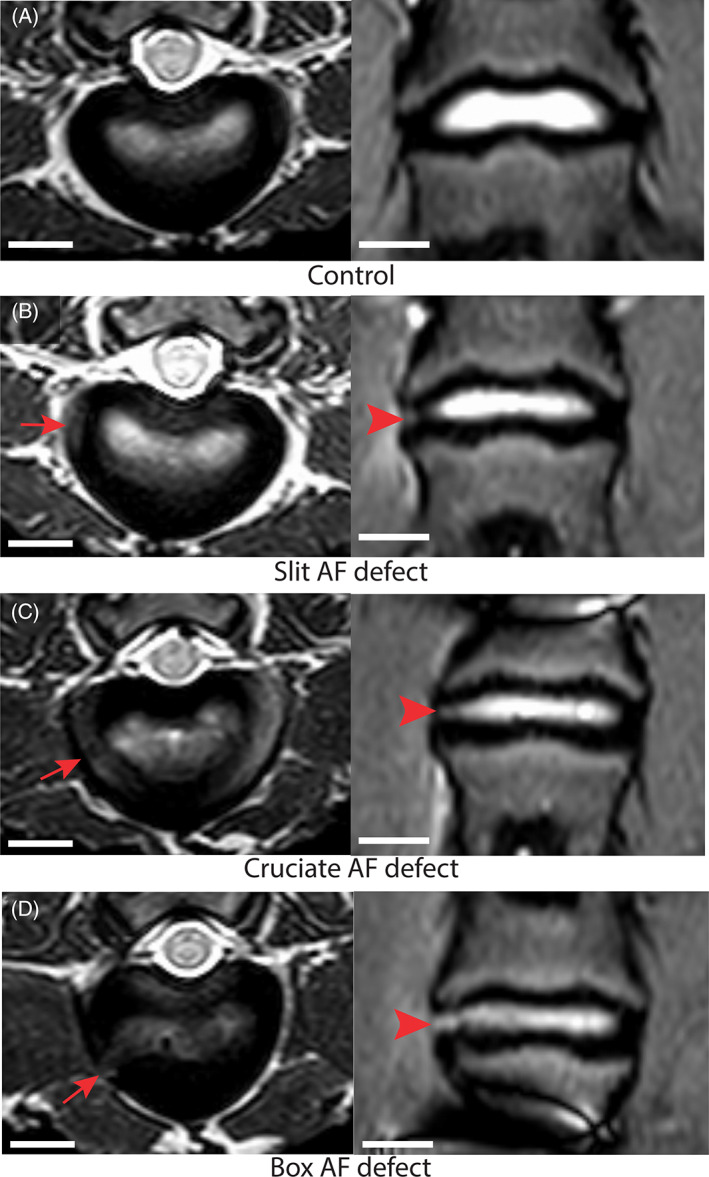
Methods: Six skeletally mature sheep were randomly assigned to one of the two observation periods (1 and 3 months) and underwent creation of 3 different AF defect types (slit, cruciate, and box-cut AF defects) in conjunction with 0.1 g NP removal in three lumbar levels using a lateral retroperitoneal surgical approach. The spine was monitored by clinical CT scans pre- and postoperatively, at 2 weeks and euthanasia, and by magnetic resonance imaging (MRI) and histology after euthanasia to determine the severity of degeneration (disc height loss, Pfirrmann grading, semiquantitative histopathology grading).
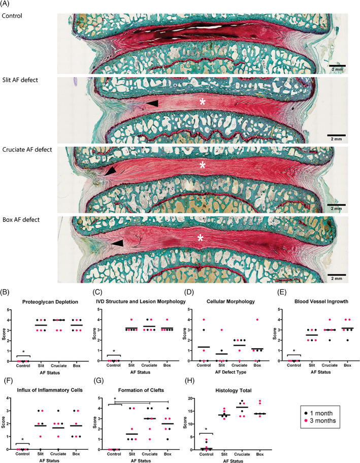
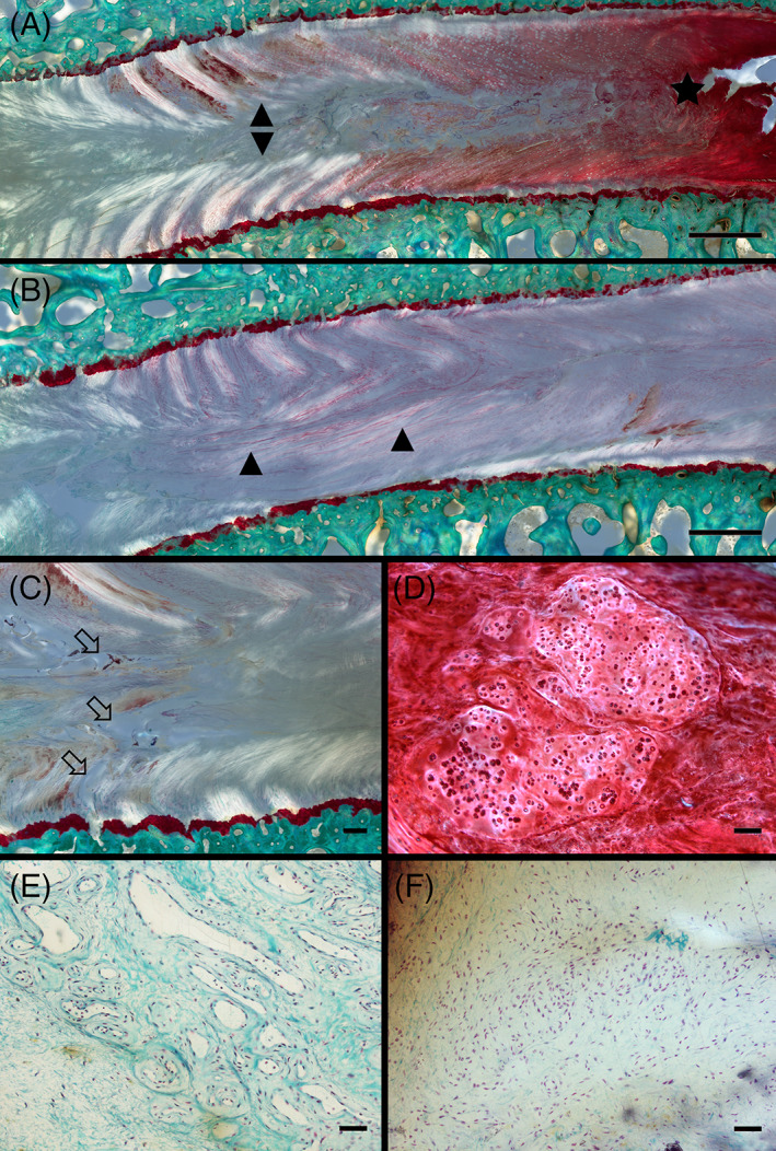
Results: All AF defects led to significant degenerative changes detectable on CT and MR images, produced bulging of disc tissue without disc herniation and led to degenerative and inflammatory histopathological changes. However, AF defects were not equal in terms of disc height loss at 3 months postoperatively; the cruciate and box-cut AF defects showed significantly decreased disc height compared to their preoperative height, with the box-cut defect creating the greatest disc height loss, while the slit AF defect showed restoration of normal preoperative disc height.
Conclusions: The tested IVD injury models do not all generate comparable disc degeneration but can be considered suitable IVD injury models to investigate new treatments. Results of the current study clearly indicate that slit AF defect should be avoided if disc height is used as one of the main outcomes; additional confirmatory studies may be warranted to generalize this finding.
Keywords: annulus fibrosus defect; discectomy; intervertebral disc degeneration; intervertebral disc injury; preclinical model; sheep.
© 2022 The Authors. JOR Spine published by Wiley Periodicals LLC on behalf of Orthopaedic Research Society.
Conflict of interest statement
The authors declare no conflict of interest
Figures
Similar articles
-
Combining adhesive and nonadhesive injectable hydrogels for intervertebral disc repair in an ovine discectomy model.JOR Spine. 2023 Dec 1;6(4):e1293. doi: 10.1002/jsp2.1293. eCollection 2023 Dec. JOR Spine. 2023. PMID: 38156055 Free PMC article.
-
Total disc replacement using a tissue-engineered intervertebral disc in vivo: new animal model and initial results.Evid Based Spine Care J. 2010 Aug;1(2):62-6. doi: 10.1055/s-0028-1100918. Evid Based Spine Care J. 2010. PMID: 23637671 Free PMC article.
-
A novel rat model of annulus fibrosus injury for intervertebral disc degeneration.Spine J. 2024 Feb;24(2):373-386. doi: 10.1016/j.spinee.2023.09.012. Epub 2023 Oct 3. Spine J. 2024. PMID: 37797841
-
Putting the Pieces in Place: Mobilizing Cellular Players to Improve Annulus Fibrosus Repair.Tissue Eng Part B Rev. 2021 Aug;27(4):295-312. doi: 10.1089/ten.TEB.2020.0196. Epub 2020 Oct 19. Tissue Eng Part B Rev. 2021. PMID: 32907498 Free PMC article. Review.
-
Biomechanics in Annulus Fibrosus Degeneration and Regeneration.Adv Exp Med Biol. 2018;1078:409-420. doi: 10.1007/978-981-13-0950-2_21. Adv Exp Med Biol. 2018. PMID: 30357635 Review.
Cited by
-
Combining adhesive and nonadhesive injectable hydrogels for intervertebral disc repair in an ovine discectomy model.JOR Spine. 2023 Dec 1;6(4):e1293. doi: 10.1002/jsp2.1293. eCollection 2023 Dec. JOR Spine. 2023. PMID: 38156055 Free PMC article.
-
Melatonin-loaded self-healing hydrogel targets mitochondrial energy metabolism and promotes annulus fibrosus regeneration.Mater Today Bio. 2023 Sep 22;23:100811. doi: 10.1016/j.mtbio.2023.100811. eCollection 2023 Dec. Mater Today Bio. 2023. PMID: 37810753 Free PMC article.
-
Animal Models of Intervertebral Disc Diseases: Advantages, Limitations, and Future Directions.Neurol Int. 2024 Dec 9;16(6):1788-1818. doi: 10.3390/neurolint16060129. Neurol Int. 2024. PMID: 39728755 Free PMC article. Review.
-
Recommendations for intervertebral disc notochordal cell investigation: From isolation to characterization.JOR Spine. 2023 Jul 9;6(3):e1272. doi: 10.1002/jsp2.1272. eCollection 2023 Sep. JOR Spine. 2023. PMID: 37780826 Free PMC article.
-
Correlation between Disc Imaging Observations and Clinical Efficacy after Percutaneous Endoscopic Lumbar Discectomy: A 1-Year Follow-up Study.Orthop Surg. 2024 Apr;16(4):851-863. doi: 10.1111/os.14013. Epub 2024 Feb 21. Orthop Surg. 2024. PMID: 38384172 Free PMC article.
References
-
- Anema JR, Schellart AJ, Cassidy JD, Loisel P, Veerman TJ, van der Beek AJ. Can cross country differences in return‐to‐work after chronic occupational back pain be explained? An exploratory analysis on disability policies in a six country cohort study. J Occup Rehabil. 2009;19(4):419‐426. - PMC - PubMed
-
- Becker A, Held H, Redaelli M, et al. Low back pain in primary care: costs of care and prediction of future health care utilization. Spine (Phila Pa 1976). 2010;35(18):1714‐1720. - PubMed
-
- Whatley BR, Wen X. Intervertebral disc (IVD): structure, degeneration, repair and regeneration. Mater Sci Eng C. 2012;32(2):61‐77.
-
- Boden SD, Davis DO, Dina TS, Patronas NJ, Wiesel SW. Abnormal magnetic‐resonance scans of the lumbar spine in asymptomatic subjects. A prospective investigation. J Bone Joint Surg Am. 1990;72(3):403‐408. - PubMed
Grants and funding
LinkOut - more resources
Full Text Sources
Miscellaneous
