

Prevalence, Etiology, and Risk Factors Associated with Occurrence of Canine Cutaneous Myiasis in Kitui County, Kenya
- PMID: 35783917
- PMCID: PMC9249507
- DOI: 10.1155/2022/5699060
Prevalence, Etiology, and Risk Factors Associated with Occurrence of Canine Cutaneous Myiasis in Kitui County, Kenya
Abstract
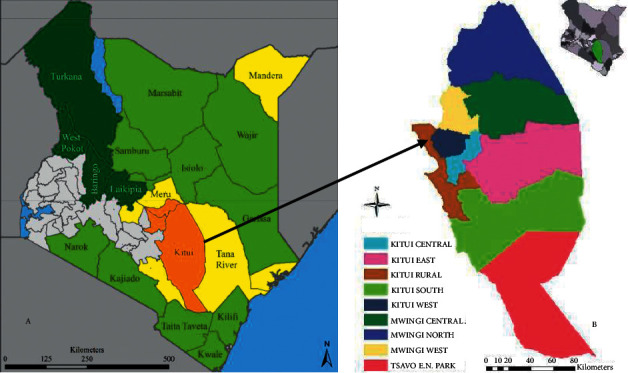
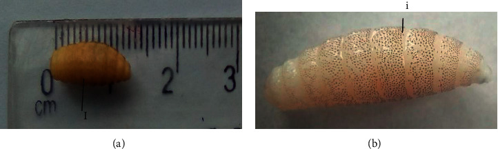
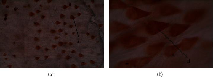
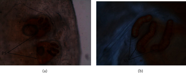
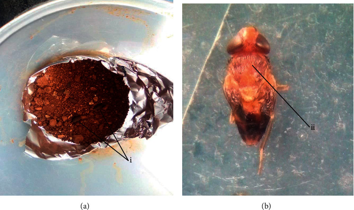
Myiasis is the infestation of living tissues of animals with dipterous larvae. In Africa, Cordylobia species (C. anthropophaga, C. rodhaini, and C. ruandae) and Dermatobia hominis are reported as the principal cause of nonmigratory cutaneous myiasis of domestic animals. None of these have been reported in dogs in Kenya. A cross-sectional study was conducted in eight subcounties of Kitui County, Kenya, from March to August 2021 to estimate the prevalence, risk factors, and etiological agents associated with canine cutaneous myiasis (CCM). A questionnaire was administered to dog owners to collect information on CCM risk factors. A total of 400 dogs were physically examined and larvae collected from myiasis skin lesions and preserved in 70% ethanol, taken to the laboratory, processed and identified using parasitological morphological features. Live larvae were incubated and emerging adults were captured and identified. The overall prevalence of CCM was 45% (180/400) (95% confidence interval: 40.0-50.0%). A total of 434 larvae were collected from 180 dogs infested with cutaneous myiasis. All larvae (100%) were identified as C. anthropophaga and hatched adults were "tumbu" flies. There were no significant differences in the prevalence of CCM at 95% confidence interval among different age and sex groups (p > 0.05), although puppies (<6 months) appeared more affected. The highest prevalence of myiasis was in Kitui Central at 65% (95% confidence interval: 51.6-76.9%), Mwingi North at 52.5% (95% confidence interval: 36.1-68.4%), Kitui South at 48.5% (95% confidence interval: 31.5-63.9%), Kitui Rural at 40% (95% confidence interval: 27.6-53.5%), Mwingi Central at 40% (95% confidence interval: 24.9-56.7%), Mwingi West at 40% (95% confidence interval: 24.9-56.7%), Kitui West at 38.3% (95% confidence interval: 26.1-51.8%), and Kitui East subcounty at 36.7% (95% confidence interval: 24.6-50.1%). Lack of housing, housing structures, and dog living area environmental hygiene were the main risk factors associated with the occurrence of CCM (p < 0.05). The CCM occurrence was significantly different among breeds (p < 0.05). Cordylobia anthropophaga larvae were the etiological agent of CCM in Kitui County. There is a need for improved dog housing and hygiene measures to prevent the occurrence of CCM, and affected dogs should be treated to prevent the spread of CCM among the dogs.
Copyright © 2022 Kamuti N. Mutinda et al.
Conflict of interest statement
The authors declare that they have no conflicts of interest regarding the publication of this paper.
Figures

References
-
- Zumpt F. Myiasis in Man and Animals in the Old World. A Textbook for Physicians, Veterinarians and Zoologists . London, UK: Butterworth and Co. (Publishers) Ltd; 1965.
-
- de Azeredo-Espin A. M. L., Lessinger A. C. Genetic approaches for studying myiasis-causing flies: molecular markers and mitochondrial genomics. Genetica . 2006;126:111–131. - PubMed
-
- Spradbery J. P. A Manual for the Diagnosis of the Screwworm Fly, Fisheries and Forestr . 2. Canberra, Australia: Department of Agriculture; 2002.
-
- Adam A. A., Rahman A. O. A., Nail A. S. M., Imam A. M. M. Cutaneous myiasis due to Dermatobia hominis: a case report from eastern Sudan. Sudan Journal of Medical Sciences . 2006;1(2):147–152.
LinkOut - more resources
Full Text Sources
