. 2022 Jun 16:13:871208.
doi: 10.3389/fpls.2022.871208. eCollection 2022.

Structural and Biophysical Characterization of Purified Recombinant Arabidopsis thaliana's Alternative Oxidase 1A (rAtAOX1A): Interaction With Inhibitor(s) and Activator

Affiliations

Structural and Biophysical Characterization of Purified Recombinant Arabidopsis thaliana's Alternative Oxidase 1A (rAtAOX1A): Interaction With Inhibitor(s) and Activator

Tadiboina Veera Sankar et al. Front Plant Sci. .

Abstract

In higher plants, alternative oxidase (AOX) participates in a cyanide resistant and non-proton motive electron transport pathway of mitochondria, diverging from the ubiquinone pool. The physiological significance of AOX in biotic/abiotic stress tolerance is well-documented. However, its structural and biophysical properties are poorly understood as its crystal structure is not yet revealed in plants. Also, most of the AOX purification processes resulted in a low yield/inactive/unstable form of native AOX protein. The present study aims to characterize the purified rAtAOX1A protein and its interaction with inhibitors, such as salicylhydroxamic acid (SHAM) and n-propyl gallate (n-PG), as well as pyruvate (activator), using biophysical/in silico studies. The rAtAOX1A expressed in E. coli BL21(DE3) cells was functionally characterized by monitoring the respiratory and growth sensitivity of E. coli/pAtAOX1A and E. coli/pET28a to classical mitochondrial electron transport chain (mETC) inhibitors. The rAtAOX1A, which is purified through affinity chromatography and confirmed by western blotting and MALDI-TOF-TOF studies, showed an oxygen uptake activity of 3.86 μmol min-1 mg-1 protein, which is acceptable in non-thermogenic plants. Circular dichroism (CD) studies of purified rAtAOX1A revealed that >50% of the protein content was α-helical and retained its helical absorbance signal (ellipticity) at a wide range of temperature and pH conditions. Further, interaction with SHAM, n-PG, or pyruvate caused significant changes in its secondary structural elements while retaining its ellipticity. Surface plasmon resonance (SPR) studies revealed that both SHAM and n-PG bind reversibly to rAtAOX1A, while docking studies revealed that they bind to the same hydrophobic groove (Met191, Val192, Met195, Leu196, Phe251, and Phe255), to which Duroquinone (DQ) bind in the AtAOX1A. In contrast, pyruvate binds to a pocket consisting of Cys II (Arg174, Tyr175, Gly176, Cys177, Val232, Ala233, Asn294, and Leu313). Further, the mutational docking studies suggest that (i) the Met195 and Phe255 of AtAOX1A are the potential candidates to bind the inhibitor. Hence, this binding pocket could be a 'potential gateway' for the oxidation-reduction process in AtAOX1A, and (ii) Arg174, Gly176, and Cys177 play an important role in binding to the organic acids like pyruvate.

Keywords: AOX structure; Arabidopsis thaliana; alternative oxidase; circular dichroism; mutational docking; recombinant protein; respiratory inhibitors; surface plasmon resonance.


Conflict of interest statement

The authors declare that the research was conducted in the absence of any commercial or financial relationships that could be construed as a potential conflict of interest.

Figures

Figure 1
Figure 1
Cloning and expression of AtAOX1A in E. coli. (A) Polymerase chain reaction (PCR) amplification of 879 bp AtAOX1A using gene-specific primers. Lane 1, 50–1000 bp DNA ladder; lane 2, PCR amplified AtAOX1A (indicated with arrow). (B) Schematic diagram of the expression vector construction. The gene sequences were cloned into restriction endonuclease EcoRI and XhoI recognition sites of the pET28a vector to produce N-terminal His6-tag recombinant AtAOX1A protein. (C) Total protein of both E. coli/pET28a and E. coli/pAtAOX1A cells were separated on 12.5% (w/v) sodium dodecyl sulfate-polyacrylamide gel electrophoresis (SDS-PAGE) for recombinant protein expression analysis. Each well was loaded with 40 μg of protein. Lane 1, 10–180 kDa protein marker; lane 2, E. coli/pET28a; lane 3, E. coli/pET28a induced with 0.1 mM IPTG; lane 4, E. coli/pAtAOX1A; lane 5, E. coli/pAtAOX1A induced with 0.1 mM IPTG. The induced rAtAOX1A is indicated with an arrow.
Figure 2
Figure 2
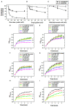
Functional characterization of rAtAOX1A in E. coli. Oxygen uptake rates (%) of E. coli/pET28a and E. coli/pAtAOX1A in the absence or presence of respiratory inhibitors: (A) KCN (0.05, 0.1, 0.25, 0.5, and 1 mM), (B) n-PG (0.025, 0.05, 0.1, 0.25, and 0.5 mM), and (C) SHAM (0.5, 1, and 2 mM). The growth pattern of E. coli cells was monitored for 5 h in the absence or presence of respiratory inhibitors: (D) Growth pattern of E. coli/pET28a and (E) E. coli/pAtAOX1A in the absence or presence of KCN; (F) Growth pattern of E. coli/pET28a and (G) E. coli/pAtAOX1A in the absence or presence of n-PG; (H) Growth pattern of E. coli/pET28a and (I) E. coli/pAtAOX1A in the absence or presence of SHAM. Each value represents the mean ± SD of three experiments. The statistical significance difference (P <0.05) was calculated for the endpoint of the growth curve and indicated with asterisks.
Figure 3
Figure 3
Purification and validation of rAtAOX1A protein using MALDI-MS-MS. (A) SDS-PAGE visualizing purification profile of rAtAOX1A from E. coli. Lane M, 10-180 kDa protein marker; lane 1, protein from E. coli/pAtAOX1A; lane 2, protein from E. coli/pAtAOX1A induced with IPTG; lane 3, flow-through; lane 4, washing fraction; lanes 5 and 6, elution fractions, arrow indicates purified rAtAOX1A with ~37 kDa molecular mass. (B) Western blot showing purified rAtAOX1A: lane M, 10-180 kDa protein marker; lane 1, 40 μg of purified rAtAOX1A. (C) A. thaliana partial AOX (gi/1872517) sequence from NCBI database. The sequence of MS peaks (1209.854, 1365.972, 1566.099, 1656.258, 2385.628, and 2476.697 Da) of rAtAOX1A are indicated in bold red font, and sequence coverage from these six major peaks is about 24%. The obtained sequence from peptide peak m/z 1656.258 (‘LPADATLRDVVMVVR') has shown 100% identity with AtAOX1A in BLAST search (D). The gel picture shown is the selective representation of three biological replicates.
Figure 4
Figure 4
Secondary structural stability analysis of rAtAOX1A. (A) CD spectra of purified rAtAOX1A at far UV (190-260 nm) in 10-mM phosphate buffer. Ellipticity in θ (mdeg) of rAtAOX1A (0.8 mg/ml) was plotted against the wavelength. Inset: High Tension profile of voltage trace. The secondary structural stability was determined at different (B) pH from 2 to 12; (C) Temperature from 4 to 90°C; (D) Temperature from 90 to 4°C. (E) Ellipticity of rAtAOX1A at 222 nm between 25°C and 90°C. (F) CD spectra of rAtAOX1A at far UV (190–260 nm), with 0.05, 0.1, and 0.5 mM SHAM and, without inhibitor; (G) CD spectra of rAtAOX1A at far UV (190-260 nm), with 0.05, 0.1, and 0.5 mM n-PG, and without inhibitor; (H) CD spectra of rAtAOX1A at far UV (190-260 nm), with 0.05, 0.1, 0.5, and 1 mM of pyruvate, and without activator. Concentration of purified rAtAOX1A used to obtain the CD spectrum (SHAM, n-PG, and pyruvate) was 0.4 mg/ml. The final spectrum is an average of three scans as described in the Materials and Methods section.
Figure 5
Figure 5
Purified rAtAOX1A is immobilized onto Series S Sensor Chip CM5. (A) Blank immobilization on flow cell Fc-3; (B) 100 μg/ml of purified rAtAOX1A (ligand) immobilization on flow cell Fc-4. Arrow indicates rAtAOX1A injection, which resulted in ~4,117 RU. SPR kinetics of SHAM and n-PG with rAtAOX1A: (C,D) Binding curves for SHAM and n-PG with rAtAOX1A at 25°C. The colored lines represent the concentrations of SHAM/n-PG (1, 2, 3, 4, and 5 mM), the black lines correspond to the fit lines to the data, and each fit line is a result of a global fit. The final sensorgram is representative of three cycles.
Figure 6
Figure 6
Molecular docking of AtAOX1A with different ligands (I-VI and IX). (a) All possible binding pockets of a specific ligand on AtAOX1A. The yellow region on AtAOX1A secondary structure indicates the best-fit binding pocket of a particular ligand. The Blue region in panel (IIa) represents the inhibitor binding pocket to compare with that of UQ, and (b) residues of AtAOX1A that are involved in forming the binding pocket of the best model (Cluster 0 Element 0) according to SwissDock results. The ligands are (I) Q1H2, (II) UQ1, (III) DQH2, (IV) DQ, (V) SHAM, (VI) n-PG, (VII) Binding pockets of inhibitor (yellow), DQH2 (green surface), Q1H2 (blue surface), and diiron binding cavity (red surface). DQH2 is shown with purple sticks. (VIII) Binding pockets of inhibitor (yellow surface), ubiquinone-1 (orange surface), and diiron cavity (red surface), (IX) Pyruvate. Q1H2, UQ1, DQH2, DQ, SHAM, n-PG, and pyruvate are shown in purple. Color scheme: carbon (red), oxygen (orange), sulfur (yellow), nitrogen (blue), and hydrogen (white).
Figure 7
Figure 7
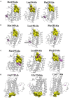
Effect of mutation on the binding affinities of DQ (A), SHAM (B), n-PG (C), and Pyruvate (D). The yellow surface indicates the location of the binding pocket in wild type (WT) AtAOX1A. The location of the ligand on the mutant AtAOX1A is shown in purple.

Similar articles

Cited by

References

    1. Affourtit C., Albury M. S., Crichton P. G., Moore A. L. (2002). Exploring the molecular nature of alternative oxidase regulation and catalysis. FEBS Lett. 510, 121–126. 10.1016/S0014-5793(01)03261-6 - DOI - PubMed
    1. Affourtit C., Moore A. L. (2004). Purification of the plant alternative oxidase from Arum maculatum: measurement, stability and metal requirement. Biochim. Biophys. Acta - Bioenerg. 1608, 181–189. 10.1016/j.bbabio.2003.12.002 - DOI - PubMed
    1. Ajayi W. U., Chaudhuri M., Hill G. C. (2002). Site-directed mutagenesis reveals the essentiality of the conserved residues in the putative diiron active site of the trypanosome alternative oxidase. J. Biol. Chem. 277, 8187–8193. 10.1074/jbc.M111477200 - DOI - PubMed
    1. Albury M. S., Elliott C., Moore A. L. (2009). Towards a structural elucidation of the alternative oxidase in plants. Physiol. Plant. 137, 316–327. 10.1111/j.1399-3054.2009.01270.x - DOI - PubMed
    1. Anraku Y., Gennis R. B. (1987). The aerobic respiratory chain of Escherichia coli. Trends Biochem. Sci. 12, 262–266. 10.1016/0968-0004(87)90131-9 - DOI

LinkOut - more resources