. 2022 Jun 15:13:911073.
doi: 10.3389/fpls.2022.911073. eCollection 2022.

Sphingolipid Long-Chain Base Phosphate Degradation Can Be a Rate-Limiting Step in Long-Chain Base Homeostasis

Affiliations

Sphingolipid Long-Chain Base Phosphate Degradation Can Be a Rate-Limiting Step in Long-Chain Base Homeostasis

Benjamin Lambour et al. Front Plant Sci. .

Abstract

Sphingolipid long-chain bases (LCBs) are building blocks for membrane-localized sphingolipids, and are involved in signal transduction pathways in plants. Elevated LCB levels are associated with the induction of programmed cell death and pathogen-derived toxin-induced cell death. Therefore, levels of free LCBs can determine survival of plant cells. To elucidate the contribution of metabolic pathways regulating high LCB levels, we applied the deuterium-labeled LCB D-erythro-sphinganine-d7 (D7-d18:0), the first LCB in sphingolipid biosynthesis, to Arabidopsis leaves and quantified labeled LCBs, LCB phosphates (LCB-Ps), and 14 abundant ceramide (Cer) species over time. We show that LCB D7-d18:0 is rapidly converted into the LCBs d18:0P, t18:0, and t18:0P. Deuterium-labeled ceramides were less abundant, but increased over time, with the highest levels detected for Cer(d18:0/16:0), Cer(d18:0/24:0), Cer(t18:0/16:0), and Cer(t18:0/22:0). A more than 50-fold increase of LCB-P levels after leaf incubation in LCB D7-d18:0 indicated that degradation of LCBs via LCB-Ps is important, and we hypothesized that LCB-P degradation could be a rate-limiting step to reduce high levels of LCBs. To functionally test this hypothesis, we constructed a transgenic line with dihydrosphingosine-1-phosphate lyase 1 (DPL1) under control of an inducible promotor. Higher expression of DPL1 significantly reduced elevated LCB-P and LCB levels induced by Fumonisin B1, and rendered plants more resistant against this fungal toxin. Taken together, we provide quantitative data on the contribution of major enzymatic pathways to reduce high LCB levels, which can trigger cell death. Specifically, we provide functional evidence that DPL1 can be a rate-limiting step in regulating high LCB levels.

Keywords: LC–MS/MS; cell death; dihydrosphingosine-1-phosphate lyase; long-chain base; metabolic flux analysis; plant sphingolipid metabolism; sphingolipid.


Conflict of interest statement

The authors declare that the research was conducted in the absence of any commercial or financial relationships that could be construed as a potential conflict of interest.

Figures

Figure 1
Figure 1
Quantification of major LCBs, LCB phosphates (LCB-Ps), and ceramides in the leaf after feeding LCB D7-d18:0. Arabidopsis leaf disks were incubated either in control solution (“control”, no feeding), or in 100 μM LCB D7-d18:0, and were harvested at indicated time points. 0 h refers to samples immediately before incubation, therefore samples for control and feeding are identical. D7-labeled and non-labeled metabolites were detected by ultraperformance liquid chromatography (UPLC)-MS/MS. Concentrations are given in nmol/g FW. Values are means of 4–6 biological replicates, with error bars indicating SE. Arrows indicate enzymatic reactions required for conversion of respective metabolites.
Figure 2
Figure 2
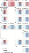
Accumulation of major labeled LCBs, LCB-Ps, and ceramides in the leaf after feeding LCB D7-d18:0. Arabidopsis leaf disks were incubated in 100 μM LCB D7-d18:0 and harvested at indicated time points. D7-labeled and non-labeled metabolites were detected by UPLC-MS/MS and relative amounts (in %) were calculated from concentrations of respective non-labeled and D7-labeled metabolites (presented in Figure 1). In case that respective non-labeled metabolites could not be detected, values for the respective limit of detection (LOD) were used for estimating the degree of labeling (light blue bars).
Figure 3
Figure 3
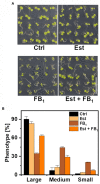
Effect of DPL1 overexpression on Fumonisin B1-induced growth inhibition in Arabidopsis seedlings. (A) Phenotypes of Arabidopsis line XVE-HA-DPL1 after 12 days growth on control medium (Ctrl), medium containing 10 μM β-estradiol (Est), 0.5 μM FB1, or a combination of both (Est + FB1). Scale bar indicates 1 cm. (B) Categorization of the seedlings of XVE-HA-DPL1, treated as depicted in (A), after 12 days of growth according to three groups: Large-, medium-, and small-sized plants. Relative frequencies of 49 seedlings per treatment were calculated. Values are means of two independent plates, with error bars indicating SD.
Figure 4
Figure 4
Effect of dihydrosphingosine-1-phosphate lyase 1 (DPL1) overexpression on Fumonisin B1-induced cell death and on LCB levels in Arabidopsis leaves. Leaf disks of Arabidopsis line XVE-HA-DPL1 were incubated in control solution (2% methanol), 10 μM β-estradiol (Est), 100 μM Fumonisin B1 (FB1), or a combination of both (Est + FB1). (A) Time-course analysis of cell death, measured as the conductivity of the solution at respective time points relative to the conductivity of the boiled sample at the end of the experiment, which was set to 100%. (B) Levels of LCBs d18:0, d18:0P, t18:0, and t18:0P in leaf disks at indicated time points. Values are means of four independent samples, each consisting of five leaf disks. Error bars indicate SD. Asterisks indicate significance levels <0.05. For cell death measurements, these levels were calculated by ANOVA for the factor “Est treatment,” with Est and FB1 treatment as fixed factors and the factor “time” as a covariate. For LCB concentrations, significance levels were calculated for each of the four LCBs in a general linear model for the factor “Est treatment,” with the factor “time” as a covariate.
Figure 5
Figure 5
Model depicting the initial capacity of sphingolipid LCB-converting enzymes. Simplified diagram of plant sphingolipid metabolism, highlighting major LCB-converting enzymatic pathways. The width of the arrows represents the capacity of respective enzymes. This capacity was estimated from levels of D7-labeled LCB d18:0 products measured at the earliest time point, 1 h after incubation. The respective values (nmol/g FW; Figure 1) are depicted on top of the arrows. Asterisks indicate those reactions requiring a previous enzymatic step, which leads to an underestimation of the capacity due to the time required for processing the metabolic precursor.

Similar articles

Cited by

References

    1. Abbas H. K., Tanaka T., Duke S. O., Porter J. K., Wray E. M., Hodges L., et al. . (1994). Fumonisin- and AAL-toxin-induced disruption of sphingolipid metabolism with accumulation of free sphingoid bases. Plant Physiol. 106, 1085–1093. doi: 10.1104/pp.106.3.1085, PMID: - DOI - PMC - PubMed
    1. Alden K. P., Dhondt-Cordelier S., McDonald K. L., Reape T. J., Ng C. K. Y., McCabe P. F., et al. . (2011). Sphingolipid long chain base phosphates can regulate apoptotic-like programmed cell death in plants. Biochem. Biophys. Res. Commun. 410, 574–580. doi: 10.1016/j.bbrc.2011.06.028, PMID: - DOI - PubMed
    1. Allen D. K. (2016). Quantifying plant phenotypes with isotopic labeling & metabolic flux analysis. Curr. Opin. Biotechnol. 37, 45–52. doi: 10.1016/j.copbio.2015.10.002, PMID: - DOI - PubMed
    1. Chen M., Cahoon E., Saucedo-García M., Plasencia J., Gavilanes-Ruiz M. (2009). “Plant Sphingolipids: Structure, Synthesis and Function,” in Lipids in Photosynthesis. Advances in Photosynthesis and Respiration. ed. Wada M. N. H. (Dordrecht: Springer; ), 77–115.
    1. Clough S. J., Bent A. F. (1998). Floral dip: a simplified method for Agrobacterium-mediated transformation of Arabidopsis thaliana. Plant J. 16, 735–743. doi: 10.1046/j.1365-313x.1998.00343.x, PMID: - DOI - PubMed

LinkOut - more resources