

The effect of AMP kinase activation on differentiation and maturation of osteoblast cultured on titanium plate
- PMID: 35784162
- PMCID: PMC9236888
- DOI: 10.1016/j.jds.2021.12.003
The effect of AMP kinase activation on differentiation and maturation of osteoblast cultured on titanium plate
Abstract
Background/purpose: 5' Adenosine monophosphate-activated protein kinase (AMPK) is known as an enzyme that maintains intracellular homeostasis and has various biological activity. The purpose of this study is evaluation effect of AMPK activation on implant prognosis.
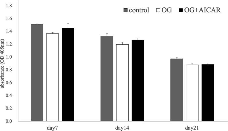
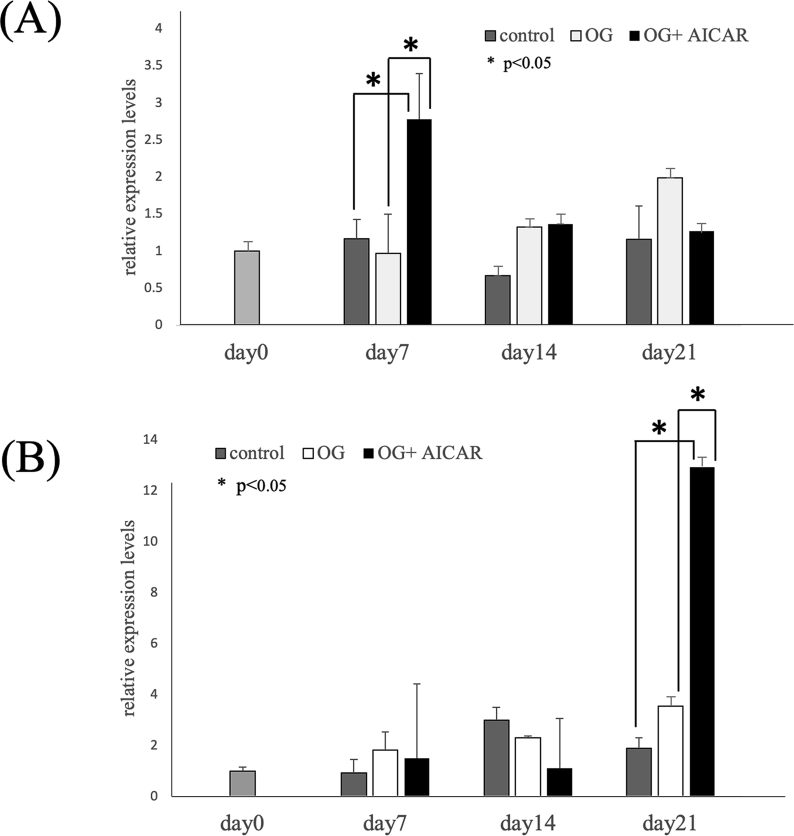
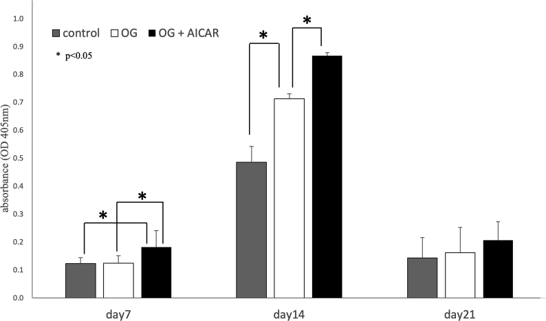
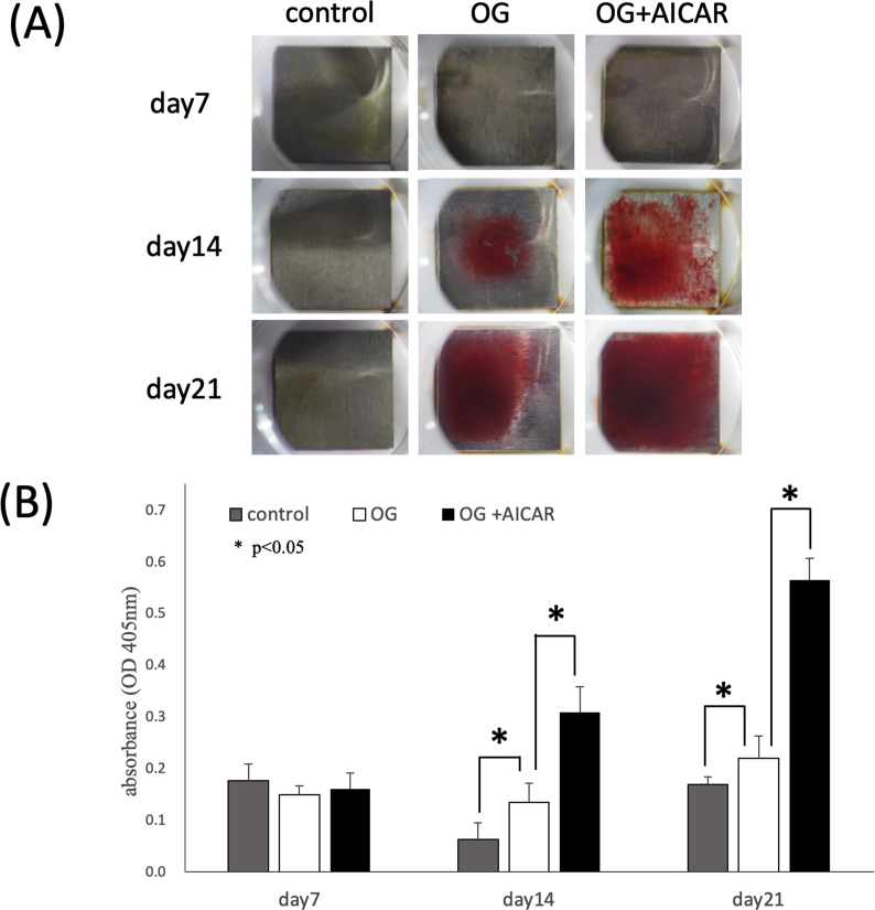
Materials & methods: MC3T3-E1 osteoblast-like cells were cultured on titanium using a 24-well plate. The experimental group was divided into the following 3 groups: (1) the normal culture group (control group), (2) the osteogenic induction group, and (3) the osteogenic induction + AMPK activation group. The cell counts were measured; real-time PCR was used to assess the expression of ALP and Osterix as osteogenic related genes at Day 0,7,14 and 21 after experiments. Additionally, ALP activity and calcification were assessed.
Results: The results of the real-time PCR assessments revealed that the expression of ALP, which is a marker for the initial stages of calcification, was significantly increased by AMPK activation compared to the normal culture or osteogenic induction. A significant increase was also observed in the expression of Osterix, which is a marker for the later stages of calcification. Because significant increases were observed in ALP activity and calcification potential, this suggested that AMPK activation could elicit an increase in osteoblast calcification potential.
Conclusion: AMPK activation promotes implant peripheral osteoblast differentiation and maturation and enhances calcification. Our results suggest that AMPK activation may help to maintain implant stability.
Keywords: 5′ adenosine monophosphate-activated protein kinase; Acadesine/AICA riboside; Dental implant; Osteoblast.
© 2021 Association for Dental Sciences of the Republic of China. Publishing services by Elsevier B.V.
Conflict of interest statement
The authors declare no conflict of interest in this study.
Figures
Similar articles
-
AMPK activation enhances osteoblast differentiation on a titanium disc via autophagy.Int J Implant Dent. 2024 Jan 29;10(1):2. doi: 10.1186/s40729-024-00525-2. Int J Implant Dent. 2024. PMID: 38286943 Free PMC article.
-
AMP-activated protein kinase stimulates osteoblast differentiation and mineralization through autophagy induction.Int J Mol Med. 2018 May;41(5):2535-2544. doi: 10.3892/ijmm.2018.3498. Epub 2018 Feb 16. Int J Mol Med. 2018. PMID: 29484369 Free PMC article.
-
Inhibition of adenosine monophosphate-activated protein kinase suppresses bone morphogenetic protein-2-induced mineralization of osteoblasts via Smad-independent mechanisms.Endocr J. 2018 Mar 28;65(3):291-298. doi: 10.1507/endocrj.EJ17-0229. Epub 2017 Dec 16. Endocr J. 2018. PMID: 29249772
-
AMP-activated protein kinase and the down-stream activated process of autophagy regulate the osteogenic differentiation of human dental follicle cells.Arch Oral Biol. 2021 Feb;122:104951. doi: 10.1016/j.archoralbio.2020.104951. Epub 2020 Nov 12. Arch Oral Biol. 2021. PMID: 33254047
-
Abnormal expression of miR-135b-5p in bone tissue of patients with osteoporosis and its role and mechanism in osteoporosis progression.Exp Ther Med. 2020 Feb;19(2):1042-1050. doi: 10.3892/etm.2019.8278. Epub 2019 Dec 4. Exp Ther Med. 2020. PMID: 32010267 Free PMC article.
Cited by
-
Influence of the Surface Topography of Titanium Dental Implants on the Behavior of Human Amniotic Stem Cells.Int J Mol Sci. 2024 Jul 6;25(13):7416. doi: 10.3390/ijms25137416. Int J Mol Sci. 2024. PMID: 39000523 Free PMC article.
-
AMPK activation enhances osteoblast differentiation on a titanium disc via autophagy.Int J Implant Dent. 2024 Jan 29;10(1):2. doi: 10.1186/s40729-024-00525-2. Int J Implant Dent. 2024. PMID: 38286943 Free PMC article.
References
-
- Brånemark P.I., Breine U., Adell R., Hansson B.O., Lindstrom J., Ohlsson A. Intra-ossous anchorage of dental prostheses. Scand J Plast ReConstr Surg Hand Surg. 1969;3:81–100. - PubMed
-
- Martin J.Y., Schwartz Z., Hummert T.W., et al. Effect of titanium surface roughness on proliferation, differentiation, and protein synthesis of human osteoblast-like cells (MG63) J Biomed Mater Res. 1995;29:389–401. - PubMed
-
- Shalabi M.M., Gortemaker A., Van't Hof M.A., Jansen J.A., Creugers N.H. Implant surface roughness and bone healing: a systematic review. J Dent Res. 2006;85:496–500. - PubMed
-
- Wennerberg A., Albrektsson T. On implant surfaces: a review of current knowledge and opinions. Int J Oral Maxillofac Implants. 2010;25:63–74. - PubMed
-
- DohanEhrenfest D.M., Vazquez L., Park Y.J., Sammartino G., Bernard J.P. Identification card and codification of the chemical and morphological characteristics of 14 dental implant surfaces. J Oral Implantol. 2011;37:525–542. - PubMed
LinkOut - more resources
Full Text Sources
