. 2022 Jun 16:13:871558.
doi: 10.3389/fimmu.2022.871558. eCollection 2022.

HBx Mediated Increase of DDX17 Contributes to HBV-Related Hepatocellular Carcinoma Tumorigenesis

Affiliations

HBx Mediated Increase of DDX17 Contributes to HBV-Related Hepatocellular Carcinoma Tumorigenesis

Mei-Ling Dong et al. Front Immunol. .

Abstract

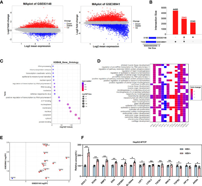
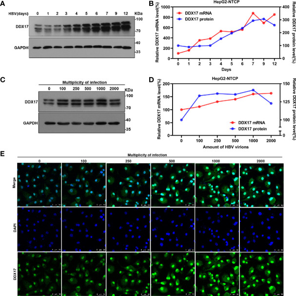
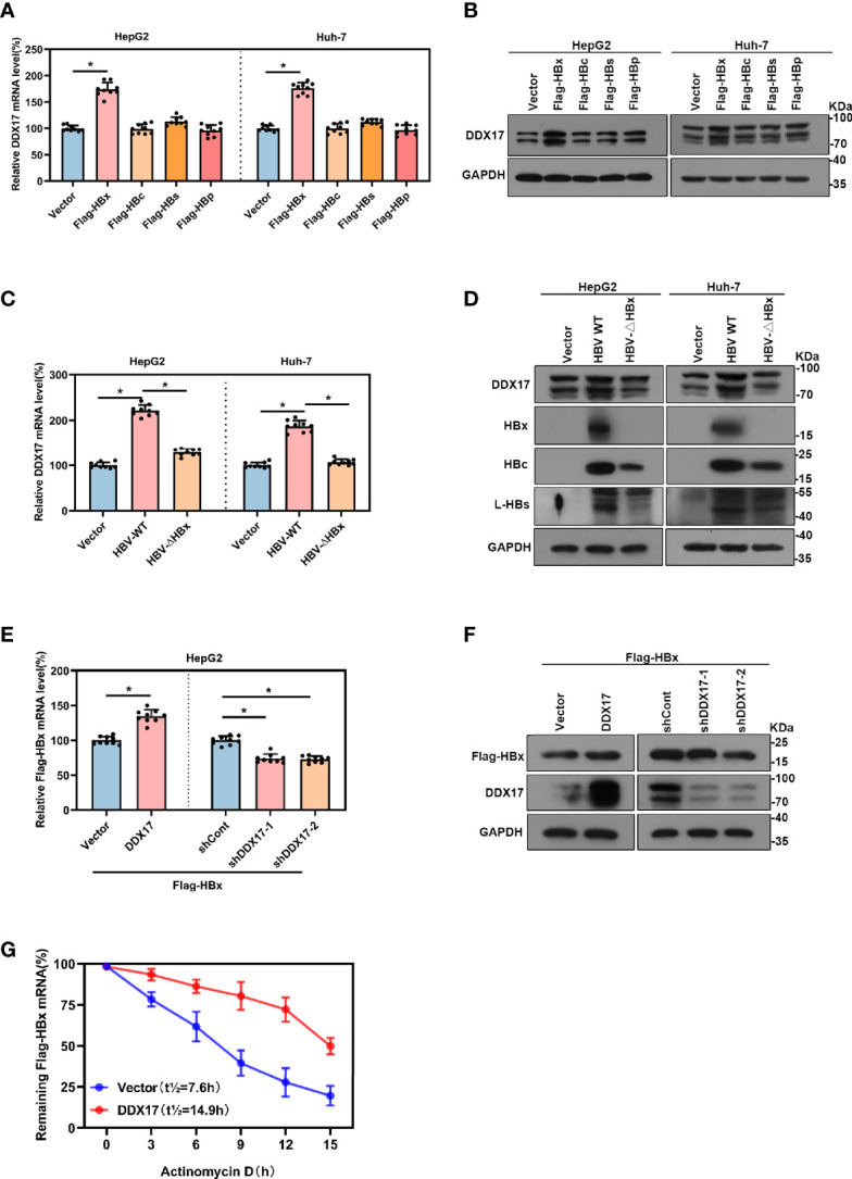
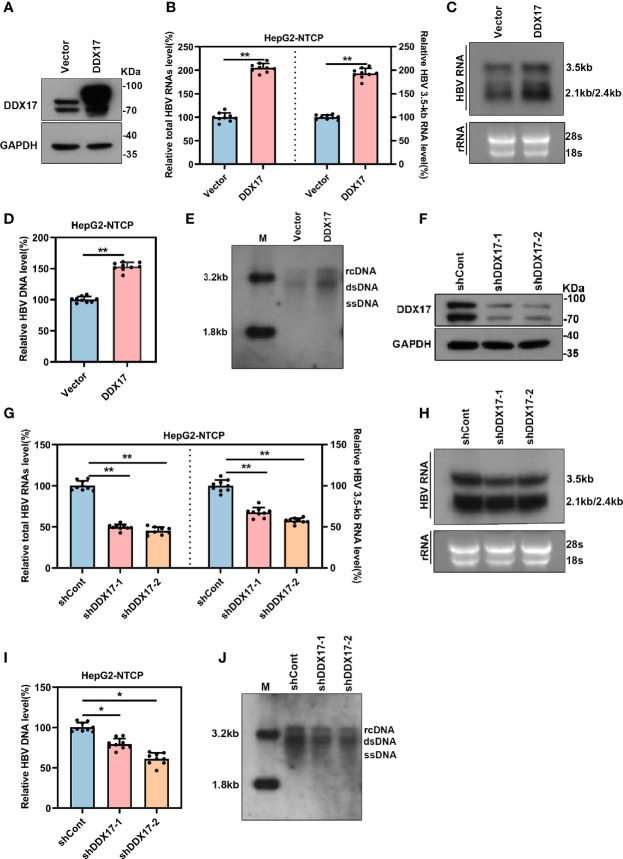
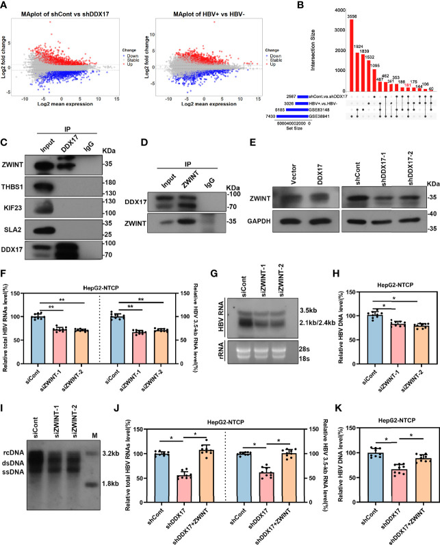
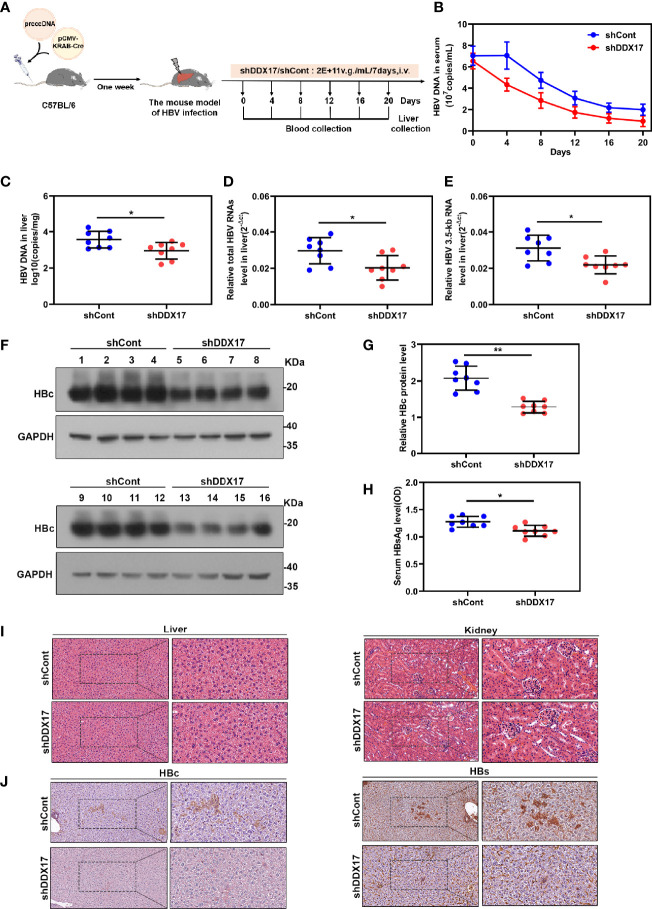
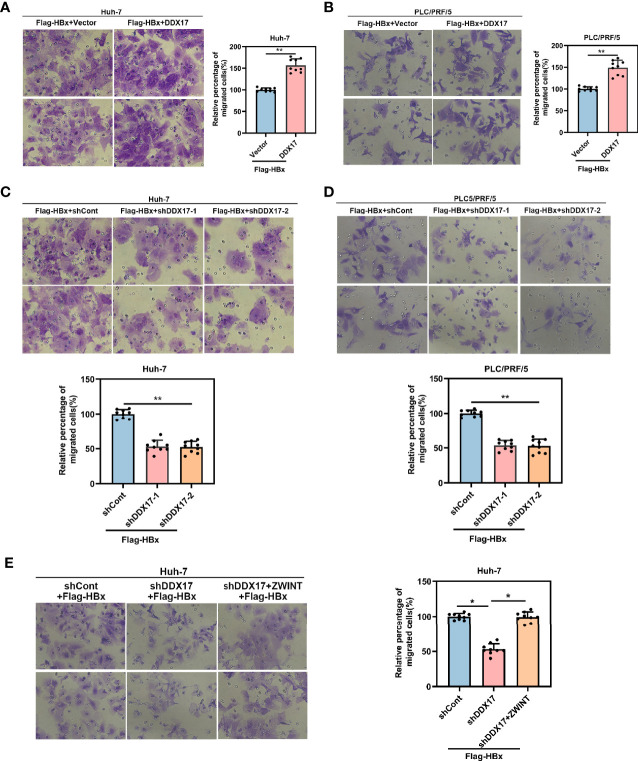
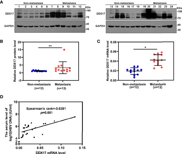
HBV is strongly associated with HCC development and DEAD-box RNA helicase 17 (DDX17) is a very important member of the DEAD box family that plays key roles in HCC development by promoting cancer metastasis. However, the important role of DDX17 in the pathogenesis of HBV-related HCC remains unclear. In this study, we investigated the role of DDX17 in the replication of HBV and the development of HBV-associated HCC. Based on data from the GEO database and HBV-infected cells, we found that DDX17 was upregulated by the HBV viral protein X (HBx). Mechanistically, increased DDX17 expression promoted HBV replication and transcription by upregulating ZWINT. Further study showed that DDX17 could promote HBx-mediated HCC metastasis. Finally, the promotive effect of DDX17 on HBV and HBV-related HCC was confirmed in vivo. In summary, the results revealed the novel role of DDX17 in the replication of HBV and the metastasis of HBV-associated HCC.

Keywords: DDX17; HBx; HCC; ZWINT; metastasis.


Conflict of interest statement

The authors declare that the research was conducted in the absence of any commercial or financial relationships that could be construed as a potential conflict of interest.

Figures

Figure 1
Figure 1
The effect of HBV on DDX17 expression were analyzed in GEO database. (A–E) Dataset GSE83148 and GSE38941 were download from GEO database and subjected to bioinformatics analysis. (A) Volcano plots show the potential differentially expressed genes in GSE83148 and GSE38941(log2FC>0.585, adjusted p < 0.05). (B) The Venn diagrams reveal the differentially expressed genes and the co-expressed genes among the 2 cohorts (fold change value >2). (C) Functional enrichment analysis shows the co-up-regulated and co-down-regulated genes. (D) Signaling pathway analysis for 14 candidate genes. (E) Co-expression of 14 genes in GSE83148 and GSE838941. (F) Real-time PCR analysis for the 13 genes expression levels in HBV-infected HepG2-NTCP cells. β-actin was used as an internal quantitative control. ns means no significance, *P < 0.05, **P < 0.01.
Figure 2
Figure 2
The effect of HBV on DDX17 expression were analyzed in HBV-infected HepG2-NTCP cells. (A, B) HepG2-NTCP cells were infected with HBV (1, 000 genome equivalents/cell) and harvested at indicated days. Real time PCR and western blot measured the mRNA and protein level of DDX17, respectively. (C, D) HepG2-NTCP cells were infected with series dose of HBV particle and harvested at 5 days post-infection. Real time PCR and western blot measured the mRNA and protein level of DDX17, respectively. (E) Endogenous DDX17 level in HepG2-NTCP cells which infected with series doses of HBV particles was determined by immunofluorescence staining.
Figure 3
Figure 3
There was a positive correlation between Viral protein HBx and DDX17. (A, B) The vector, Flag-HBx, Flag-HBc, Flag-HBs or Flag-HBp plasmids were transfected into HepG2 cells and Huh-7 cells. Real-time PCR and Western blot examined the mRNA and protein levels of DDX17. β-actin and GAPDH were used as the internal quantitative controls respectively. (C, D) The vector, HBV WT or HBV-ΔHBx plasmids were transfected into HepG2 and Huh-7 cells. Real-time PCR examined the mRNA evels of DDX17 and Western blot examined the protein levels of DDX17, HBx, HBc and HBs. β-actin and GAPDH were used as the internal quantitative controls, respectively. (E, F) The vector or DDX17 plasmids were co-transfected with Flag-HBx into the HepG2 cells; the short hairpin RNAs targeting DDX17 (shDDX17-1 and shDDX17-2) or nontargeting shRNA (shCont) were co-transfected with Flag-HBx into the HepG2 cells. Real-time PCR and Western blot examined the mRNA and protein levels of Flag-HBx. (G) DDX17 overexpression in HepG2 cells followed by treatment with 5ug/mL actinomycin D at the indicated times. β-actin and GAPDH were used as the internal quantitative controls, respectively. *P < 0.05.
Figure 4
Figure 4
The expression of DDX17 regulates HBV transcription and replication. The vector or DDX17 plasmids and lentivirus expressing shRNA targeting DDX17 or shCont transfected into HepG2-NTCP cells that were infected with 1,000 genome equivalents/cell of HBV particles in the presence of 4% PEG8000 for 24 h. Total HBV RNAs, 3.5-kb RNA, and HBV DNA were analyzed at day 5 after plasmid transduction. (A) The expression of DDX17 after transfected with plasmid expressing DDX17 and empty vector in HBV-infected HepG2-NTCP cells were access by using Western blot assay. GAPDH was used as the internal quantitative control. (B) Real-time PCR was used to analyze the levels of total HBV RNAs and 3.5-kb RNA. β-actin was used as the internal quantitative control. (C) Northern blot assay hybridization of HBV RNAs by using DIG-labeled plus- or minus-strand-specific HBV riboprobe with a length of 1000 bp. Ribosomal RNAs (28S and 18S) were used as loading control. (D, E) Real-time PCR and Southern blot assay examined the levels of HBV DNA replicative intermediates. (F) The expression of DDX17 after transfected with negative control (shCont) or shRNA (shDDX17-1 and shDDX17-2) in HBV-infected HepG2-NTCP cells were accessed using Western blot assay. GAPDH was used as the internal quantitative control. (G, H) Real-time PCR and Northern blot assay were used to analyze the effect of DDX17 knockdown on HBV RNAs in HBV-infected HepG2-NTCP cells. (I, J) Real-time PCR and Southern blot assay were used to analysis the effect of DDX17 knockdown on HBV DNA replicative intermediates in HBV-infected HepG2-NTCP cells. *P < 0.05, **P < 0.01.
Figure 5
Figure 5
DDX17 promotes HBV transcription and replication by upregulating ZWINT. (A) Volcano plot for potential DEGs in two RNA-seq datasets (HBV-infected HepG2-NTCP cells with or without DDX17 knockout; liver tissue from HCC patients with or without HBV infection) (log2FC>0.585, p value<0.05). (B) The Venn diagram shows 181 co-expressed genes were identified among GSE83148, GSE38941 and two RNA-seq data. (C) The co-immunoprecitation assay (Co-IP) analysis showing the interactions between DDX17 and its candidate interacting proteins. (D) Co-IP assay was performed with anti-DDX17 antibody in HBV-infected HepG2-NTCP cells transfected plasmid expressing ZWINT. (E) DDX17 regulated ZWINT protein expression in HBV-infected HepG2-NTCP cells measured by western blot assay. GAPDH was used as an internal quantitative control. (F, G) Real-time PCR and Northern blot assay were used to analysis the effect of ZWINT knockdown on HBV RNAs in HBV-infected HepG2-NTCP cells. (H, I) Real-time PCR and Southern blot assay were used to analysis the effect of ZWINT knockdown on HBV DNA replicative intermediates in HBV-infected HepG2-NTCP cells. (J, K) Reintroduction of ZWINT antagonized the effect of DDX17 knockdown on HBV transcription and replication. Total HBV RNAs, 3.5-kb RNA (J) and HBV DNA replicative intermediates (K) were extracted and examined by real-time PCR. *P < 0.05, **P < 0.01.
Figure 6
Figure 6
DDX17 promotes HBV transcription and replication in vivo. (A) Flow chart explaining the way and concentration of AAV-shDDX17 administration as well as the intervals of vein blood collection. The mouse model established by injecting prcccDNA and pCMV-KRAB-Cre from tail vein. The mouse model of HBV infection involving HBV recombinant (r) cccDNA constructed successfully were randomly assigned to 2 groups (n = 8 per group). (B) Real-time PCR was subjected to detect the serum level of HBV DNA. (C) Real-time PCR was subjected to detect the level of HBV DNA in liver tissue. (D, E) Real-time PCR was subjected to detect the total HBV RNAs and 3.5-kb RNA levels in liver tissue, β-actin was used as the internal quantitative control. (F, G) The levels of HBc protein were measured by Western blot assay. (H) ELISA assay was subjected to determine the level of HBsAg in serum during the treatment. (I) Abnormal pathological changes in the two groups were determined by HE staining. (J) The expression of HBs and HBc in liver tissues were analyzed by immunohistochemistry. *P < 0.05, **P < 0.01.
Figure 7
Figure 7
DDX17 could facilitate HBx-mediated HCC migration. (A, B) Migratory ability was assessed by transwell assays in Huh-7 and PLC/PRF/5 cells with DDX17 overexpression. (C, D) Migratory ability was assessed by transwell assays in Huh-7 and PLC/PRF/5 cells with DDX17 knockout. (E) The effects of decreased DDX17 meanwhile upregulation of ZWINT in Huh-7 cells for migration analyzed by transwell assays. *P < 0.05, **P < 0.01.
Figure 8
Figure 8
DDX17 play an important role in HBV-related HCC metastasis. (A, B) Western blot assay was subjected to examined the protein level of DDX17 in HBV-related HCC tissue samples, GAPDH was used as internal quantitative controls. (C) Real-time PCR was subjected to examined the mRNA level of DDX17 in HBV-related HCC tissue samples, β-actin was used as the internal quantitative controls. (D) Correlation between the serum HBV DNA levels and the liver DDX17 levels in HBV-related HCC patients. HBV DNA levels was log10-transformed. Correlation coefficients (r) and two-tailed P values were evaluated by Pearson’s test. *P < 0.05, **P < 0.01.

Similar articles

Cited by

References

    1. Chu PY, Tung SL, Tsai KW, Shen FP, Chan SH. Identification of the Novel Oncogenic Role of SAAL1 and Its Therapeutic Potential in Hepatocellular Carcinoma. Cancers (Basel) (2020) 12(7):1843. doi: 10.3390/cancers12071843 - DOI - PMC - PubMed
    1. Ghavimi S, Apfel T, Azimi H, Persaud A, Pyrsopoulos NT. Management and Treatment of Hepatocellular Carcinoma With Immunotherapy: A Review of Current and Future Options. J Clin Transl Hepatol (2020) 8(2):168–76. doi: 10.14218/JCTH.2020.00001 - DOI - PMC - PubMed
    1. Renzulli M, Brandi N, Argalia G, Brocchi S, Farolfi A, Fanti S, et al. . Morphological, Dynamic and Functional Characteristics of Liver Pseudolesions and Benign Lesions. Radiol Med (2022) 127(2):129–44. doi: 10.1007/s11547-022-01449-w - DOI - PubMed
    1. Seo W, Gao Y, He Y, Sun J, Xu H, Feng D, et al. . ALDH2 Deficiency Promotes Alcohol-Associated Liver Cancer by Activating Oncogenic Pathways via Oxidized DNA-Enriched Extracellular Vesicles. J Hepatol (2019) 71(5):1000–11. doi: 10.1016/j.jhep.2019.06.018 - DOI - PMC - PubMed
    1. Arfianti A, Pok S, Barn V, Haigh WG, Yeh MM, Ioannou GN, et al. . Exercise Retards Hepatocarcinogenesis in Obese Mice Independently of Weight Control. J Hepatol (2020) 73(1):140–8. doi: 10.1016/j.jhep.2020.02.006 - DOI - PubMed

Publication types

Substances