. 2022 Jun 21:20:418-433.
doi: 10.1016/j.bioactmat.2022.06.010. eCollection 2023 Feb.

Dental plaque-inspired versatile nanosystem for caries prevention and tooth restoration

Affiliations

Dental plaque-inspired versatile nanosystem for caries prevention and tooth restoration

Yue Xu et al. Bioact Mater. .

Abstract

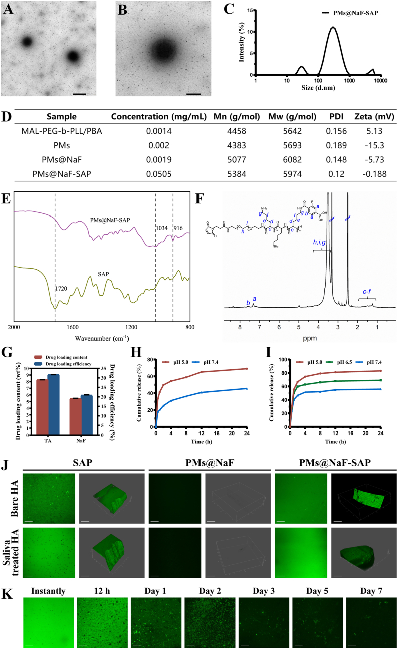
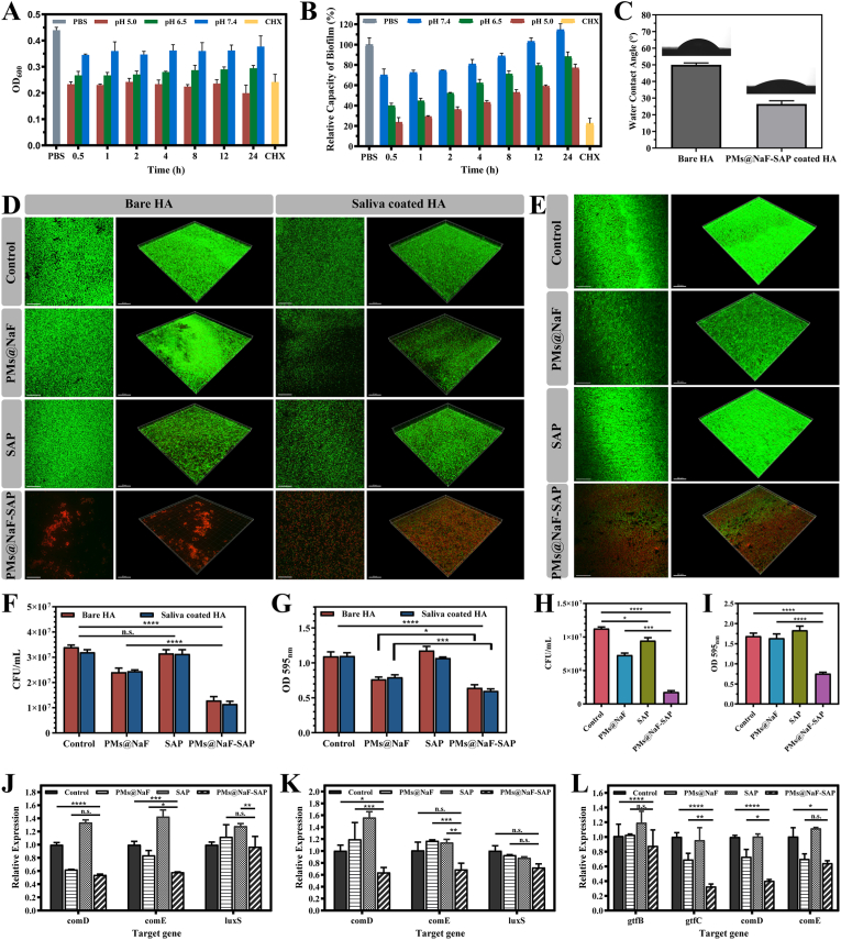
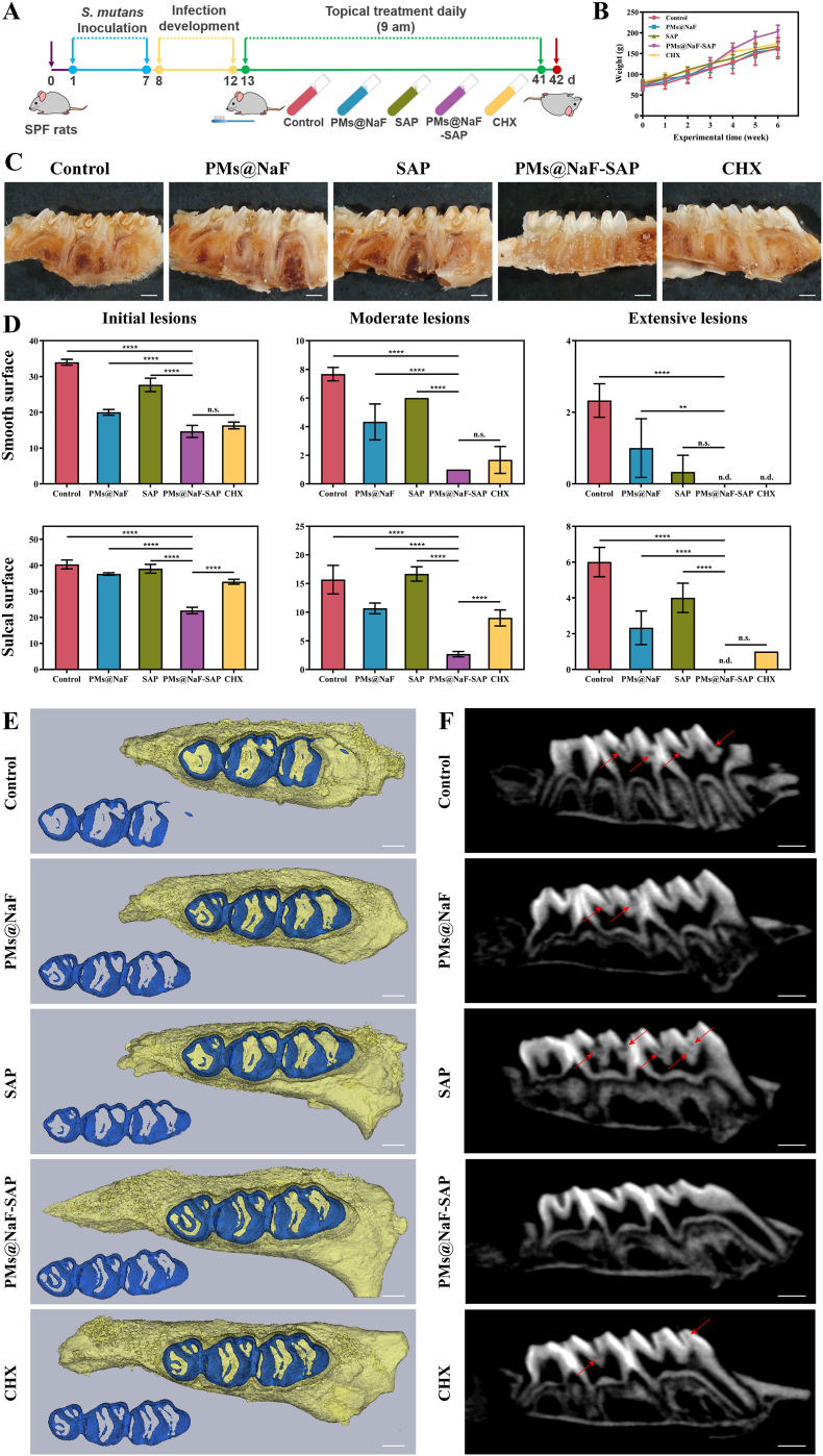
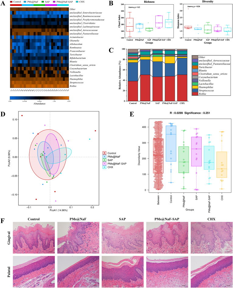
Dental caries is one of the most prevalent human diseases resulting from tooth demineralization caused by acid production of bacteria plaque. It remains challenges for current practice to specifically identify, intervene and interrupt the development of caries while restoring defects. In this study, inspired by natural dental plaque, a stimuli-responsive multidrug delivery system (PMs@NaF-SAP) has been developed to prevent tooth decay and promote enamel restoration. Classic spherical core-shell structures of micelles dual-loaded with antibacterial and restorative agents are self-assembled into bacteria-responsive multidrug delivery system based on the pH-cleavable boronate ester bond, followed by conjugation with salivary-acquired peptide (SAP) to endow the nanoparticle with strong adhesion to tooth enamel. The constructed PMs@NaF-SAP specifically adheres to tooth, identifies cariogenic conditions and intelligently releases drugs at acidic pH, thereby providing antibacterial adhesion and cariogenic biofilm resistance, and restoring the microarchitecture and mechanical properties of demineralized teeth. Topical treatment with PMs@NaF-SAP effectively diminishes the onset and severity of caries without impacting oral microbiota diversity or surrounding mucosal tissues. These findings demonstrate this novel nanotherapy has potential as a promising biomedical application for caries prevention and tooth defect restoration while resisting biofilm-associated diseases in a controlled manner activated by pathological bacteria.

Keywords: Biofilms; Dental caries; Micelles; Peptide; Stimuli responsive polymers; Tooth remineralization.


Conflict of interest statement

The authors declare that they have no known competing financial interests or personal relationships that could have appeared to influence the work reported in this paper.

Figures

Image 1
Graphical abstract
Fig. 1
Fig. 1
Proposed concept of the bacteria-responsive micellar multidrug delivery system (PMs@NaF-SAP) targeting cariogenic biofilm. (A) Illustration of formulation and the bacteria-responsive activity of PMs@NaF. (B) The modification of SAP and the formulation of PMs@NaF-SAP. (C) Schematic illustration of topical application of PMs@NaF-SAP on dental plaque biofilm and proposed mechanism for caries prevention and enamel restoration.
Fig. 2
Fig. 2
Characterization of PMs@NaF-SAP and its binding to the enamel surface. (A) and (B) TEM images of PMs@NaF-SAP (scale bar for A: 500 nm, B: 200 nm). (C) Size distribution of PMs@NaF-SAP determined by DLS. (D) Concentrations, molecular weights (Mn and Mw), PDI and zeta potentials of stage products. (E) FT-IR spectra of SAP and PMs@NaF-SAP. (F) 1H NMR of MAL-PEG-b-PLL/PBA in DMSO-d6. (G) Drug loading content (%) and efficiency (%) from PMs@NaF-SAP, the results were reported as mean ± s.d (n = 3). Cumulative in vitro release of (H) TA and (I) NaF from PMs@NaF-SAP at different pH values. (J) Adsorption capacity assay and (K) adsorption time assay of PMs@NaF-SAP when monitoring the oral salivary environment (scale bar: 50 μm).
Fig. 3
Fig. 3
Antibacterial adhesion abilities and cariogenic biofilm resistance of topical PMs@NaF-SAP treatment in vitro. (A) OD600 value of S. mutans and (B) relative capacity of biofilm (%) as a function of incubation with PMs@NaF-SAP at pH 7.4, 6.5 or 5.0, with CHX as positive control and PBS as blank control. (C) Water contact angle (°) of bare HA and PMs@NaF-SAP coated HA. (D) Adhesion of S. mutans on HA and sHA discs evaluated by CLSM (live cells are labeled in green by SYTO 9 and dead cells in red by propidium iodide, scale bar: 50 μm), (F) CFU counting and (G) crystal violet staining after pre-treated with sterile PBS (control), PMs@NaF, SAP or PMs@NaF-SAP solutions. (E) CLSM images (scale bar: 50 μm), (H) CFU counting and (I) crystal violet staining evaluating the penetration and antibiofilm activity of sterile PBS (control), PMs@NaF, SAP or PMs@NaF-SAP solution treating on the pre-established S. mutans biofilm. Relative expressions of genes related to S.mutans (J) on HA or (K) sHA when treated with sterile PBS (control), PMs@NaF, SAP or PMs@NaF-SAP in antibacterial adhesion assay and (L) in cariogenic biofilm resistance assay, respectively. Each experiment was performed in triplicate, and the data were presented as mean ± s.d. (n = 3). The quantitative data were subjected to Student's t-test for a pairwise comparison. *p < 0.5, **p < 0.01, ***p < 0.001, ****p < 0.0001.
Fig. 4
Fig. 4
Demineralization and remineralization effects, mechanical recovery properties and cytotoxicity of PMs@NaF-SAP. (A) FE-SEM micrographs of the demineralization and remineralization surfaces in sterile PBS (control), PMs@NaF, SAP or PMs@NaF-SAP and normal enamel (scale bar: 1 μm). (B) Amount of Ca/P loss from enamel surfaces in demineralization solutions measured by ICP-AES after sterile PBS (control), PMs@NaF, SAP or PMs@NaF-SAP coating. The red bar denotes Ca loss and the blue bar indicates P loss. (C) Amount of Ca/P gain of enamel surfaces after 24 h in remineralization solutions by ICP-AES after sterile PBS (control), PMs@NaF, SAP or PMs@NaF-SAP coating. The red bar indicates the Ca gain and the blue bar denotes the P gain. (D) The XRD spectra for the mineral phase evolution on the demineralization or remineralization enamel window: the normal enamel (line a), sterile PBS coated (line b), PMs@NaF coated (line c), SAP coated (line d) and PMs@NaF-SAP coated (line e); (E) AFM images (scale bar: 4 μm) and (F) Ra of demineralization or remineralization surfaces in sterile PBS (control), PMs@NaF, SAP or PMs@NaF-SAP. (G) The % SHL of the four groups after demineralization. (H) The % SMHRR of the four groups after remineralization. (I) Cell viability of MC3T3 cells at various dilutions of medium containing PMs@NaF-SAP or CHX by CCK-8 assay. (J) Cytoskeletion staining and fluorescence quantitative analysis of MC 3T3 cells at various dilutions of medium containing PMs@NaF-SAP or CHX, with cells untreated as the blank control (scale bar: 100 μm). Each experiment was performed in triplicate, and the data were presented as mean ± s.d. (n = 3). The quantitative data were subjected to Student's t-test for a pairwise comparison. *p < 0.5, **p < 0.01, ***p < 0.001, ****p < 0.0001.
Fig. 5
Fig. 5
Therapeutic efficacy of topical PMs@NaF-SAP treatment against tooth decay in vivo as evaluated by Keyes' scoring and micro-CT analysis. In this model, tooth enamel progressively develops caries lesions (similar to those observed in humans), proceeding from initial lesions of demineralization to moderate lesions and on to extensive lesions characterized by enamel structure damage and cavitation. (A) Experimental design and treatment regimen of the animal model, the treatments (control, PMs@NaF, SAP, PMs@NaF-SAP and CHX) are represented in the centrifuge tubes. (B) Body weights of rats during the experimental period. (C) Images of rat teeth after treatment of the five groups (scale bar: 1 mm). (D) Caries onset and severity of smooth and sulcal surfaces. Caries scores are recorded as stages and extent of carious lesion severity according to Larson's modification of Keyes' scoring system. Data were presented as the mean ± s.d. (n = 6); ordinary one-way ANOVA, p value < 0.01, 0.001 and 0.0001 are indicated by **, *** and ****, respectively; n.s. nonsignificant; n.d. nondetectable. (E) Three-dimensional reconstruction of micro-CT images of maxillary molars in five groups, separated enamel (blue) by setting the density threshold above 4500 Hounsfield units (scale bar: 1 mm). (F) 2D scale sagittal images of the maxillary molars analyzed by micro-CT (red arrows, caries lesion sites, scale bar: 1 mm).
Fig. 6
Fig. 6
Effects of topical PMs@NaF-SAP on oral microflora and soft tissue in vivo after 28 days of treatment (n = 6 per group). (A) The heatmap indicated main bacterial genera found across all samples according to treatment groups. (B) Chao 1 richness index and Shannon α-diversity index. The whiskers (bars from the box) represent the upper and lower quartiles of the data. (C) Bacteria relative abundances within rodent body sites for each treatment group. (D) Weighted Unifrac PCoA, which indicated the β-diversity in each group; the analysis revealed that the PMs@NaF-SAP group has similar composition between samples. (E) Bray-Curtis ANOSIM test had R value close to 0 and p value > 0.05, which indicated that there were no significant differences between and within groups based on their Bray–Curtis distances. (F) Histopathology of gingival and palatal tissues in rats treated with PMs@NaF-SAP was similar to control (scale bar: 50 μm).

References

    1. Bernabe E., Marcenes W., Hernandez C.R., Bailey J., Abreu L.G., Alipour V., Amini S., Arabloo J., Arefi Z., Arora A., Ayanore M.A., Bärnighausen T.W., Bijani A., Cho D.Y., Chu D.T., Crowe C.S., Demoz G.T., Demsie D.G., Dibaji Forooshani Z.S., Du M., El Tantawi M., Fischer F., Folayan M.O., Futran N.D., Geramo Y.C.D., Haj-Mirzaian A., Hariyani N., Hasanzadeh A., Hassanipour S., Hay S.I., Hole M.K., Hostiuc S., Ilic M.D., James S.L., Kalhor R., Kemmer L., Keramati M., Khader Y.S., Kisa S., Kisa A., Koyanagi A., Lalloo R., Le Nguyen Q., London S.D., Manohar N.D., Massenburg B.B., Mathur M.R., Meles H.G., Mestrovic T., Mohammadian-Hafshejani A., Mohammadpourhodki R., Mokdad A.H., Morrison S.D., Nazari J., Nguyen T.H., Nguyen C.T., Nixon M.R., Olagunju T.O., Pakshir K., Pathak M., Rabiee N., Rafiei A., Ramezanzadeh K., Rios-Blancas M.J., Roro E.M., Sabour S., Samy A.M., Sawhney M., Schwendicke F., Shaahmadi F., Shaikh M.A., Stein C., Tovani-Palone M.R., Tran B.X., Unnikrishnan B., Vu G.T., Vukovic A., Warouw T.S.S., Zaidi Z., Zhang Z.J., Kassebaum N.J. Global, regional, and national levels and trends in burden of oral conditions from 1990 to 2017: a systematic analysis for the global burden of disease 2017 study. J. Dent. Res. 2020;99:362–373. doi: 10.1177/0022034520908533. - DOI - PMC - PubMed
    1. Kassebaum N.J., Smith A.G.C., Bernabé E., Fleming T.D., Reynolds A.E., Vos T., Murray C.J.L., Marcenes W. Global, regional, and national prevalence, incidence, and disability-adjusted life years for oral conditions for 195 countries, 1990-2015: a systematic analysis for the global burden of diseases, injuries, and risk factors. J. Dent. Res. 2017;96:380–387. doi: 10.1177/0022034517693566. - DOI - PMC - PubMed
    1. G.D.a.I.I.a.P. Collaborators Global, regional, and national incidence, prevalence, and years lived with disability for 310 diseases and injuries 1990-2015: a systematic analysis for the Global Burden of Disease Study 2015. Lancet. 2016;388:1545–1602. doi: 10.1016/S0140-6736(16)31678-6. - DOI - PMC - PubMed
    1. Petersen P.E., Bourgeois D., Ogawa H., Estupinan-Day S., Ndiaye C. The global burden of oral diseases and risks to oral health. Bull. World Health Organ. 2005;83:661–669. - PMC - PubMed
    1. Kassebaum N.J., Bernabé E., Dahiya M., Bhandari B., Murray C.J.L., Marcenes W. Global burden of untreated caries: a systematic review and metaregression. J. Dent. Res. 2015;94:650–658. doi: 10.1177/0022034515573272. - DOI - PubMed