. 2022 Jun 15:13:867477.
doi: 10.3389/fphar.2022.867477. eCollection 2022.

Chinese Medicine, Succinum, Ameliorates Cognitive Impairment of Carotid Artery Ligation Rats and Inhibits Apoptosis of HT22 Hippocampal Cells via Regulation of the GSK3β/β-Catenin Pathway

Affiliations

Chinese Medicine, Succinum, Ameliorates Cognitive Impairment of Carotid Artery Ligation Rats and Inhibits Apoptosis of HT22 Hippocampal Cells via Regulation of the GSK3β/β-Catenin Pathway

Chongqi Wei et al. Front Pharmacol. .

Abstract

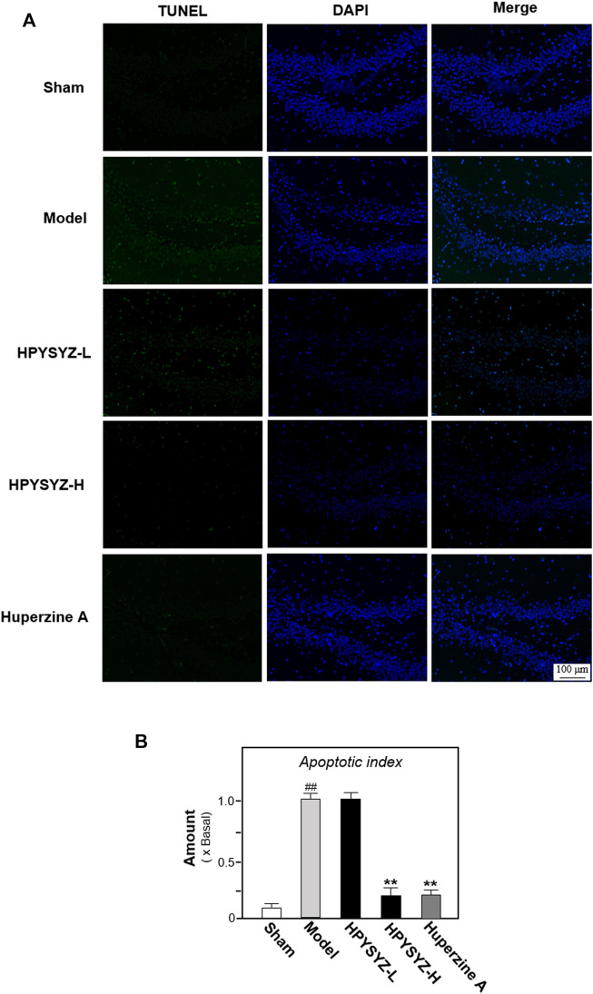
Succinum is an organic mineral formed from the resin of ancient coniferous and leguminous plants, which is applied for tranquilizing mood, promoting blood circulation, and removing blood stasis in Chinese medicine. For quite a long time, the modern research of succinum mainly focuses on the study of physical and chemical properties and authenticity identification while few reports on its medicinal mechanism. In current study, we evaluated different solvent extracts of succinum on carotid artery ligation rats mimicking vascular dementia. It was found that ethyl acetate extracts of succinum significantly improved the learning and memory abilities of model rats and inhibited neuronal apoptosis in the hippocampus. On a mice hippocampal neuronal cell line (HT22), ethyl acetate extracts of succinum also exerted better action trend in inhibiting cell apoptosis induced by oxygen glucose deprivation (OGD). By using XAV-939 on both in vivo and in vitro studies, it was found that ethyl acetate extracts of succinum might exert these functions by regulating the GSK3β/β-catenin pathway. These studies revealed the neuronal function of succinum, which explained the traditional effects of succinum and provided more modern scientific basis for its clinical application.

Keywords: Wnt/GSK3β; apoptosis; mineral medicine; succinum; vascular dementia.


Conflict of interest statement

The authors declare that the research was conducted in the absence of any commercial or financial relationships that could be construed as a potential conflict of interest.

Figures

FIGURE 1
FIGURE 1
Extracts of succinum ameliorated learning and memory impairment on carotid artery ligation rats. The effect of succinum extracts on the latency time, platform crossing number, and average speed of carotid artery ligation rats. Values are expressed as mean ± SEM (n = 8). Comparisons between groups were carried out by a one-way ANOVA followed by a post hoc Bonferroni test. ## p < 0.01 (compared with the sham control group), *p < 0.05, and **p < 0.01 (compared with the carotid artery ligation model group).
FIGURE 2
FIGURE 2
Ethyl acetate extracts of succinum reduced the apoptosis in the hippocampus of carotid artery ligation rats. (A) Apoptosis of the hippocampal dentate gyrus of rats in the sham control group, model group, low dose and high dose treatment of ethyl acetate extracts of succinum groups, and positive control huperzine A group were determined by TUNEL assays. (B) Percentage of TUNEL staining positive cells to DAPI-staining positive cells was calculated for apoptosis evaluation. The low dose treatment of ethyl acetate extracts of succinum (HPYSYZ-L) was set as 0.21 g/kg/d, and the high dose treatment (HPYSYZ-H) was set as 0.42 g/kg/d (n = 3). The analysis was performed from two brain sections of one rat and three rats from one group (mean ± SD, *p < 0.05, and **p < 0.01).
FIGURE 3
FIGURE 3
Ethyl acetate extracts of succinum regulated the caspase-dependent pathway in the hippocampus of carotid artery ligation rats. (A) Effects of ethyl acetate extracts of succinum at low dosage (HPYSYZ-L) and high dosage (HPYSYZ-H) on the expressions of caspase-3, cleaved-caspase-3, and Bax and Bcl-2 of hippocampus of carotid artery ligation rats were evaluated by Western blotting analysis. The representative images are displayed (n = 5). (B) Expression of caspase-3 was analyzed and the expression of β-actin was applied for normalization. Expression of cleave-caspase-3 was analyzed and the expression of β-actin was applied for normalization. (D) Expressions of Bax and Bcl-2 were analyzed and the expression of β-actin was also used for normalization. The expression ratio of Bcl-2 to Bax was calculated. Values are expressed as the percentage of the control group (no drug treatment normal mice) as mean ± SEM (n = 5). Comparisons between groups were analyzed by a one-way ANOVA followed by a post hoc Bonferroni test. ## p < 0.01 (compared with control group), *p < 0.05, and **p < 0.05 (compared with the model group).
FIGURE 4
FIGURE 4
Ethyl acetate extracts of succinum regulated the expression of neuronal marker MAP2 in the hippocampus on carotid artery ligation rats. (A) Expressions of neuronal cell marker MAP2 were determined in the hippocampus of carotid artery ligation rats by Western blotting analysis. Values are expressed as mean ± SEM (n = 5). Comparisons between groups were carried out by a one-way ANOVA followed by a post hoc Bonferroni test. ## p < 0.01 (compared with the normal control group), * p < 0.05, and ** p < 0.01 (compared with the carotid artery ligation model group). (B) Expressions of neuronal cell marker MAP2 were determined in the hippocampus of carotid artery ligation rats by immunohistochemistry analysis. The low dose treatment of ethyl acetate extracts of succinum (HPYSYZ-L) was set as 0.21 g/kg/d, and the high dose treatment (HPYSYZ-H) was set as 0.42 g/kg/d (n = 3).
FIGURE 5
FIGURE 5
Ethyl acetate extracts of succinum regulated the GSK3β/β-catenin signaling pathway in the hippocampus of carotid artery ligation rats. (A) Effects of ethyl acetate extracts of succinum at low dosage (HPYSYZ-L) and high dosage (HPYSYZ-H) on the expressions of GSK3β, p-GSK3β (Ser9), β-catenin, p-β-catenin, and β-actin of hippocampus of carotid artery ligation rats were evaluated by Western blotting analysis. The representative images are displayed (n = 5). (B) Expression ratio of p-β-catenin to β-catenin was calculated, and the expression of β-actin was applied for normalization. Expression ratio of p-GSK3β (Ser9) to GSK3β was calculated, and the expression of β-actin was applied for normalization. Values are expressed as the percentage of the control group (no drug treatment normal mice) as mean ± SEM (n = 5). Comparisons between groups were analyzed by a one-way ANOVA followed by a post hoc Bonferroni test. ## p < 0.01 (compared with the control group), *p < 0.05, and **p < 0.05 (compared with the model group).
FIGURE 6
FIGURE 6
Effect of co-treatment of XAV-939 and ethyl acetate extracts of succinum on learning and memory abilities of carotid artery ligation rats. The effect of co-treatment of XAV-939 and ethyl acetate extracts of succinum on learning and memory abilities on carotid artery ligation rats were evaluated by determining the latency time and platform crossing number of water maze test. Values are expressed as mean ± SEM (n = 8). Comparisons between groups were carried out by a one-way ANOVA followed by a post hoc Bonferroni test. ## p < 0.01, * p < 0.05, and ** p < 0.01.
FIGURE 7
FIGURE 7
Effect of co-treatment of XAV-939 and ethyl acetate extracts of succinum on the caspase-dependent pathway of hippocampus of carotid artery ligation rats. (A) Effect of ethyl acetate extracts of succinum (HPYSYZ) on the expressions of caspase-3, cleaved-caspase-3, Bcl-2, Bax, and β-actin in hippocampus of carotid artery ligation rats after co-treatment of XAV-939 were evaluated by Western blotting analysis. The representative images are displayed (n = 5). (B) Expression ratio of caspase-3 to β-actin, cleaved-caspase-3 to β-actin and Bcl-2 to Bax were calculated and the expression of β-actin was applied for normalization. Values are expressed as mean ± SEM (n = 5). Comparisons between groups were carried out by a one-way ANOVA followed by a post hoc Bonferroni test. ## p < 0.01, * p < 0.05, and ** p < 0.01.
FIGURE 8
FIGURE 8
Effect of co-treatment of XAV-939 and ethyl acetate extracts of succinum on the GSK3β/β-catenin pathway of hippocampus of carotid artery ligation rats. (A) Effect of ethyl acetate extracts of succinum (HPYSYZ) on the expressions of GSK3β, p-GSK3β (Ser9), β-catenin, p-β-catenin, and β-actin in hippocampus of carotid artery ligation rats after co-treatment of XAV-939 were evaluated by Western blotting analysis. The representative images are displayed (n = 5). (B) Expression ratio of p-GSK3β to GSK3β was calculated, and the expression of β-actin was applied for normalization. Expression ratio of p-β-catenin to β-catenin was calculated, and the expression of β-actin was applied for normalization. Values are expressed as mean ± SEM (n = 5). Comparisons between groups were carried out by a one-way ANOVA followed by a post hoc Bonferroni test. ## p < 0.01, * p < 0.05, and ** p < 0.01.
FIGURE 9
FIGURE 9
Ethyl acetate extracts of succinum decreased the apoptosis of HT22 cells under OGD condition. (A) HT22 cells were treated with ethyl acetate extracts of succinum (HPYSYZ) from 0.1 μg/ml to 10.0 μg/ml for 48 h under OGD condition, and cell viability was determined by MTT assay. (B) Treatment of HT22 cells was same as that in (B) and the TUNEL assay was applied for evaluation of cell apoptosis. The representative images of the control group, OGD model group, and HPYSYZ treatment (10 μg/ml) are displayed. (C) Percentage of TUNEL-staining positive cells to DAPI-staining positive cells was calculated for apoptosis evaluation. Values are expressed as the percentage of the control group as mean ± SEM (n = 8). Comparisons between groups were carried out by a one-way ANOVA followed by a post hoc Bonferroni test. #p < 0.05 (compared with control group); *p < 0.05, and **p < 0.01 (compared with the OGD model group).
FIGURE 10
FIGURE 10
Ethyl acetate extracts of succinum regulated the caspase-dependent pathway of HT22 cells under OGD condition. (A) Effects of ethyl acetate extracts of succinum (HPYSYZ) on the expressions of caspase-3, Bcl-2, and Bax of HT22 cells under OGD condition were evaluated by Western blotting analysis. The representative images are displayed (n = 5). (B) Expression of caspase-3 was analyzed, and the expression of β-actin was applied for normalization. Expressions of Bax and Bcl-2 were analyzed, and the expression of β-actin was also used for normalization. The expression ratio of Bcl-2 to Bax was calculated. Values are expressed as the percentage of the control group (no drug treatment group) as mean ± SEM (n = 5). Comparisons between groups were analyzed by a one-way ANOVA followed by a post hoc Bonferroni test. ## p < 0.01 (compared with control group), *p < 0.05, and **p < 0.05 (compared with the OGD model group).
FIGURE 11
FIGURE 11
Ethyl acetate extracts of succinum regulated the GSK3β/β-catenin signaling pathway of HT22 cells under OGD condition. (A) Effect of ethyl acetate extracts of succinum (HPYSYZ) on the expressions of GSK3β, p-GSK3β (Ser9), β-catenin, p-β-catenin, and β-actin of HT22 cells under OGD condition were evaluated by Western blotting analysis. The representative images are displayed (n = 5). (B) Expression ratio of p-β-catenin to β-catenin was calculated, and the expression of β-actin was applied for normalization. Expression ratio of p-GSK3β (Ser9) to GSK3β was calculated, and the expression of β-actin was applied for normalization. Values are expressed as the percentage of the control group (no drug treatment group) as mean ± SEM (n = 5). Comparisons between groups were analyzed by a one-way ANOVA followed by a post hoc Bonferroni test. ## p < 0.01 (compared with the control group), *p < 0.05, and **p < 0.05 (compared with the OGD model group).
FIGURE 12
FIGURE 12
Effect of co-treatment of ethyl acetate extracts of succinum and XAV-939 on regulating the caspase-dependent pathway of HT22 cells under OGD condition. (A) Effect of ethyl acetate extracts of succinum (HPYSYZ) on the expressions of caspase-3, Bcl-2, Bax, and β-actin of HT22 cells under OGD condition after the co-treatment of XAV-939 was evaluated by Western blotting analysis. The representative images are displayed (n = 5). (B) Expression ratio of caspase-3 to β-actin was calculated, and the expression of β-actin was applied for normalization. (C) The expression ratio of Cleaved-Caspase 3 to β-actin was calculated and the expression of β-actin was applied for normalization. (D) Expression ratio of Bcl-2 to Bax was calculated, and the expression of β-actin was applied for normalization. Values are expressed as mean ± SEM (n = 5). Comparisons between groups were carried out by a one-way ANOVA followed by a post hoc Bonferroni test. ## p < 0.01, * p < 0.05, and ** p < 0.01.
FIGURE 13
FIGURE 13
Effect of co-treatment of XAV-939 and ethyl acetate extracts of succinum on regulating the GSK3β/β-catenin pathway of HT22 cells under OGD condition. (A) Effect of ethyl acetate extracts of succinum (HPYSYZ) on the expressions of GSK3β, p-GSK3β (Ser9), β-catenin, and β-actin of HT22 cells under OGD condition were evaluated by Western blotting analysis. The representative images are displayed (n = 5). (B) Expression ratio of p-GSK3β to GSK3β was calculated, and the expression of β-actin was applied for normalization. Expression ratio of p-β-catenin to β-catenin was calculated, and the expression of β-actin was applied for normalization. Values are expressed as mean ± SEM (n = 5). Comparisons between groups were carried out by a one-way ANOVA followed by a post hoc Bonferroni test. ## p < 0.01, * p < 0.05, and ** p < 0.01.

Similar articles

Cited by

References

    1. Abe T., Kobayashi M., Okawa Y., Inui T., Yoshida J., Higashio H., et al. (2016). Yeast Ca(2+)-Signal Transduction Inhibitors Isolated from Dominican Amber Prevent the Degranulation of RBL-2H3 Cells through the Inhibition of Ca(2+)-Influx. Fitoterapia 113, 188–194. 10.1016/j.fitote.2016.07.018 - DOI - PubMed
    1. Bai X., Zhang M. (2021). Traditional Chinese Medicine Intervenes in Vascular Dementia: Traditional Medicine Brings New Expectations. Front. Pharmacol. 12, 689625. 10.3389/fphar.2021.689625 - DOI - PMC - PubMed
    1. Chen J., Li J., Miao Z., Xu X., Liu C. F. (2014). XAV939, a Small Molecular Inhibitor, Provides Neuroprotective Effects on Oligodentrocytes. J. Neurosci. Res. 92, 1252–1258. 10.1002/jnr.23415 - DOI - PubMed
    1. Cheng J., Shen W., Jin L., Pan J., Zhou Y., Pan G., et al. (2020). Treadmill Exercise Promotes Neurogenesis and Myelin Repair via Upregulating Wnt/β-catenin S-ignaling P-athways in the J-uvenile B-rain F-ollowing F-ocal C-erebral I-schemia/reperfusion. Int. J. Mol. Med. 45, 1447–1463. 10.3892/ijmm.2020.4515 - DOI - PMC - PubMed
    1. Damar U., Gersner R., Johnstone J. T., Schachter S., Rotenberg A. (2016). Huperzine A as a Neuroprotective and Antiepileptic Drug: a Review of Preclinical Research. Expert Rev. Neurother. 16, 671–680. 10.1080/14737175.2016.1175303 - DOI - PubMed

LinkOut - more resources