. 2022 Jul;9(3):035016.
doi: 10.1088/2053-1583/ac7339. Epub 2022 Jun 10.

Aerosol-jet-printed graphene electrochemical immunosensors for rapid and label-free detection of SARS-CoV-2 in saliva

Affiliations

Aerosol-jet-printed graphene electrochemical immunosensors for rapid and label-free detection of SARS-CoV-2 in saliva

Cícero C Pola et al. 2d Mater. 2022 Jul.

Abstract

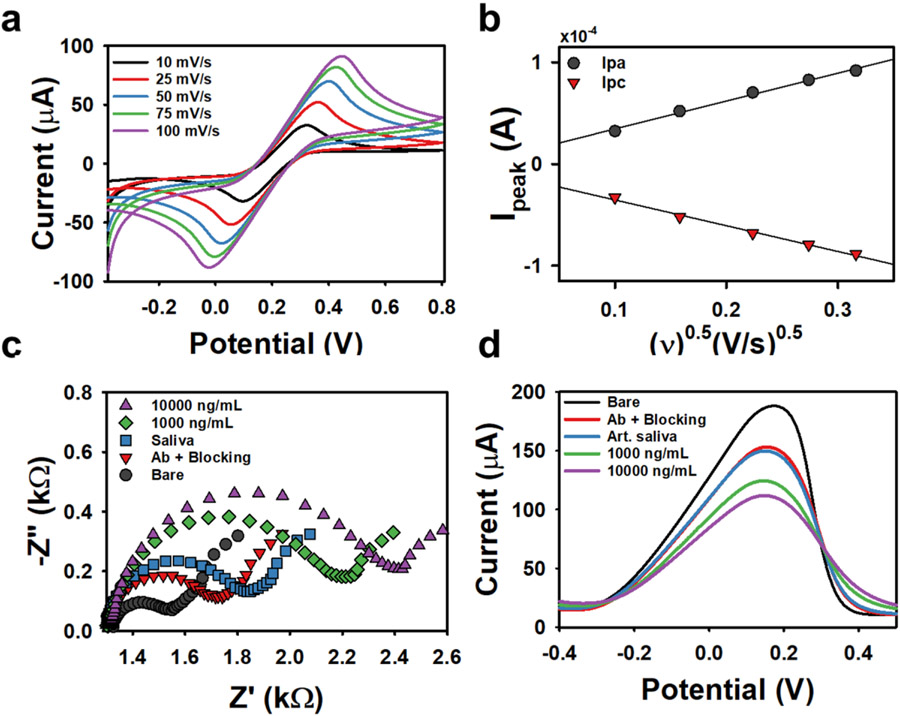
Rapid, inexpensive, and easy-to-use coronavirus disease 2019 (COVID-19) home tests are key tools in addition to vaccines in the world-wide fight to eliminate national and local shutdowns. However, currently available tests for SARS-CoV-2, the virus that causes COVID-19, are too expensive, painful, and irritating, or not sufficiently sensitive for routine, accurate home testing. Herein, we employ custom-formulated graphene inks and aerosol jet printing (AJP) to create a rapid electrochemical immunosensor for direct detection of SARS-CoV-2 Spike Receptor-Binding Domain (RBD) in saliva samples acquired non-invasively. This sensor demonstrated limits of detection that are considerably lower than most commercial SARS-CoV-2 antigen tests (22.91 ± 4.72 pg/mL for Spike RBD and 110.38 ± 9.00 pg/mL for Spike S1) as well as fast response time (~30 mins), which was facilitated by the functionalization of printed graphene electrodes in a single-step with SARS-CoV-2 polyclonal antibody through the carbodiimide reaction without the need for nanoparticle functionalization or secondary antibody or metallic nanoparticle labels. This immunosensor presents a wide linear sensing range from 1 to 1000 ng/mL and does not react with other coexisting influenza viruses such as H1N1 hemagglutinin. By combining high-yield graphene ink synthesis, automated printing, high antigen selectivity, and rapid testing capability, this work offers a promising alternative to current SARS-CoV-2 antigen tests.

Keywords: COVID-19; aerosol jet printing; biosensor; electrochemical impedance spectroscopy; graphene.


Conflict of interest statement

Conflict of interest The authors declare no competing financial interest.

Figures

Figure 1.
Figure 1.
Schematic representation of the fabrication, biofunctionalization, and proposed implementation of the aerosol-jet-printed (AJP) graphene biosensor. (a) AJP of graphene ink in a dipstick pattern on a polyimide substrate. (b) Immobilization of SARS-CoV-2 antibodies on the AJP graphene dipstick surface via carbodiimide cross-linking chemistry. (c) Prevention of nonspecific adsorption onto the sensor in subsequent steps by treating the remaining unfunctionalized areas of the AJP graphene dipstick with blocking agent (Superblock™). (d) Proposed sample collection paradigm in which the saliva sample can be noninvasively obtained from a patient and promptly tested. (e) Incubation of the AJP graphene dipstick biosensor with SARS-CoV-2 virions in a real saliva sample or with SARS-CoV-2 Spike S1 or RBD in artificial saliva. (f-g) Graphical representation of the resulting changes in the differential pulse voltammetry (DPV) and electrochemical impedance spectroscopy (EIS) signals, respectively, with background signal from artificial saliva (black) and output signal from SARS-CoV-2 Spike RBD (red).
Figure 2.
Figure 2.
Microscopic and spectroscopic characterization of AJP dipstick electrodes on a polyimide substrate. (a) Optical micrograph at 6.3x magnification. (b) AFM height map across the cleaned region. (c) Height profile extracted from AFM displaying an average thickness of ~225 nm. (d) SEM image revealing the surface topography at 15,000x magnification. (e) Representative Raman spectrum with the characteristic D, G, and 2D modes of graphene. (f) Raman map (30 x 30 μm region) displaying ID/IG peak ratio with an average value of 0.33 ± 0.025. (g) XPS spectrum revealing surface functional groups.
Figure 3.
Figure 3.
Electrochemical characterization of the AJP graphene dipstick electrodes. (a) Cyclic voltammetry (CV) at scan rates of 10, 25, 50, 75, 100 mV/s. (b) Randles-Sevcik plot showing a linear variation of the peak anodic (Ipa) and cathodic (Ipc) currents with square root of the scan rate with the resulting slope being used to calculate the ESA. (c, d) Nyquist plot and DPV plot of the electrode after each biosensor experimental step: as-prepared AJP graphene electrode (bare), functionalization with SARS-CoV-2 Spike Rabbit polyclonal antibody (Ab) and surface blocking using Superblock™, incubation with artificial saliva, and incubation with 1000 and 10000 ng/mL SARS-CoV-2 Spike RBD, respectively.
Figure 4.
Figure 4.
SARS-CoV-2 Spike RBD detection in artificial saliva using AJP graphene sensors. (a) Representative Nyquist plots for each Spike RBD concentration added to artificial saliva. (b) Representative Nyquist plot for the baseline over time – i.e., subsequent incubations of a functionalized electrode in artificial saliva without addition of Spike RBD. (c) Calibration plot showing the percentage change of charge transfer resistance (ΔRct) with respect to Spike RBD concentration ranging from 1 ng/mL to 10 μg/mL in artificial saliva. (d) Calibration plot showing the percentage change of charge transfer resistance (ΔRct) for the subsequent incubations in artificial saliva. Error bars represent the standard deviation calculated from three independently biofunctionalized electrodes (n = 3).
Figure 5.
Figure 5.
SARS-CoV-2 Spike S1 and Spike RBD detection in artificial saliva and against interferents using the AJP graphene sensor (a) Representative Nyquist plots for each Spike S1 concentration added to artificial saliva using the same concentration range tested previously for SARS-CoV-2 Spike RBD. (b) Representative Nyquist plots for each concentration of Middle East Respiratory Syndrome (MERS) Spike S1 added to artificial saliva. (c) Representative Nyquist plot for each concentration of Influenza H1N1 hemagglutinin (HA) protein added to artificial saliva. (d) Calibration plot showing the percentage change of charge transfer resistance (ΔRct) observed at different concentrations of each protein tested, including Spike RBD. Error bars represent the standard deviation calculated from three independently biofunctionalized electrodes (n = 3).

References

    1. WHO. WHO Coronavirus (COVID-19) Dashboard [Internet]. 2022. [cited 2022 Mar 30]. Available from: https://covid19.who.int/
    1. Hristov D, Rijal H, Gomez-Marquez J, Hamad-Schifferli K. Developing a Paper-Based Antigen Assay to Differentiate between Coronaviruses and SARS-CoV-2 Spike Variants. Anal Chem. 2021; - PubMed
    1. Kaushik AK, Dhau JS, Gohel H, Mishra YK, Kateb B, Kim NY, et al. Electrochemical SARS-CoV-2 Sensing at Point-of-Care and Artificial Intelligence for Intelligent COVID-19 Management. ACS Appl Bio Mater. 2020; - PubMed
    1. International Monetary Fund. World Economic Outlook Update, January 2021. World Econ Outlook. 2021;(January 2021):1–8.
    1. UNESCO. Adverse consequences of school closures [Internet]. Vol. 10, Unesco.org. 2020. [cited 2021 Nov 19]. p. 1–2. Available from: https://en.unesco.org/covid19/educationresponse/consequences

LinkOut - more resources