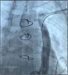
Active fixation of bipolar left ventricular lead through a persistent left superior vena cava
- PMID: 35785375
- PMCID: PMC9237318
- DOI: 10.1002/joa3.12699
Active fixation of bipolar left ventricular lead through a persistent left superior vena cava
Abstract
A left superior vena cava persistence was found in a 61 year-old patient affected by dilated and hypokinetic cardiopathy with severe dysfunction of the left ventricle (left ventricular ejection fraction of 32%) and valvular disease. After a negative coronary angiography, he was implanted with a cardiac resynchronization therapy with defibrillation function device (CRT-D). The present case describes the successful implantation of a biventricular defibrillator in this challenging congenital abnormality of cardiac venous system.
Keywords: biventricular; cardiac resynchronization; defibrillator; persistent left superior vena cava.
© 2022 The Authors. Journal of Arrhythmia published by John Wiley & Sons Australia, Ltd on behalf of the Japanese Heart Rhythm Society.
Conflict of interest statement
No conflict of interest to be declared.
Figures


References
-
- McDonagh TA, Metra M, Adamo M, Gardner RS, Baumbach A, Bohm M, et al. 2021 ESC guidelines for the diagnosis and treatment of acute and chronic heart failure. Eur Heart J. 2021;42:3599–726. - PubMed
-
- Batouty NM, Sobh DM, Gadelhak B, Sobh HM, Mahmoud W, Tawfik AM. Left superior vena cava: cross‐sectional imaging overview. Radiol Med. 2020;125:237–46. - PubMed
-
- Bontempi L, Aboelhassan M, Cerini M, Salghetti F, Fabbricatore D, Maiolo V, et al. Technical considerations for CRT‐D implantation in different varieties of persistent left superior vena cava. J Interv Card Electrophysiol. 2021;61:517–24. - PubMed
LinkOut - more resources
Full Text Sources
Research Materials

