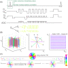
Ultrahigh Resolution fMRI at 7T Using Radial-Cartesian TURBINE Sampling
- PMID: 35785429
- PMCID: PMC9546489
- DOI: 10.1002/mrm.29359
Ultrahigh Resolution fMRI at 7T Using Radial-Cartesian TURBINE Sampling
Abstract
Purpose: We investigate the use of TURBINE, a 3D radial-Cartesian acquisition scheme in which EPI planes are rotated about the phase-encoding axis to acquire a cylindrical k-space for high-fidelity ultrahigh isotropic resolution fMRI at 7 Tesla with minimal distortion and blurring.
Methods: An improved, completely self-navigated version of the TURBINE sampling scheme was designed for fMRI at 7 Telsa. To demonstrate the image quality and spatial specificity of the acquisition, thin-slab visual and motor BOLD fMRI at 0.67 mm isotropic resolution (16 mm slab, TRvol = 2.32 s), and 0.8 × 0.8 × 2.0 mm (whole-brain, TRvol = 2.4 s) data were acquired. To prioritize the high spatial fidelity, we employed a temporally regularized reconstruction to improve sensitivity without any spatial bias.
Results: TURBINE images provide high structural fidelity with almost no distortion, dropout, or T2 * blurring for the thin-slab acquisitions compared to conventional 3D EPI owing to the radial sampling in-plane and the short echo train used. This results in activation that can be localized to pre- and postcentral gyri in a motor task, for example, with excellent correspondence to brain structure measured by a T1 -MPRAGE. The benefits of TURBINE (low distortion, dropout, blurring) are reduced for the whole-brain acquisition due to the longer EPI train. We demonstrate robust BOLD activation at 0.67 mm isotropic resolution (thin-slab) and also anisotropic 0.8 × 0.8 × 2.0 mm (whole-brain) acquisitions.
Conclusion: TURBINE is a promising acquisition approach for high-resolution, minimally distorted fMRI at 7 Tesla and could be particularly useful for fMRI in areas of high B0 inhomogeneity.
Keywords: 7T; TURBINE; fMRI; high-resolution fMRI; radial-Cartesian; ultrahigh-field MRI.
© 2022 The Authors. Magnetic Resonance in Medicine published by Wiley Periodicals LLC on behalf of International Society for Magnetic Resonance in Medicine.
Figures


Similar articles
-
Enhancement of temporal resolution and BOLD sensitivity in real-time fMRI using multi-slab echo-volumar imaging.Neuroimage. 2012 May 15;61(1):115-30. doi: 10.1016/j.neuroimage.2012.02.059. Epub 2012 Feb 28. Neuroimage. 2012. PMID: 22398395 Free PMC article.
-
Fast 3D fMRI acquisition with high spatial resolutions over a reduced FOV.Magn Reson Med. 2024 Nov;92(5):1952-1964. doi: 10.1002/mrm.30191. Epub 2024 Jun 18. Magn Reson Med. 2024. PMID: 38888135
-
Simultaneous pure T2 and varying T2'-weighted BOLD fMRI using Echo Planar Time-resolved Imaging for mapping cortical-depth dependent responses.Neuroimage. 2021 Dec 15;245:118641. doi: 10.1016/j.neuroimage.2021.118641. Epub 2021 Oct 13. Neuroimage. 2021. PMID: 34655771 Free PMC article.
-
Multi-echo fMRI: A review of applications in fMRI denoising and analysis of BOLD signals.Neuroimage. 2017 Jul 1;154:59-80. doi: 10.1016/j.neuroimage.2017.03.033. Epub 2017 Mar 29. Neuroimage. 2017. PMID: 28363836 Review.
-
Tradeoffs in pushing the spatial resolution of fMRI for the 7T Human Connectome Project.Neuroimage. 2017 Jul 1;154:23-32. doi: 10.1016/j.neuroimage.2016.11.049. Epub 2016 Nov 25. Neuroimage. 2017. PMID: 27894889 Free PMC article. Review.
Cited by
-
3D MERMAID: 3D Multi-shot enhanced recovery motion artifact insensitive diffusion for submillimeter, multi-shell, and SNR-efficient diffusion imaging.Magn Reson Med. 2025 Jun;93(6):2311-2330. doi: 10.1002/mrm.30436. Epub 2025 Mar 4. Magn Reson Med. 2025. PMID: 40035173 Free PMC article.
-
Non-Cartesian 3D-SPARKLING vs Cartesian 3D-EPI encoding schemes for functional Magnetic Resonance Imaging at 7 Tesla.PLoS One. 2024 May 13;19(5):e0299925. doi: 10.1371/journal.pone.0299925. eCollection 2024. PLoS One. 2024. PMID: 38739571 Free PMC article.
-
SNAKE: A modular realistic fMRI data simulator from the space-time domain to k-space and back.Imaging Neurosci (Camb). 2025 Sep 2;3:IMAG.a.121. doi: 10.1162/IMAG.a.121. eCollection 2025. Imaging Neurosci (Camb). 2025. PMID: 40909357 Free PMC article.
-
The United States Department of Energy and National Institutes of Health Collaboration: Medical Care Advances via Discovery in Physical Sciences.Med Phys. 2023 Mar;50(3):e53-e61. doi: 10.1002/mp.16252. Epub 2023 Feb 22. Med Phys. 2023. PMID: 36705550 Free PMC article.
References
-
- Kok P, Bains LJ, Van Mourik T, Norris DG, De Lange FP. Selective activation of the deep layers of the human primary visual cortex by top‐down feedback. Curr Biol. 2016;26:371‐376. - PubMed
Publication types
MeSH terms
Grants and funding
LinkOut - more resources
Full Text Sources
Medical

