. 2022 Dec;227(9):3043-3061.
doi: 10.1007/s00429-022-02526-6. Epub 2022 Jul 4.

Mapping lesion, structural disconnection, and functional disconnection to symptoms in semantic aphasia

Affiliations

Mapping lesion, structural disconnection, and functional disconnection to symptoms in semantic aphasia

Nicholas E Souter et al. Brain Struct Funct. 2022 Dec.

Abstract

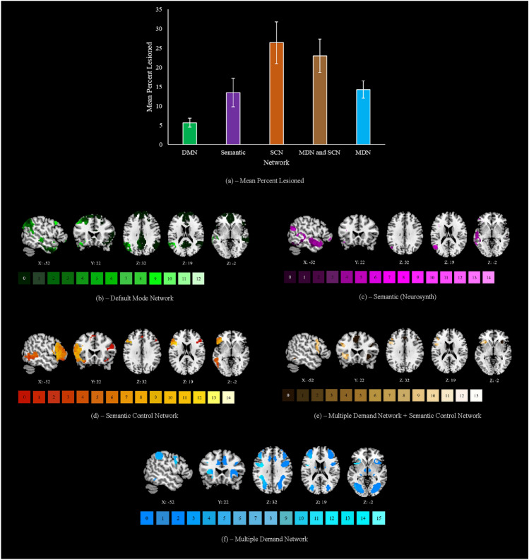
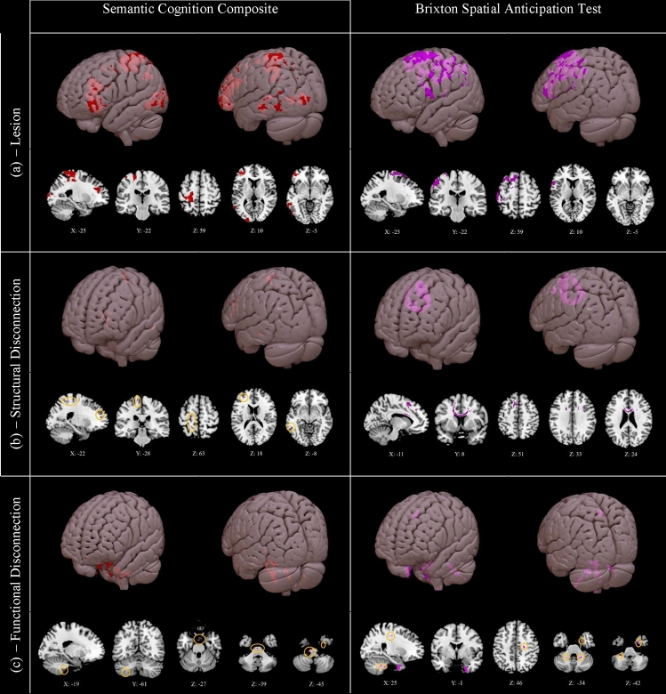
Patients with semantic aphasia have impaired control of semantic retrieval, often accompanied by executive dysfunction following left hemisphere stroke. Many but not all of these patients have damage to the left inferior frontal gyrus, important for semantic and cognitive control. Yet semantic and cognitive control networks are highly distributed, including posterior as well as anterior components. Accordingly, semantic aphasia might not only reflect local damage but also white matter structural and functional disconnection. Here, we characterise the lesions and predicted patterns of structural and functional disconnection in individuals with semantic aphasia and relate these effects to semantic and executive impairment. Impaired semantic cognition was associated with infarction in distributed left-hemisphere regions, including in the left anterior inferior frontal and posterior temporal cortex. Lesions were associated with executive dysfunction within a set of adjacent but distinct left frontoparietal clusters. Performance on executive tasks was also associated with interhemispheric structural disconnection across the corpus callosum. In contrast, poor semantic cognition was associated with small left-lateralized structurally disconnected clusters, including in the left posterior temporal cortex. Little insight was gained from functional disconnection symptom mapping. These results demonstrate that while left-lateralized semantic and executive control regions are often damaged together in stroke aphasia, these deficits are associated with distinct patterns of structural disconnection, consistent with the bilateral nature of executive control and the left-lateralized yet distributed semantic control network.

Keywords: Aphasia; Disconnection; Functional; Semantic; Stroke; Structural.


Conflict of interest statement

The authors have no relevant financial or non-financial interests to disclose.

Figures

Fig. 1
Fig. 1
a Lesion overlap between 4 and 16 cases. Lesions are confined to the left hemisphere and are frontoparietal, peaking in the precentral and middle frontal gyri. b Structural disconnection overlap between 19 and 23 cases, generated using the BCB Toolkit. Structural disconnection is left lateralised at this threshold and shows maximal overlap with the superior longitudinal fasciculus and inferior fronto-occipital fasciculus. c Functional disconnection overlap between 19 and 23 cases, generated using CONN. Functional disconnection is bilateral and extensive, peaking in the temporooccipital part of the left inferior temporal gyrus and the right inferior frontal gyrus pars triangularis. 3D renderings generated in SurfIce. d A line graph displaying the numbers of cases showing overlapping lesions, structural disconnection (confined to white matter) and functional disconnection (confined to grey matter), expressed as a percentage of the total number of voxels damaged or disconnected for at least one participant. This figure demonstrates the homogeneity of patterns of anticipated structural and functional disconnection, relative to the lesion overlap map. N = 23
Fig. 2
Fig. 2
a The mean percentage lesioned for each network of interest (confined to the left hemisphere). DMN default mode network, SCN semantic control network, MDN multiple demand network. This peaks in the semantic control network at 26%, followed by areas shared between the multiple demand and semantic control networks at 23%, core semantic regions and regions exclusive to the multiple demand network both at 14%, and the default mode network at 6%. Locations of most frequent damage are displayed for each network in following sections. Any right hemisphere aspects of each network are visualised but were never impacted by lesion. b The default mode network, lesion peaks in the angular gyrus and insular cortex at a threshold of 12 cases. c Core semantic regions, lesion peaks in the inferior frontal gyrus pars opercularis at a threshold of 14 cases. d The semantic control network, lesion peaks in the inferior frontal gyrus pars opercularis at a threshold of 14 cases. e Regions shared by the semantic control and multiple demand networks, lesion peaks in the middle frontal and precentral gyri at 13 cases. f The multiple demand network, lesion peaks in the precentral gyrus at a threshold of 15 cases. Keys under each map reflect the number of patients with lesion to a given voxel. N = 23
Fig. 3
Fig. 3
Clusters associated with lower semantic cognition composite scores (left) and lower scores on the Brixton Spatial Anticipation Test (right), for a lesion, b structural disconnection, and c functional disconnection. Generated using non-parametric permutation tests in Randomise with threshold-free cluster enhancement. Highlighted voxels have a t value of 2.6 or higher. Small clusters are highlighted in orange circles. 3D rendering generated in Surface. Lesioned clusters associated with poorer semantic and Brixton performance are large and left lateralised, and reflect the regions listed in Section “Lesion-symptom mapping”. Structurally disconnected clusters are small and left lateralised for semantic cognition, reflecting regions listed in Section “Structural disconnection-symptom mapping”, while for poorer Brixton performance one large cluster is observed across the corpus callosum. Functionally disconnected clusters are small for both measures, and reflect the regions listed in Section “Functional disconnection-symptom mapping”. N=20

References

    1. Agosta F, Henry RG, Migliaccio R, Neuhaus J, Miller BL, Dronkers NF, Brambati SM, Filippi M, Ogar JM, Wilson SM, Gorno-Tempini ML. Language networks in semantic dementia. Brain. 2010;133(1):286–299. doi: 10.1093/brain/awp233. - DOI - PMC - PubMed
    1. Almairac F, Herbert G, Moritz-Gasser S, Menjot de Champfleur N, Duffau H. The left inferior fronto-occipital fasciculus subserves language semantics: a multilevel lesion study. Brain Struct Funct. 2015;220:1983–1995. doi: 10.1007/s00429-014-0773-1. - DOI - PubMed
    1. Andreottia J, Dierks T, Wahlund L-O, Grieder M. Diverging progression of network disruption and atrophy in Alzheimer’s disease and semantic dementia. J Alzheimer’s Dis. 2017;55:981–993. doi: 10.3233/JAD-160571. - DOI - PMC - PubMed
    1. Avants BB, Tustison NJ, Song G, Cook PA, Klein A, Gee JC. A reproducible evaluation of ANTs similarity metric performance in brain image registration. Neuroimage. 2011;54(3):2033–2044. doi: 10.1016/j.neuroimage.2010.09.025. - DOI - PMC - PubMed
    1. Avants BB, Tustison NJ, Stauffer M, Song G, Wu B, Gee JC. The Insight ToolKit image registration framework. Front Neurosci. 2014;8:44. doi: 10.3389/fninf.2014.00044. - DOI - PMC - PubMed