. 2022 Jul 20;144(28):12934-12941.
doi: 10.1021/jacs.2c04824. Epub 2022 Jul 5.

TF-DUBTACs Stabilize Tumor Suppressor Transcription Factors

Affiliations

TF-DUBTACs Stabilize Tumor Suppressor Transcription Factors

Jing Liu et al. J Am Chem Soc. .

Abstract

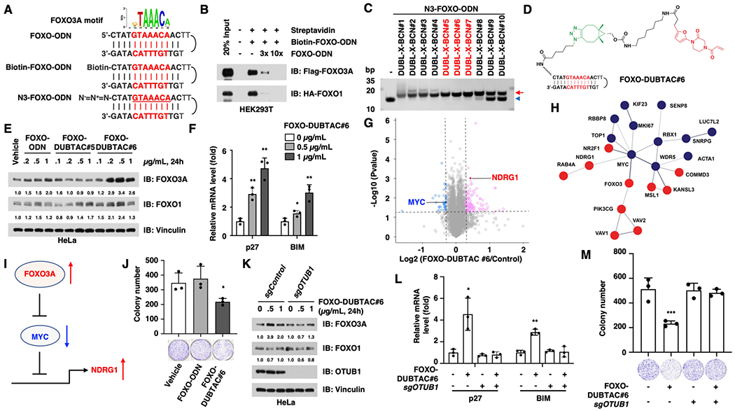
Targeted protein degradation approaches have been widely used for degrading oncogenic proteins, providing a potentially promising therapeutic strategy for cancer treatment. However, approaches to targeting tumor suppressor proteins are very limited, and only a few agonists have been developed to date. Here, we report the development of a platform termed TF-DUBTAC, which links a DNA oligonucleotide to a covalent ligand of the deubiquitinase OTUB1 via a click reaction, to selectively stabilize tumor suppressor transcription factors. We developed three series of TF-DUBTACs, namely, FOXO-DUBTAC, p53-DUBTAC, and IRF-DUBTAC, which stabilize FOXO3A, p53, and IRF3 in cells, respectively, in an OTUB1-dependent manner. These results suggest that TF-DUBTAC is a generalizable platform to achieve selective stabilization of tumor suppressor transcription factors as a therapeutic means to suppress tumorigenesis.


Conflict of interest statement

The authors declare the following competing financial interest(s): W.W. is a co-founder and stockholder of the Rekindle Therapeutics. J.J. is a co-founder and equity shareholder in Cullgen, Inc. and a consultant for Cullgen, Inc., EpiCypher, Inc., and Accent Therapeutics, Inc. The Jin laboratory received re-search funds from Celgene Corporation, Levo Therapeutics, Cullgen, Inc. and Cullinan Oncology. All other authors declare no competing interests.

Figures

Figure 1.
Figure 1.
Schematic diagram of the TF-DUBTAC platform. The BCN-linked OTUB1 binder [DUBL-X-BCN, X = (CH2)n, n = 2–11] was conjugated onto an azide-modified DNA oligomer (N3-ODN) via a copper-free SPAAC reaction, resulting in a TF-DUBTAC that recruits the DUB OTUB1 to remove polyubiquitin chain from the targeted TF.
Figure 2.
Figure 2.
FOXO-DUBTAC #6 stabilizes FOXO3A in an OTUB1-dependent manner. (A) Schematic diagram for the FOXO3A motif and FOXO-ODN. (B) FOXO-ODN binds both FOXO3A and its close family member FOXO1, and the binding can be antagonized by free FOXO-ODN. Cell lysates with Flag-FOXO3A or HA-FOXO1 protein were extracted from HEK293T cells that expressed indicated constructs for 48 h. (C) Linkers with six–eight methylene groups are optimal for the in vitro SPAAC reactions between BCN-modified OTUB1 ligands and N3-FOXO-ODN to form FOXO-DUBTACs. The SPAAC reaction products were separated by 20% native polyacrylamide gel electrophoresis (PAGE). The red arrow indicates the click reaction products, FOXO-DUBTACs, and the blue arrowhead indicates N3-FOXO-ODN. (D) Chemical structure of FOXO-DUBTAC #6. (E) FOXO-DUBTAC #6 stabilizes FOXO3A protein in HeLa cells. HeLa cells were treated with the indicated concentrations of FOXO-ODN, FOXO-DUBTAC #5, or #6 for 24 h, followed by immunoblot analysis. (F) FOXO-DUBTAC #6 treatment increases the expression of p27 and BIM. *, p < 0.05; **, p < 0.01. (G) FOXO-DUBTAC #6 treatment changes the proteome profile of HeLa cells. (H) Enrichment of the MYC-related protein network in FOXO-DUBTAC #6-treated cells. Red indicates increased proteins, and blue indicates decreased proteins. (I) Schematic diagram to show that FOXO3A represses the MYC expression and function, resulting in increase in NDRG1. (J) FOXO-DUBTAC #6 suppresses the tumorigenesis of HeLa cells. HeLa cells were treated with 1 μ/mL FOXO-DUBTAC #6, followed by assessment in a colony formation assay. *, p < 0.05. (K) FOXO-DUBTAC #6 increases FOXO3A protein abundance in OTUB1+/+ cells but not in OTUB1−/− cells. HeLa cells were infected with the sgControl or sgOTUB1 virus to knock out endogenous OTUB1 and selected with puromycin for 72 h, followed by the treatment with FOXO-DUBTAC #6 for 24 h and were further analyzed with immunoblotting. (L) FOXO-DUBTAC #6 treatment increases the expression of p27 and BIM in an OTUB1-dependent manner. *, p < 0.05; **, p < 0.01. (M) FOXO-DUBTAC #6 treatment represses colony formation in an OTUB1-dependent manner. ***, p < 0.001.
Figure 3.
Figure 3.
p53-DUBTAC #6 and p53-DUBTAC #7 stabilize p53 in an OTUB1-dependent manner. (A) Schematic diagram for the p53 motif, p53-ODN, biotin-modified p53-ODN (biotin-p53-ODN), and azide-modified p53-ODN (N3-p53-ODN). (B) Biotin-p53-ODN binds Flag-tagged p53. The cell lysates with p53 protein were extracted from HEK293T cells that expressed the Flag-p53 construct for 48 h. (C) Chemical structure of p53-DUBTAC #6. (D) p53-DUBTACs #5, #6, and #7 (1 μ/mL, 24 h) increase the p53 protein level in HeLa cells. (E) p53-DUBTACs #5, #6, and #7 (0.1, 0.2, and 0.4 μ/mL, 24 h) increase the p53 protein level in HeLa cells. Vehicle stands for the treatment with transfection reagents, and control stands for the treatment without transfection reagents. (F) p53-DUBTAC #6 treatment changes the proteome profile of HeLa cells. (G) Enrichment of the AURKA-related protein network in p53-DUBTAC #6-treated cells. Red indicates increased proteins, and blue indicates decreased proteins. (H) p53-DUBTACs #6 and #7 (1 μ/mL, 24 h) increase the p53 protein level in an OTUB1-dependent manner in HeLa cells. HeLa cells infected with either the sgControl or sgOTUB1 virus and selected with puromycin, followed by treatment with the indicated compound for 24 h and immunoblotting analysis of p53 protein abundance.
Figure 4.
Figure 4.
IRF-DUBTAC #7 stabilizes IRF3 in HeLa cells. (A) Schematic diagram for the IRF3 motif, IRF-ODN, biotin-modified IRF-ODN (biotin-IRF-ODN), and azide-modified IRF-ODN (N3-IRF-ODN). (B) Biotin-IRF-ODN binds multiple IRF family members, except IRF9. The cell lysates were extracted from HEK293T cells that overexpressed indicated Flag-IRF constructs. (C) DUBL-X-BCNs #5–7 are most effective in forming IRF-DUBTACs via in vitro SPAAC reactions with N3-IRF-ODN. The SPAAC reaction products were separated by 20% native PAGE. The red arrow indicates the click reaction products, IRF-DUBTACs, and the blue arrowhead indicates IRF-ODN. (D) IRF-DUBTAC #7 (2 μ/mL, 24 h) is most effective in increasing the IRF3 protein level in HeLa cells. Vehicle stands for the treatment with transfection reagents.

References

    1. Vogelstein B; Kinzler KW Cancer genes and the pathways they control. Nat. Med 2004, 10, 789–799. - PubMed
    1. Hanahan D; Weinberg RA Hallmarks of cancer: the next generation. Cell 2011, 144, 646–674. - PubMed
    1. Lehmann JM; Moore LB; Smith-Oliver TA; Wilkison WO; Willson TM; Kliewer SA An Antidiabetic Thiazolidinedione Is a High Affinity Ligand for Peroxisome Proliferator-activated Receptor γ (PPARγ). J. Biol. Chem 1995, 270, 12953–12956. - PubMed
    1. Willson TM; Cobb JE; Cowan DJ; Wiethe RW; Correa ID; Prakash SR; Beck KD; Moore LB; Kliewer SA; Lehmann JM The Structure–Activity Relationship between Peroxisome Proliferator-Activated Receptor γ Agonism and the Antihyperglycemic Activity of Thiazolidinediones. J. Med. Chem 1996, 39, 665–668. - PubMed
    1. Momose Y; Maekawa T; Yamano T; Kawada M; Odaka H; Ikeda H; Sohda T Novel 5-substituted 2,4-thiazolidinedione and 2,4-oxazolidinedione derivatives as insulin sensitizers with antidiabetic activities. J. Med. Chem 2002, 45, 1518–1534. - PubMed

Publication types

MeSH terms

Substances