. 2022 Jul 12;119(28):e2118182119.
doi: 10.1073/pnas.2118182119. Epub 2022 Jul 5.

A long noncoding RNA influences the choice of the X chromosome to be inactivated

Affiliations

A long noncoding RNA influences the choice of the X chromosome to be inactivated

Andreas Hierholzer et al. Proc Natl Acad Sci U S A. .

Abstract

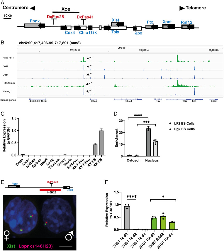
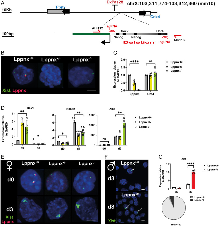
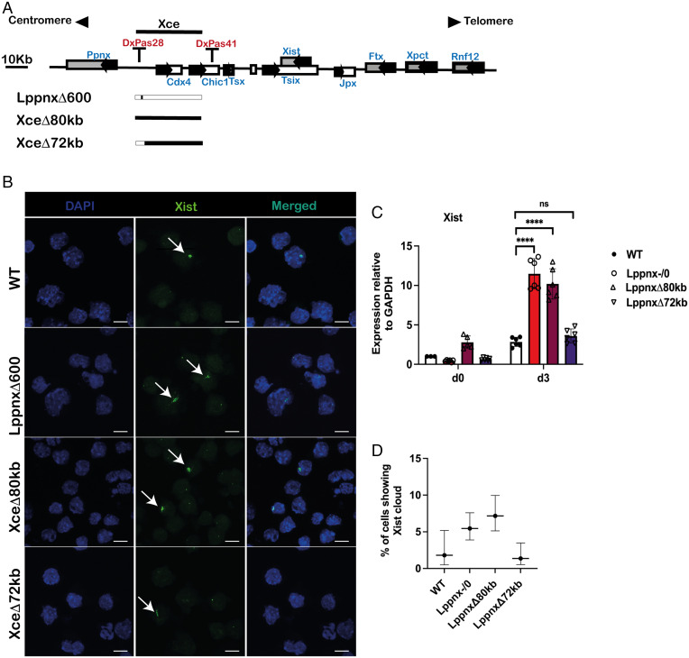
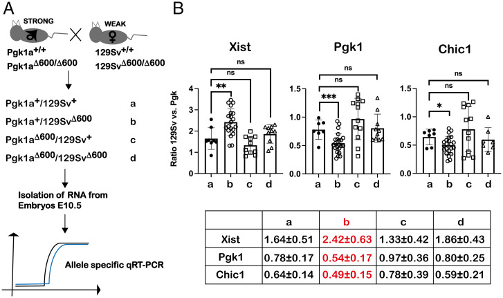
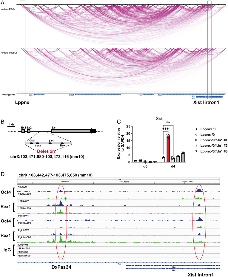
X chromosome inactivation (XCI) is the process of silencing one of the X chromosomes in cells of the female mammal which ensures dosage compensation between the sexes. Although theoretically random in somatic tissues, the choice of which X chromosome is chosen to be inactivated can be biased in mice by genetic element(s) associated with the so-called X-controlling element (Xce). Although the Xce was first described and genetically localized nearly 40 y ago, its mode of action remains elusive. In the approach presented here, we identify a single long noncoding RNA (lncRNA) within the Xce locus, Lppnx, which may be the driving factor in the choice of which X chromosome will be inactivated in the developing female mouse embryo. Comparing weak and strong Xce alleles we show that Lppnx modulates the expression of Xist lncRNA, one of the key factors in XCI, by controlling the occupancy of pluripotency factors at Intron1 of Xist. This effect is counteracted by enhanced binding of Rex1 in DxPas34, another key element in XCI regulating the activity of Tsix lncRNA, the main antagonist of Xist, in the strong but not in the weak Xce allele. These results suggest that the different susceptibility for XCI observed in weak and strong Xce alleles results from differential transcription factor binding of Xist Intron 1 and DxPas34, and that Lppnx represents a decisive factor in explaining the action of the Xce.

Keywords: X chromosome inactivation; X-controlling element; female mouse embryo; noncoding RNA; pluripotency factors.


Conflict of interest statement

The authors declare no competing interest.

Figures

Fig. 1.
Fig. 1.
A long noncoding RNA is transcribed from the predicted X-controlling element. Schematic view of the X-inactivation center with the predicted Xce indicated by a black bar (A). ChIPseq analysis shows a clear signal, marked with a black arrow, for the pluripotency factors Oct4, Nanog and Sox2 within the Xce. RNA-Pol II and H3K79m2 indicate initiation and maintenance of active transcription (B). The transcript identified in B is mainly expressed in mouse male and female ES cells but not in differentiated cells, as exemplified by organ analysis of adult mice. Relative expression levels of the transcript were measured by qRT-PCR in mouse organs, embryos, and cell lines. XX Fibro: Female fibroblast cell line, XY Fibro: Male fibroblast cell line. XX ES: Female embryonic stem cell line XY ES: Male embryonic stem cell line. (C). Fractionation of cytosol and nuclei in two ES cell lines, LF2 and Pgk, reveals the localization of the transcript to be mainly in the nucleus, indicating a bona fide lncRNA transcript. Enrichment in the two compartments was measured using qRT-PCR. Two-sample t test: P < 0.0001(LF2), P = 0.0006 (Pgk) (D). Visualization of the transcript in the nucleus of male and female ES cells using RNA-FISH. The position of the BAC used as FISH probe is highlighted as a red bar (E). The expression of the transcript depends on Oct4. Oct4-deficient ES cell line (ZHBT) which carries a tetracycline-repressible Oct4 transgene was differentiated either in the presence (RA) or absence (TC) of Oct4. In the absence of Oct4 (Tc) the transcript is rapidly down-regulated during differentiation, whereas the maintenance of Oct4 during differentiation (RA) maintains also the transcription. Transcript levels are shown relative to glyceraldehyde-3-phosphate dehydrogenase (GAPDH). Two-sample t test: P < 0.0001. One-way ANOVA: P = 0.0143 (F).
Fig. 2.
Fig. 2.
Deletion of the putative promoter sequence of Lppnx in 129Sv mice. CRISPR/Cas9 mediated genome editing was used to delete the 595-bp promoter fragment in mice. Schematic view of the position of the sgRNAs and the primer used for subsequent genotyping of the founder animals (A). FISH analysis of female ES cells generated from LppnxΔ600 mice show the absence of a signal for Lppnx indicating that the predicted promoter fragment drives Lppnx expression (B). Lppnx expression is down-regulated in heterozygous ES cells and absent in homozygous cells carrying the deletion of the putative promoter region. Two-sample t test: P < 0.0001 (comparing WT with homozygous cells). The absence of Lppnx expression does not affect pluripotency in these ES cells as shown by a comparison of Oct4 expression levels. One-way ANOVA: P = 0.4998 (C). Female ES cells homo- and heterozygous for the deletion of Lppnx show different levels of key markers compared to WT cells upon differentiation. Cells were analyzed at day 3 after onset of differentiation (see Materials and Methods section). Rex1 levels are elevated in both, homozygous and heterozygous undifferentiated cells. Two-sample t test: P = 0.0023. After differentiation Rex is down-regulated in all three lines. One-way ANOVA: P = 0.008. Nestin was used as a differentiation marker. The degree of differentiation was calculated as fold change in expression of Nestin. The fold change is given above the bars in d3. All three lines show comparable fold changes during differentiation. Two-sample t test: P = 0.0242 (d0) P = 0.0032 (d3). Xist expression is markedly increased after differentiation in homozygous cells. d0: One-way ANOVA: P = 0.4959. d3:Ttwo-sample t test between WT and homozygous deletion P = 0.0064 (D). RNA-FISH analysis of differentiated female ES cells shows an increased intensity of Xist associated clouds upon Lppnx depletion, in agreement with the analysis in D (E). Accordingly, male ES cells deficient for Lppnx express higher Xist levels after differentiation as shown by qRT-PCR and the elevated number of Xist clouds indicated by arrows (F, G). Two-sample t test: P < 0.0001. The increase of Xist clouds in ES cells deficient for Lppnx compared to WT cells is visualized in a pie chart (G).
Fig. 3.
Fig. 3.
Comparison of the number of Xist clouds in male ES cells. Schematic view of the X inactivation center and the CRISPR mediated deletion. 129Sv male ES cells were used to introduce a deletion of the entire predicted Xce (XceΔ80kb) or a deletion which leaves intact the Lppnx region (LppnxΔ72kb). These cell lines were compared with a WT and the previously shown LppnxΔ600 (corresponding to Lppnx-/0) cell line (A). After differentiation XceΔ80kb and LppnxΔ600 show elevated numbers of Xist clouds whereas LppnxΔ72kb and WT show comparable levels (B). qRT-PCR analysis of the cells in B showed elevated Xist expression levels in XceΔ80kb and LppnxΔ600 but not in LppnxΔ72kb compared to WT. Two-sample t test: P < 0.0001 (C). Quantification of Xist clouds shown in B are given in percentage of Xist clouds within the different cell lines. Remarkably, only XceΔ80kb and LppnxΔ600 show elevated ratio of clouds, whereas LppnxΔ72kb and WT cells show comparable levels. Percentages are given with an upper and lower level and are shown with a CI of 95% (Wilson/Brown) (D).
Fig. 4.
Fig. 4.
Deletion of Lppnx promotes a shift in X-linked expression ratio in heterozygous embryos. Schematic view of the breeding strategy used to analyze X-linked expression ratios. From heterozygous Xce crosses between Xcea (weak, 129Sv) and Xcec (strong, Pgk1a), indicated from ad, E10.5 female embryos were isolated. Δ600 corresponds to the 600-bp promoter deletion shown before. We used allele-specific qRT-PCR to define the X-linked expression ratio as follows: 2Ct(Pgk)-Ct(129) (A). Reciprocal crosses of Xcea with Xcec with or without a depletion of Lppnx show a significant shift of Xist ratio as well as of X-linked genes. Pgk1 and Chic1 in E10.5 embryos only in Xcea-LppnxΔ600/XcecWT embryos, whereas all other combinations are comparable to WT. Two-sample t test Xist: P = 0.0048, Pgk1: P = 0.0042, Chic1: P = 0.0379. Each dot in the plots represents the value of a single embryo measured in three technical replicates. Expression ratios are summarized in a table, only crossing b (marked in red) shows significant difference to WT (B).
Fig. 5.
Fig. 5.
The Lppnx locus may interact with Xist Intron1 in mouse ES cells and controls the loading of Oct4 and Rex1 in XI1. Screenshot of a Hi-C analysis showing a part of the X inactivation center including Xist and Lppnx. The green rectangles indicate the transcription start site of Lppnx and Xist Intron1 showing a possible interaction between these two regions in both, male and female ES cells (A). Schematic view of the pluripotency factor binding region of Xist intron 1 which was deleted in 129Sv and 129SvΔ600 male ES cells, respectively, using CRISPR (B). Elevated Xist expression observed in 129SvΔ600 male ES cells (Lppnx-/0) after differentiation is completely reversed after the deletion of Xist Intron1. Three independent lines (#1, #2, #3) are shown. Two-sample t test P = 0.001 (C). Lppnx regulates the loading of pluripotency factors in Xist Intron1 and DxPas34. ChIPseq experiments show that upon deletion of Lppnx the loading of Oct4 and Rex1 is reduced at Xist Intron1 (red circle, Right) in male 129Sv and Pgk1a ES cells. In contrast, Rex1 binding at DxPas34, a regulatory region of Tsix, is reduced in Lppnx-deficient 129Sv ES cells, whereas in Pgk1a cells Rex1 binding is increased (red circle, Left) (D).

Comment in

References

    1. Mank J. E., The W, X, Y and Z of sex-chromosome dosage compensation. Trends Genet. 25, 226–233 (2009). - PMC - PubMed
    1. Ercan S., Mechanisms of x chromosome dosage compensation. J Genomics 3, 1–19 (2015). - PMC - PubMed
    1. Kuroda M. I., Hilfiker A., Lucchesi J. C., Dosage compensation in Drosophila-a model for the coordinate regulation of transcription. Genetics 204, 435–450 (2016). - PMC - PubMed
    1. Payer B., Lee J. T., X chromosome dosage compensation: How mammals keep the balance. Annu. Rev. Genet. 42, 733–772 (2008). - PubMed
    1. Leeb M., Wutz A., Mechanistic concepts in X inactivation underlying dosage compensation in mammals. Heredity 105, 64–70 (2010). - PubMed

LinkOut - more resources