Review
. 2022 Jul 4;24(1):42.
doi: 10.1186/s12968-022-00872-2.

Society for Cardiovascular Magnetic Resonance 2021 cases of SCMR and COVID-19 case collection series

Affiliations
Review

Society for Cardiovascular Magnetic Resonance 2021 cases of SCMR and COVID-19 case collection series

Jason N Johnson et al. J Cardiovasc Magn Reson. .

Abstract

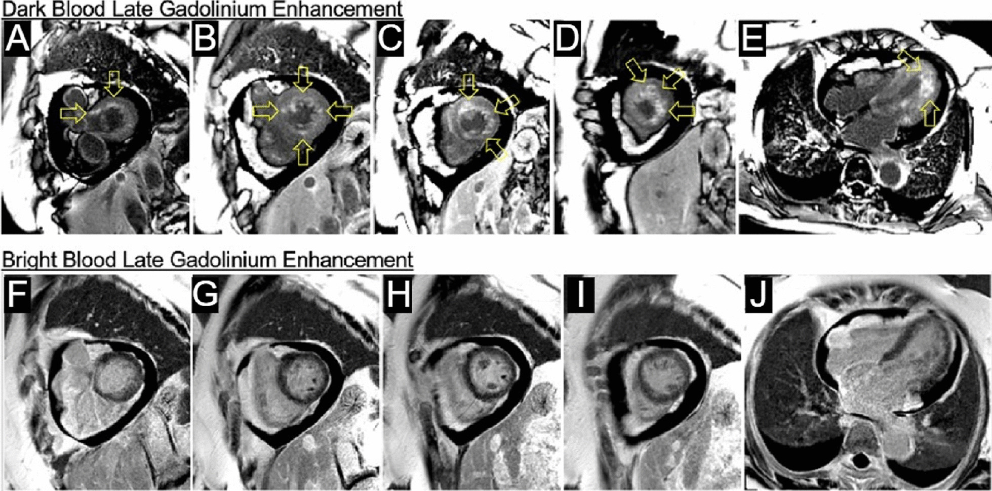
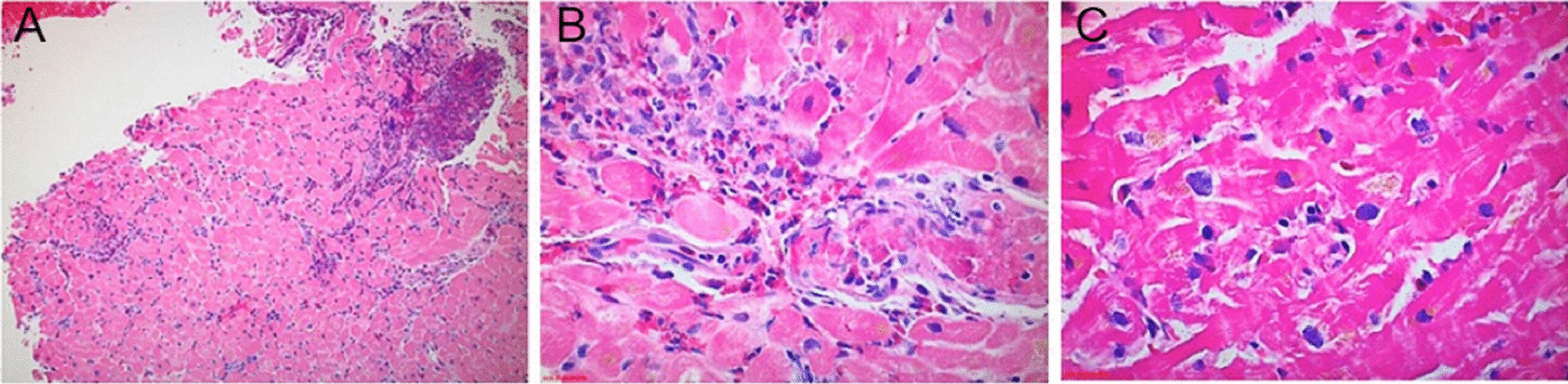
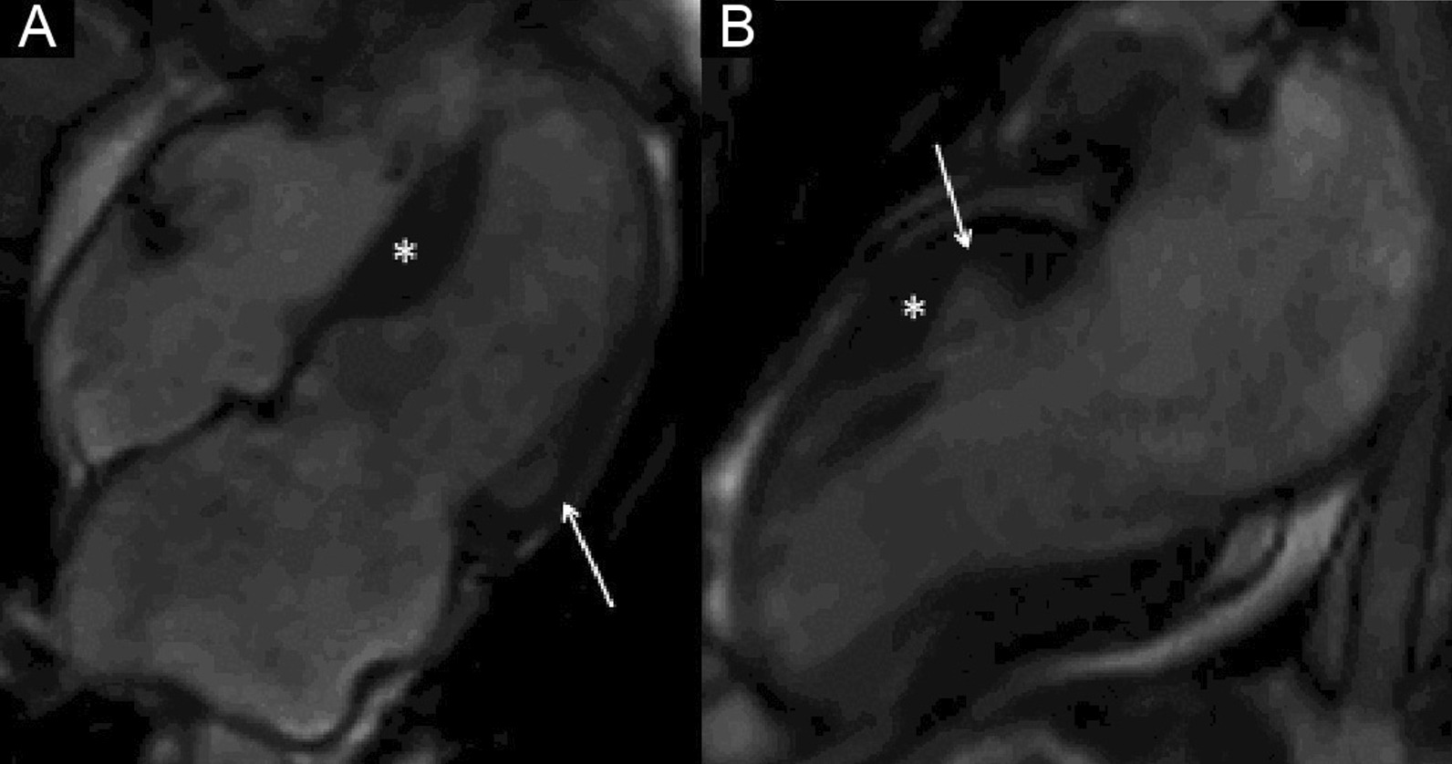
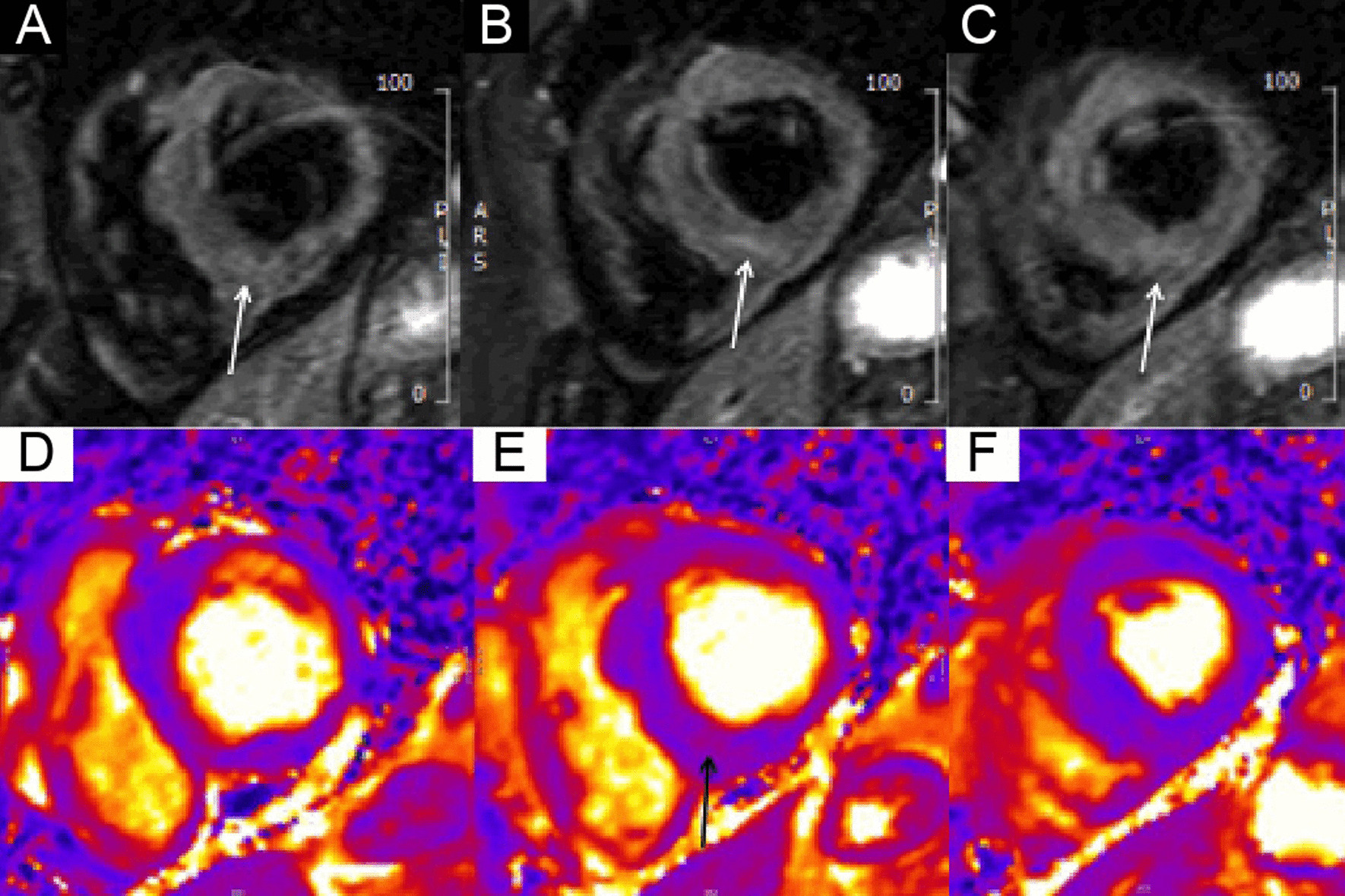
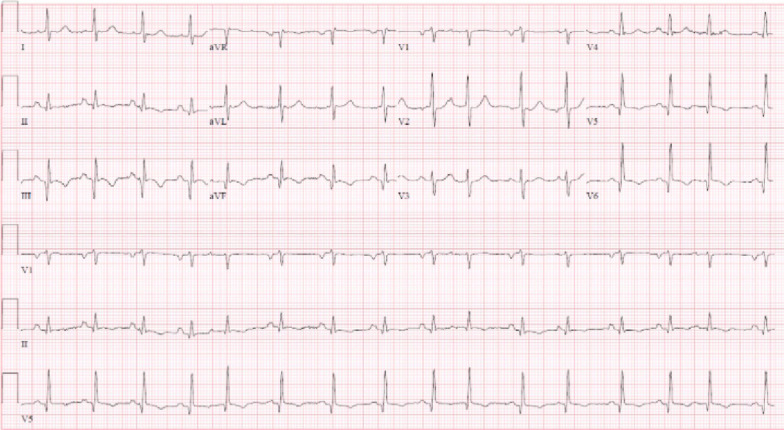
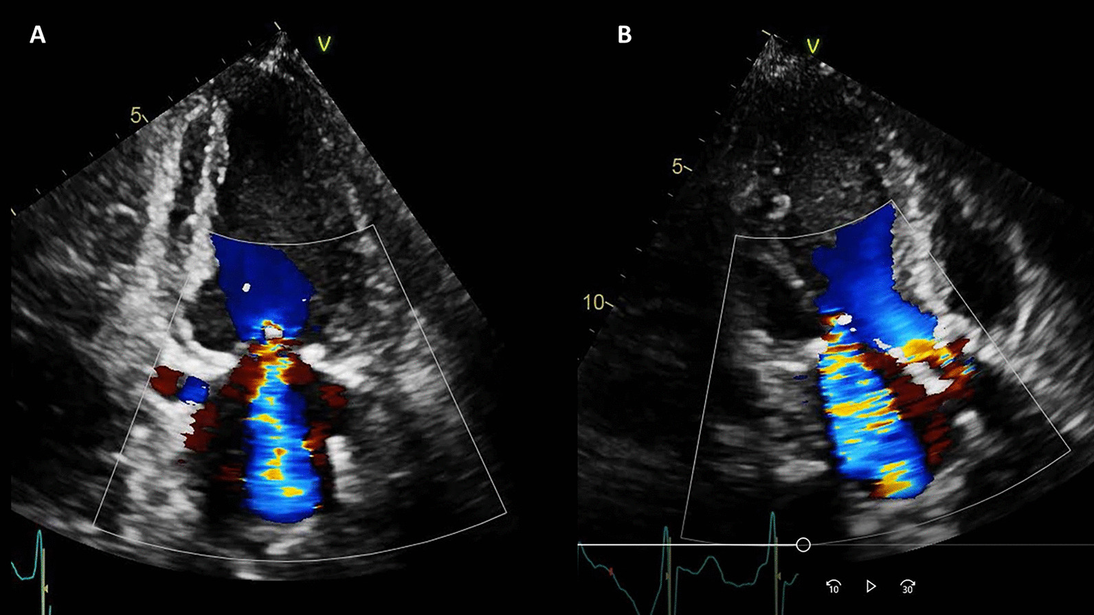
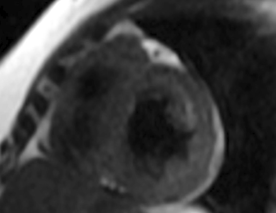
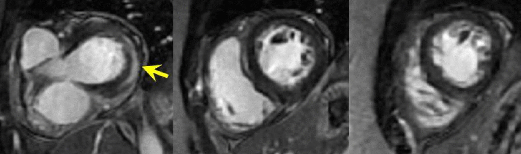
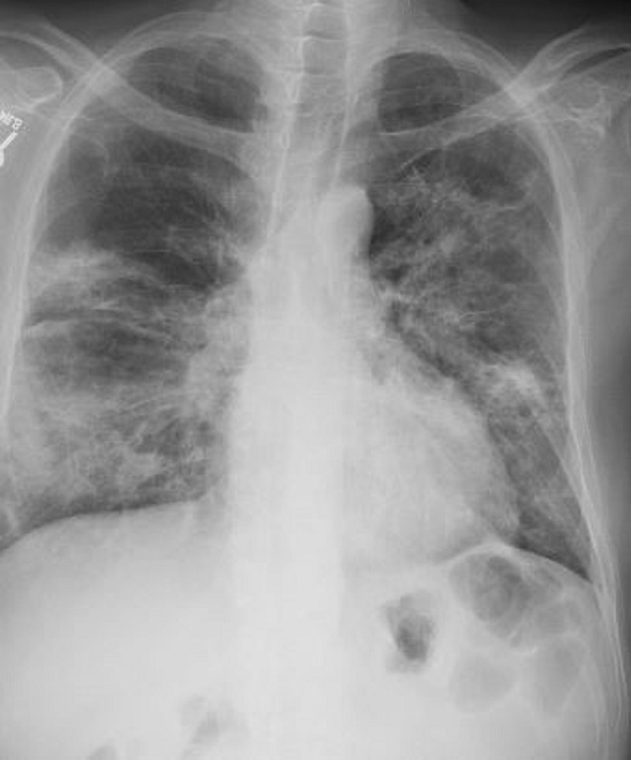
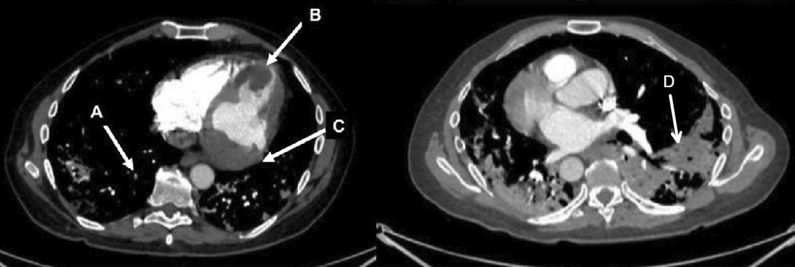
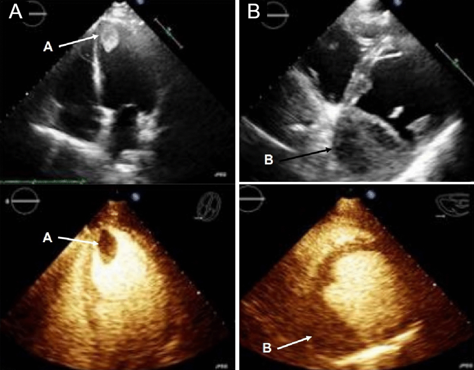
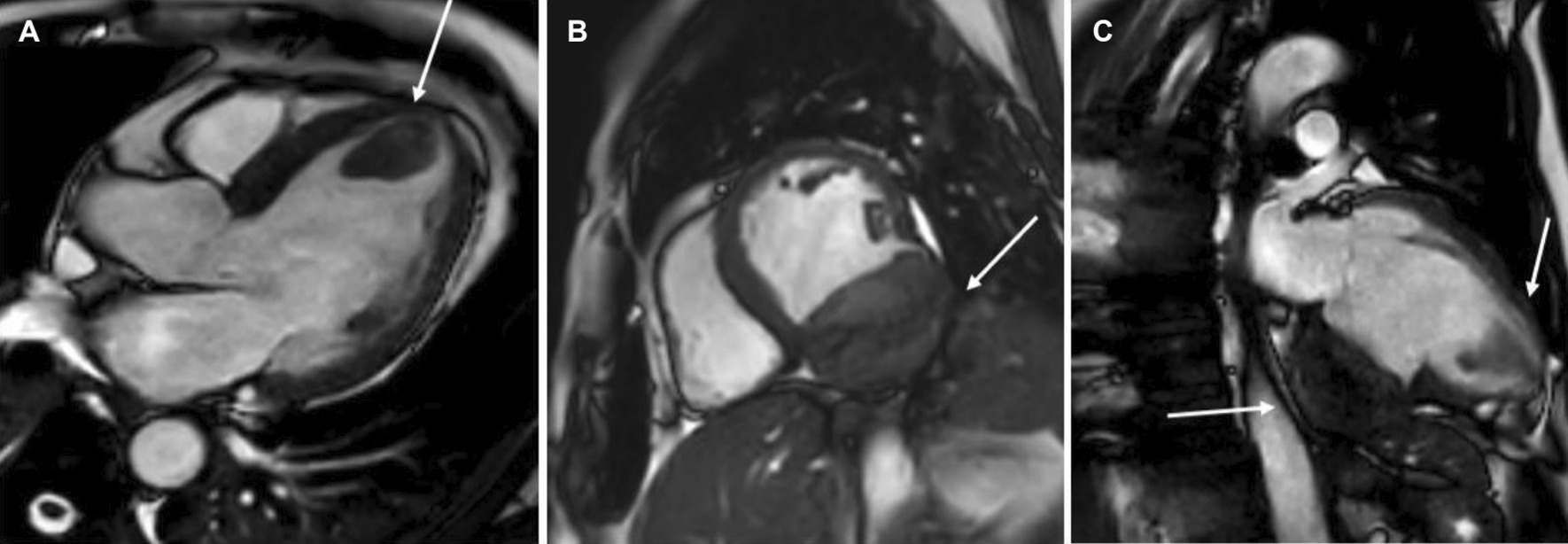
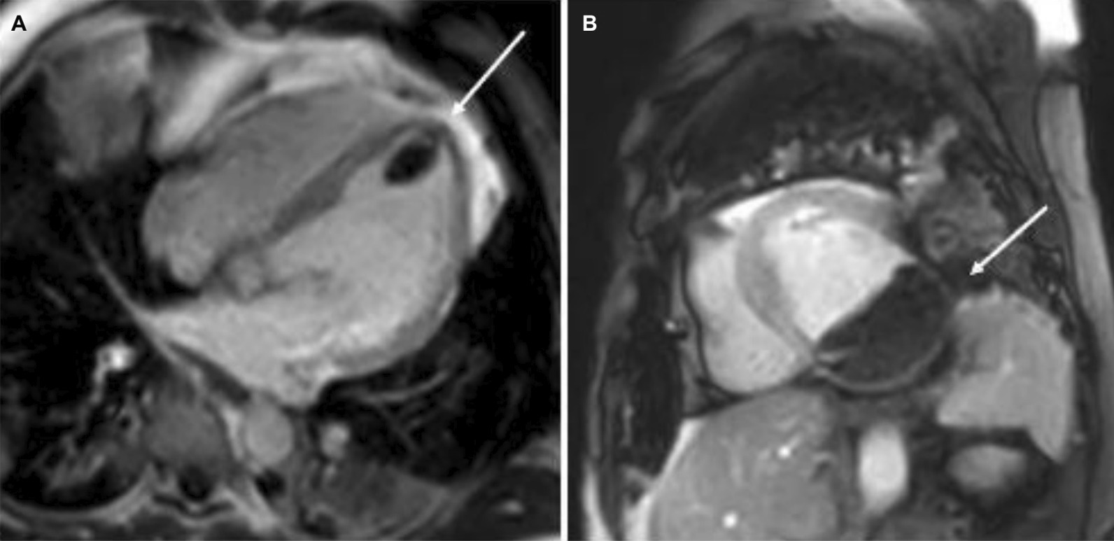
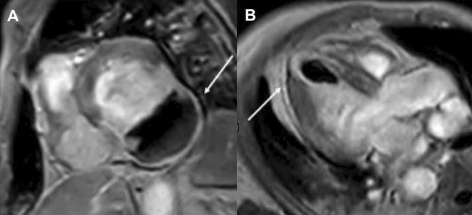
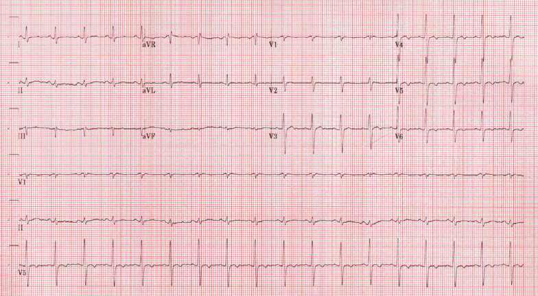
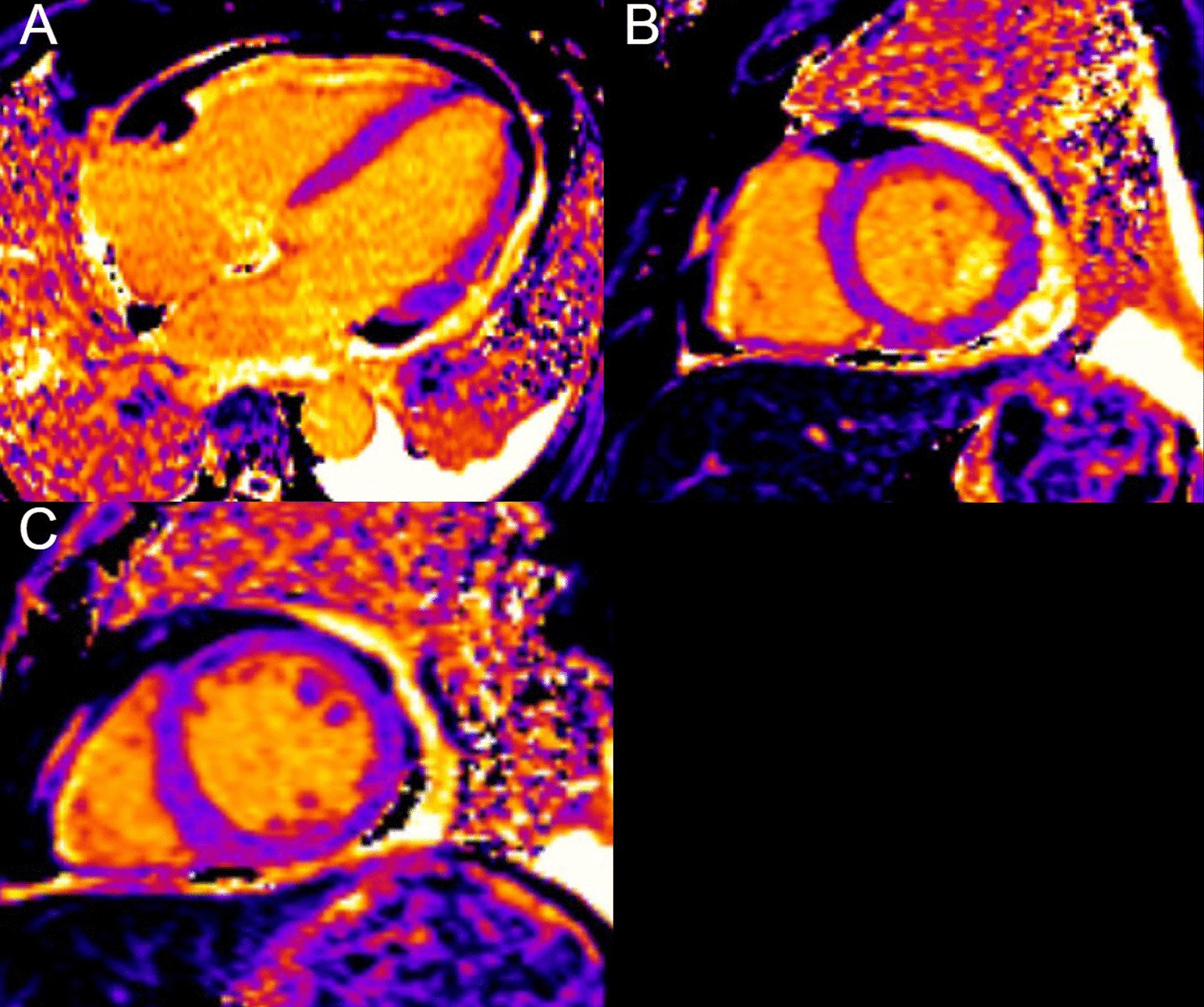
The Society for Cardiovascular Magnetic Resonance (SCMR) is an international society focused on the research, education, and clinical application of cardiovascular magnetic resonance (CMR). "Cases of SCMR" is a case series hosted on the SCMR website ( https://www.scmr.org ) that demonstrates the utility and importance of CMR in the clinical diagnosis and management of cardiovascular disease. The COVID-19 Case Collection highlights the impact of coronavirus disease 2019 (COVID-19) on the heart as demonstrated on CMR. Each case in series consists of the clinical presentation and the role of CMR in diagnosis and guiding clinical management. The cases are all instructive and helpful in the approach to patient management. We present a digital archive of the 2021 Cases of SCMR and the 2020 and 2021 COVID-19 Case Collection series of nine cases as a means of further enhancing the education of those interested in CMR and as a means of more readily identifying these cases using a PubMed or similar literature search engine.


Conflict of interest statement

There are no competing interests.

Figures

Fig. 1
Fig. 1
Case 1. Twelve lead electrocardiogram (ECG). Sinus tachycardia with diffuse ST depression
Fig. 2
Fig. 2
Case 1. Posteroanterior A and lateral B chest X-ray. Enlarged cardiomediastinal silhouette and small bilateral pleural effusions
Fig. 3
Fig. 3
Case 1. Cine balanced steady state free precession (bSSFP) 4 chamber at end-diastole. There is brief right atrial (RA) compression (arrow) present
Fig. 4
Fig. 4
Case 1. Short axis post contrast phase sensitive inversion recovery with prolonged inversion time. There are multiple intracardiac thrombi (arrows)
Fig. 5
Fig. 5
Case 1. Short axis and four chamber dark blood AE and bright blood FJ late gadolinium enhancement (LGE) images. Comparison of bright and dark blood LGE imaging (FIDDLE technique). Dark blood LGE imaging reveals endocardial hyperenhancement not evident on conventional bright blood imaging due to superior contrast between scar and the blood pool
Fig. 6
Fig. 6
Case 1. Endomyocardial biopsy at 100 power (A), 200 power (B), and 400 power (C). Perivascular and interstitial inflammatory infiltrate with prominent eosinophils
Fig. 7
Fig. 7
Case 2. Figure 1. Four A and two chamber B bSSFP cine sequences. There are septal and anterior left ventricular (LV) hypertrophy (LVH) (white asterisk) and myocardial crypts of the basal anterior and anterolateral walls (white arrow) present
Fig. 8
Fig. 8
Case 2. Figure 2. Basal short axis T2-weighted sequences (AC). Basal short axis T2-mapping images (DF). The T2-weighted images show mid-wall increased signal intensity in the basal inferoseptum, consistent with myocardial oedema (white arrows). The T2-mapping images show increased T2 values in the basal inferoseptum (black arrow)
Fig. 9
Fig. 9
Case 2. Figure 3. Basal to mid-wall short axis phase sensitive inversion recovery (PSIR) post-contrast sequences (AD). Short axis native T1 mapping images (EJ). The post-contrast images show patchy mid-wall myocardial LGE of the inferoseptum (white arrows) and in the mid-cavity anterior wall (black arrows). The native T1 mapping images show increased native T1 values in the basal inferoseptum (black arrow)
Fig. 10
Fig. 10
Case 2. Figure 4. Endomyocardial biopsy. Hypertrophic myocytes with interstitial fibrosis (black asterisk) are present
Fig. 11
Fig. 11
Case 3. Figure 1. 12 lead ECG demonstrating non-sustained ventricular tachycardia originating from the right ventricular outflow tract (A). Baseline ECG (B)
Fig. 12
Fig. 12
Case 3. Figure 2. Black blood T1-weighted imaging without A and with B fat saturation. Possible epicardial fat infiltration of the lateral LV (arrows)
Fig. 13
Fig. 13
Case 3. Figure 3. LGE on 4-chamber A and short axis B views. Epicardial to mid-myocardial LGE of the LV apical lateral wall
Fig. 14
Fig. 14
Case 4. Figure 1. ECG on admission. Sinus tachycardia with old inferior myocardial infarction and non-specific T-wave abnormalities in the inferior and lateral leads
Fig. 15
Fig. 15
Case 4. Figure 2. Color Doppler echocardiography of A apical 2 chamber and B apical 3 chamber views. Severe mitral regurgitation and turbulent flow at the aortic valve
Fig. 16
Fig. 16
Case 4. Figure 3. Invasive coronary angiography in anterior–posterior cranial view A and let anterior oblique caudal view (B). Severe multi-vessel coronary disease was present. There was retrograde filling of the right posterior descending artery through contralateral collaterals (yellow arrow) indicating chronic occlusion of right coronary artery (RCA). The left circumflex coronary artery (LCx) is a small caliber vessel with 99% thrombotic subtotal occlusion in its mid segment (yellow arrowhead)
Fig. 17
Fig. 17
Case 4. Figure 4. LGE imaging demonstrating a large area of myocardial infarction with > 75% transmural extension in the territory of RCA and LCx
Fig. 18
Fig. 18
Case 5. Figure 1. T2 spin echo mid short axis. Increased signal in the lateral wall is present
Fig. 19
Fig. 19
Case 5. Figure 2. Short axis stack myocardial delayed enhancement. There is basal lateral wall epicardial LGE (yellow arrow)
Fig. 20
Fig. 20
Case 6. Figure 1. Axial chest computed tomography (CT). Bilateral pulmonary consolidations (red arrows) present
Fig. 21
Fig. 21
Case 6. Figure 2. Twelve lead ECG. Inverted T-waves in inferior leads and ventricular ectopy present
Fig. 22
Fig. 22
Case 6. Figure 3. CT coronary angiogram. LV inferolateral myocardial wall thinning (red arrows) present
Fig. 23
Fig. 23
Case 6. Figure 4. T2 weighted mid short axis image. Semi-quantitative analysis showing a mean global myocardial T2 signal of 40 ms
Fig. 24
Fig. 24
Case 6. Figure 5. Base A and mid B short axis and four chambr C) LGE. Epicardial enhancement involving the anterior, inferior, lateral and mid septal walls
Fig. 25
Fig. 25
Case 7. Figure 1. Twelve lead ECG. Normal sinus rhythm without ST-T abnormalities and frequent premature ventricular contractions present
Fig. 26
Fig. 26
Case 7. Figure 2. Basal to apical (panels A-basal, B-mid, and C-apical respectively) shot axis short tau inversion recovery images. No hyperintensity regions present indicating absent myocardial edema
Fig. 27
Fig. 27
Case 7. Figure 3. Four chamber (A), two chamber (B), three chamber (C), and mid short axis D LGE images. Likely patchy mid-myocardial LGE affecting the mid anterior and mid to apical anteroseptal walls. No pericardial enhancement present
Fig. 28
Fig. 28
Case 8. Figure 1. Twelve lead ECG. Sinus rhythm present with Q waves in the inferior leads (II, III and aVF)
Fig. 29
Fig. 29
Case 8. Figure 2. Chest X-ray. Multifocal interstitial and patchy alveolar airspace opacities noted throughout the mid right lower lung and mid left lung
Fig. 30
Fig. 30
Case 8. Figure 3. CT scan of the chest. Contrast axial view showing a sub-segmental pulmonary embolism of the right lower lobe (A), a mass near in the inferior border of the heart B and in LV apex (C), and multifocal pulmonary infiltrates (D)
Fig. 31
Fig. 31
Case 8. Figure 4. Transthoracic echocardiogram (TTE) four chamber A and short axis B views. Non contrast and contrast enhanced views showed the presence of the masses in the LV apex A and inferior wall (B)
Fig. 32
Fig. 32
Case 8. Figure 5. Cine bSSFP three chamber (A), short axis (B), and two chamber (C) views. Masses (arrows) present in the LV apex and inferior wall
Fig. 33
Fig. 33
Case 8. Figure 6. First pass perfusion four chamber A and short axis B views. No uptake of contrast present in either masses (arrows)
Fig. 34
Fig. 34
Case 8. Figure 7. LGE phase sensitive inversion recovery short axis (A), four chamber (B), and two chamber C views. Large transmural infarct present in the basal and mid inferior, inferoseptal and inferolateral walls as well as the inferior apical wall (white arrows) and 50% subendocardial enhancement consistent with infarct of the entire LV apex and the right ventricular (RV) inferior wall and apex (white arrows). No enhancement present of the masses (black arrows) of the inferior and apical walls
Fig. 35
Fig. 35
Case 8. Figure 8. LGE phase sensitive inversion recovery with long inversion time (600 ms) short axis A and three chamber B views. Thrombus is present in the inferior wall and apex (arrows)
Fig. 36
Fig. 36
Case 9. Figure 1. Twelve lead ECG. Sinus tachycardia and non-specific ST abnormalities present
Fig. 37
Fig. 37
Case 9. Figure 2. Four chamber A and basal B and mid C short axis T2 maps. No evidence of myocardial inflammation or edema present
Fig. 38
Fig. 38
Case 9. Figure 3. Four chamber A and basal B and mid C short axis native T1 maps. Native T1 values were within normal reference institutional ranges
Fig. 39
Fig. 39
Case 9. Figure 4. Four A and three B chamber T2 short tau inversion recovery. Pericardial thickening and inflammation and edema present
Fig. 40
Fig. 40
Case 9. Figure 5. Four chamber (A), short axis (B), and three chamber C post contrast inversion recovery gradient echo images. Marked LGE of the pericardium present

References

    1. Dahiya A, Chao C, Younger J, Kar J, Baldwin BM, Cohen MV, et al. Society for cardiovascular magnetic resonance 2019 case of the week series. J Cardiovasc Magn Reson. 2021;23(1):44. doi: 10.1186/s12968-020-00671-7. - DOI - PMC - PubMed
    1. Johnson JN, Mandell JG, Christopher A, Olivieri LJ, Loke YH, Campbell MJ, et al. Society for cardiovascular magnetic resonance 2020 case of the week series. J Cardiovasc Magn Reson. 2021;23(1):108. doi: 10.1186/s12968-021-00799-0. - DOI - PMC - PubMed
    1. Restrepo CS, Lemos DF, Lemos JA, Velasquez E, Diethelm L, Ovella TA, et al. Imaging findings in cardiac tamponade with emphasis on CT. Radiographics. 2007;27(6):1595–1610. doi: 10.1148/rg.276065002. - DOI - PubMed
    1. Mubarik A, Iqbal AM. Loeffler Endocarditis. StatPearls. Treasure Island (FL): StatPearls Publishing Copyright © 2022, StatPearls Publishing LLC.; 2022.
    1. Sen T, Gungor O, Akpinar I, Cetin M, Tufekcioglu O, Golbasi Z. Cardiac involvement in hypereosinophilic syndrome. Tex Heart Inst J. 2009;36(6):628–629. - PMC - PubMed