. 2022 Jul 4;13(1):3850.
doi: 10.1038/s41467-022-31544-5.

Sex differences in heart mitochondria regulate diastolic dysfunction

Affiliations

Sex differences in heart mitochondria regulate diastolic dysfunction

Yang Cao et al. Nat Commun. .

Abstract

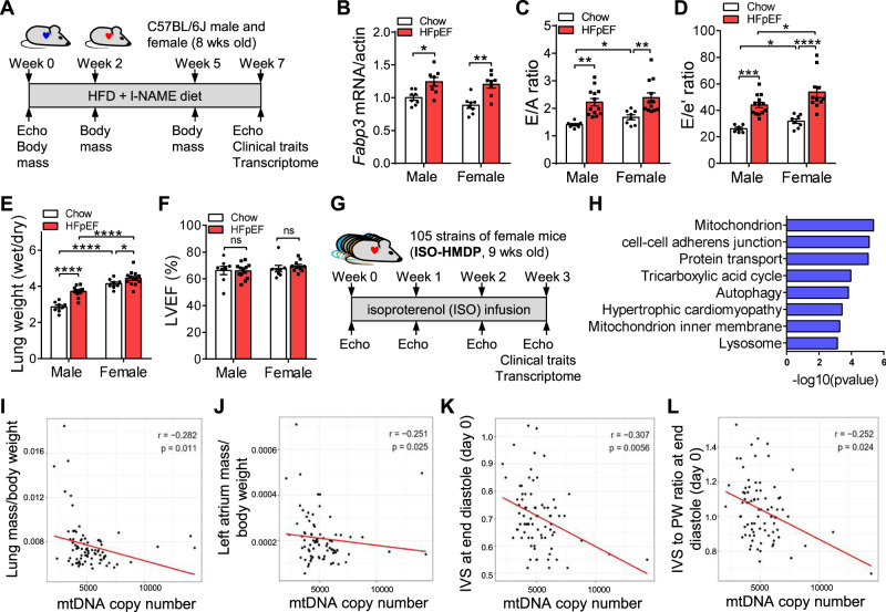
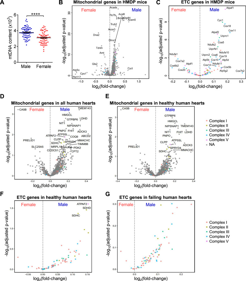
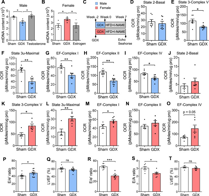
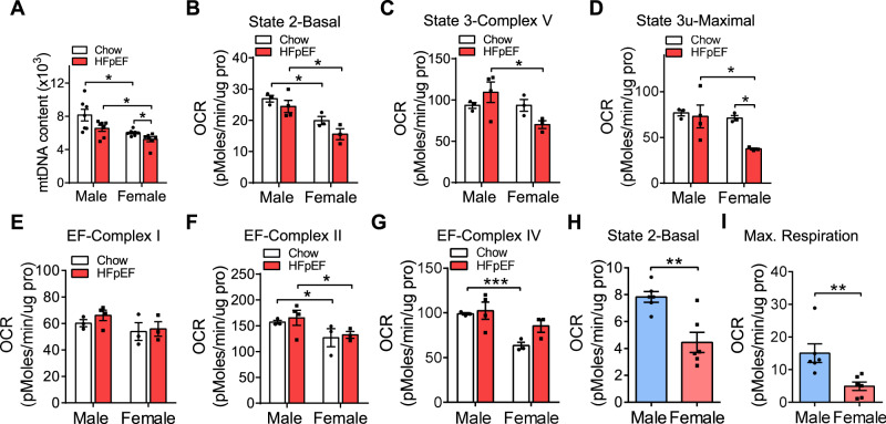
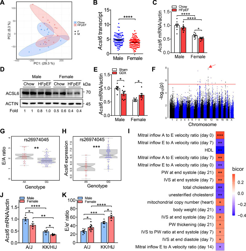
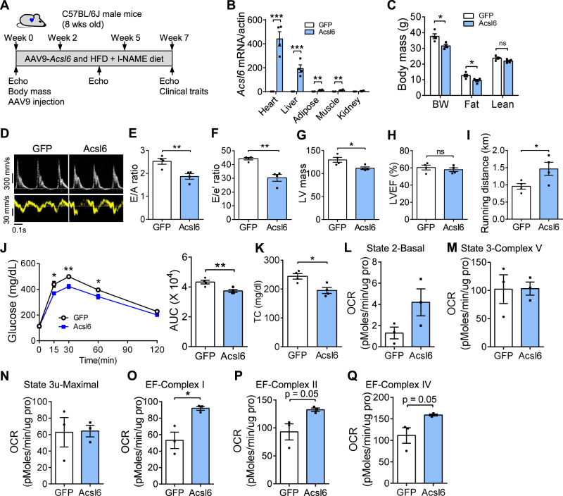
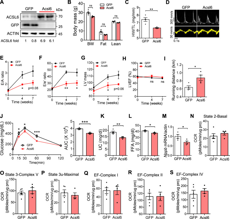
Heart failure with preserved ejection fraction (HFpEF) exhibits a sex bias, being more common in women than men, and we hypothesize that mitochondrial sex differences might underlie this bias. As part of genetic studies of heart failure in mice, we observe that heart mitochondrial DNA levels and function tend to be reduced in females as compared to males. We also observe that expression of genes encoding mitochondrial proteins are higher in males than females in human cohorts. We test our hypothesis in a panel of genetically diverse inbred strains of mice, termed the Hybrid Mouse Diversity Panel (HMDP). Indeed, we find that mitochondrial gene expression is highly correlated with diastolic function, a key trait in HFpEF. Consistent with this, studies of a "two-hit" mouse model of HFpEF confirm that mitochondrial function differs between sexes and is strongly associated with a number of HFpEF traits. By integrating data from human heart failure and the mouse HMDP cohort, we identify the mitochondrial gene Acsl6 as a genetic determinant of diastolic function. We validate its role in HFpEF using adenoviral over-expression in the heart. We conclude that sex differences in mitochondrial function underlie, in part, the sex bias in diastolic function.


Conflict of interest statement

The authors declare no competing interests.

Figures

Fig. 1
Fig. 1. Sex-biased diastolic dysfunction in the progress to HFpEF and mtDNA level was associated with diastolic function.
A Experimental timeline for the development of HFpEF in C57BL/6 J mice (8 weeks old). B Fabp3 mRNA level in C57BL/6 J male and female heart after 7 weeks of HFD + l-NAME diet. N = 8. CF E/A ratio (C), E/e’ ratio (D), wet/dry ratio of lung weight (E), and left ventricle ejection fraction (F) of C57BL/6 J male and female mice after 7 weeks of HFD + l-NAME diet. Male-chow, n = 7 (C), 6 (D), or 8 (E, F); male-HFpEF, n = 13 (C, D, F) or 12 (E); female-chow, n = 8; female-HFpEF, n = 12 (C), 11 (D), 13 (E), or 10 (F). B, p (male) = 0.016; p (female) = 0.001. C p (male) = 0.002; p (female) = 0.007; p (male–female) = 0.03. D p (male) = 0.001; p (female) < 0.0001; p (male–female) = 0.03 (chow) and 0.04 (HFpEF). E p (male) < 0.0001; p (female) = 0.04; p (male–female) < 0.0001 (chow) and < 0.0001 (HFpEF). G. Experimental design for the development of isoproterenol (ISO) induced cardiomyopathy across HMDP mice. Nine-week-old female mice from 105 of the HMDP inbred strains were treated with isoproterenol (30 mg/kg/day) via an intra-abdominally implanted osmotic pump for 21 days. Heart function at baseline (prior to ISO pump implantation), week 1, week 2, and week 3 were obtained with echocardiography. After 3 weeks of ISO infusion, mice were sacrificed and left ventricle transcriptome was performed. N = 3–12 for each strain. H Enriched pathways of genes that were significantly associated with mitral inflow E to A velocity ratio (E/A ratio) across female ISO-HMDP cohort. Pathway analysis was performed with The Database for Annotation, Visualization, and Integrated Discovery (DAVID). Scores from DAVID are modified Fisher tests, corrected for false discovery rate (FDR) using the Benjamini–Hochberg (BH) method. IL mtDNA copy number significantly correlated with lung mass/body weight (I), left atrium mass/body weight (J), IVS at end-diastole (day 0, K), and IVS to PW ratio at end-diastole (day 0, L) in ISO-HMDP mice. Each point represents a mouse from an inbred strain. IVS interventricular septal thickness, PW posterior wall thickness. p-values are from biweight midcorrelation (bicor) tests. Each point represents a mouse. All data are presented as the mean ± SEM. ns not significant, *P < 0.05, **P < 0.01, ***P < 0.001, and ****p < 0.0001 by two-way ANOVA (BF). Source data are provided as a Source Data file.
Fig. 2
Fig. 2. Mitochondrial gene expression in human and mouse hearts.
A Heart mtDNA copy number in male and female HMDP mice (about 10 weeks old). Each point represents a mouse from an inbred strain. N = 72. P < 0.0001. B, C Differential expressed genes encoding mitochondrial proteins (B) and electron transport chain (ETC) proteins (C) in 100 strains of HMDP mice. D, E Differential expressed mitochondrial genes in all human hearts (D) and healthy human hearts (E). F, G Differential expressed ETC genes in healthy (F) and failing (G) human hearts. Genes of each complex were denoted. All data are presented as the mean ± SEM. ****P < 0.0001 by two-sided Student’s t-test (A). DESeq2 results use the Wald test, corrected for false discovery rate (FDR) using the Benjamini–Hochberg (BH) method (BG). Source data are provided as a Source Data file.
Fig. 3
Fig. 3. Mitochondrial and diastolic function in gonadectomized mice.
A, B Heart mtDNA copy number in C57BL/6 J male (A) and female (B) mice subjected to sham, gonadectomy or hormone replacement after gonadectomy under chow diet. mtDNAs were normalized to nuclear DNA. N = 4. A p = 0.019 (Sham/GDX) and 0.013 (GDX/Testosterone). B p = 0.036 (Sham/GDX), and 0.04 (GDX/Estrogen). C Experimental timeline for the gonadectomy and induction of HFpEF in C57BL/6 J male and female mice. DI Oxygen consumption rates (OCR) of isolated mitochondria from C57BL/6 J male hearts were measured by Seahorse assay. The coupling assay measures basal respiration in presence of palmitoyl-carnitine, state 3 (ADP) and state 3 u (FCCP). The electron flow measures complex I (palmitoyl-carnitine), complex II (succinate), and complex IV (TMPD) respiration. N = 7. E p = 0.012. F p = 0.001. G p = 0.008. H p = 0.001. I p = 0.017. JO Same Seahorse assays were performed in C57BL/6 J female mice after gonadectomy and 7 weeks of HFD + l-NAME feeding. Sham, n = 5; GDX, n = 6. K p = 0.013. L p = 0.006. M p = 0.049. P, Q E/e’ ratio (P) and LVEF (Q) of C57BL/6 J male mice after gonadectomy and 7 weeks of HFD + l-NAME diet. N = 11. P p = 0.027. RT E/e’ ratio (R), E/A ratio (S), and LVEF (T) of C57BL/6 J female mice after gonadectomy and 7 weeks of HFD + l-NAME diet. Sham, n = 6; GDX, n = 7. R p = 0.0001. S p = 0.016. Each point represents a mouse. All data are presented as the mean ± SEM. ns not significant, *P < 0.05, **P < 0.01, and ***P < 0.001, by one-way ANOVA (A, B) or by two-sided Student’s t-test (DT). Source data are provided as a Source Data file.
Fig. 4
Fig. 4. Female mice exhibited reduced mitochondrial function.
A Heart mtDNA content in C57BL/6 J male and female mice (8 weeks old) fed with chow diet or HFD + l-NAME for 7 weeks. N = 7. p (male-chow/female-chow) = 0.011; p (male-HFpEF/female-HFpEF) = 0.015; p (female) = 0.03. BG Oxygen consumption rates (OCR) of isolated mitochondria from C57BL/6 J male and female hearts were measured by Seahorse assay. The coupling assay measures basal respiration in presence of palmitoyl-carnitine, state 3 (ADP), and state 3 u (FCCP). The electron flow measures complex I (palmitoyl-carnitine), complex II (succinate), and complex IV (TMPD) respiration. Chow, n = 3; HFpEF, n = 4 (male) or 3 (female). B p (male-chow/female-chow) = 0.014; p (male-HFpEF/female-HFpEF) = 0.022. C p (male-HFpEF/female-HFpEF) = 0.046. D p (male-HFpEF/female-HFpEF) = 0.048; p (female-chow/female-HFpEF) = 0.01. F p (male-chow/female-chow) = 0.026; p (male-HFpEF/female-HFpEF) = 0.043. G p (male-chow/female-chow) = 0.0009. HI Adult cardiomyocytes were isolated from male and female mice after 7 weeks of HFD + l-NAME feeding. OCR was measured before and after the sequential injection of 1.5 µM oligomycin, 1.5 µM FCCP, and 4 µM of rotenone/myxothiazol. N = 6. H p = 0.002. I p = 0.009. Each point represents a mouse. All data are presented as the mean ± SEM. *P < 0.05, **P < 0.01, and ***P < 0.001, by two-way ANOVA (AG) or by two-sided Student’s t-test (HI). Source data are provided as a Source Data file.
Fig. 5
Fig. 5. Identification of Acsl6 as a sex-biased cis-regulator of diastolic dysfunction.
A Principal component analysis (PCA) plot of male and female C57BL/6 J mice under chow diet and 7 weeks of HFD + l-NAME diet. DEGs were identified with a false discovery rate (FDR) < 0.05. B Acsl6 mRNA levels in the hearts of male (n = 96) and female (n = 87) HMDP mice. p < 0.0001. C, D qRT-PCR (C) and western blotting (D) showing Acsl6 mRNA and protein in C57BL/6 J male and female hearts under chow and HFpEF condition. mRNA and protein levels were normalized to actin. N = 8. C p (male-chow/female-chow) < 0.0001; p (male-HFpEF/female-HFpEF) < 0.0001; p (female-chow/female-HFpEF) = 0.014. E qRT-PCR showing mRNA levels of Acsl6 in C57BL/6 J male and female heart after gonadectomy and 7 weeks of HFD + l-NAME diet. mRNA levels were normalized to actin. Male, n = 8; female, n = 6 (sham) or 7 (GDX). p (male) = 0.019; p (female) = 0.024. F Manhattan plot showing the significance (−log10 of p) of all SNPs and eQTL of Acsl6 in female mice after 3 weeks of isoproterenol infusion (female ISO-HMDP). The significant threshold (red line) of p = 4.1 × 10−6 is indicated. Association p-values from FaST-LMM are from Wald tests uncorrected for FDR. G, H E/A ratio distribution (G) and Acsl6 heart expression (H) based on genotype distribution at peak SNP associated with Acsl6 on chromosome 11 (rs26974045). Box and whisker plot depicting mean and distribution. Boxplots are shown centered on medians with upper and lower quartiles indicated by the box boundaries; whiskers enclose the box ± 1.5× the interquartile range (IQR). AA allele, n = 30; GG allele, n = 75. G p = 0.005. H p = 0.0002. I The correlation between local genetic variation in Acsl6 expression (cis-eQTL) and parameters of diastolic function and metabolism in ISO-HMDP. p-values are from biweight midcorrelation (bicor) tests. J, K Acsl6 expression (J) and E/e’ ratio (K) in male and female A/J and KK/HIJ strains of mice after 7 weeks of HFD + l-NAME feeding. J n = 4. K A/J-male, n = 7; A/J-female, n = 8; KK/HIJ-male, n = 6; KK/HIJ-female, n = 4. J p (A/J-male/A/J-female) = 0.025; p (A/J-male/KK/HIJ-male) < 0.0001; p (A/J-female/KK/HIJ-female) = 0.001; p (KK/HIJ-male/KK/HIJ-female) = 0.038. K, p (A/J-male/A/J-female) = 0.025; p (A/J-male/KK/HIJ-male) = 0.0001; p (A/J-female/KK/HIJ-female) < 0.0001; p (KK/HIJ-male/KK/HIJ-female) = 0.021. Each point represents a mouse. All data are presented as the mean ± SEM. *P < 0.05, **P < 0.01, ***P < 0.001, and ****p < 0.0001, by two-way ANOVA (C, E, J, K) or by two-sided Student’s t-test (B, G, H). Source data are provided as a Source Data file.
Fig. 6
Fig. 6. Acsl6 improves diastolic function and mitochondrial function in C57BL/6 J male mice in HFpEF model.
C57BL/6 J male mice (8 weeks old) were injected with AAV9-GFP and AAV9-Acsl6. Mice were fed with HFD + l-NAME for 7 weeks to induce HFpEF. After feeding, diastolic function and mitochondrial function were examined. A Experimental timeline. B qRT-PCR showing Acsl6 expression in the heart, liver, adipose tissue, skeletal muscle, and kidney after AAV injection. N = 4. p (heart) = 0.0003; p (liver) = 0.0008; p (adipose) = 0.002; p (muscle) = 0.004. C Body composition. BW body weight, Fat fat mass, Lean lean mass. N = 4. p (BW) = 0.015; p (fat) = 0.033. DH Representative images of echo echocardiography (D), E/A ratio (E), E/e’ ratio (F), LV mass (G), and LVEF (H) were examined. N = 4. E p = 0.009. F p = 0.002. G p = 0.025. IK Running distance (I), glucose-tolerance test and area under curve (AUC, J), and plasma total cholesterol (K) were examined after the feeding. N = 4. I p = 0.049. J p = 0.009. K p = 0.012. LQ OCR of isolated heart mitochondria were measured by Seahorse assay. The coupling assay measures basal respiration in presence of palmitoyl-carnitine, state 3 (ADP) and state 3 u (FCCP). The electron flow measures complex I (palmitoyl-carnitine), complex II (succinate), and complex IV (TMPD) respiration. N = 3. O p = 0.018. Each point represents a mouse. All data are presented as the mean ± SEM. ns not significant, *P < 0.05, **P < 0.01, and ***P < 0.001, by two-sided Student’s t-test. Source data are provided as a Source Data file.
Fig. 7
Fig. 7. Acsl6 improves diastolic function in C57BL/6 J female mice in HFpEF model.
C57BL/6 J female mice (8 weeks old) were injected with AAV9-GFP and AAV9-Acsl6. Mice were fed with HFD + l-NAME for 7 weeks to induce HFpEF. After feeding, diastolic function and mitochondrial function were examined. A Western blotting showing protein levels of ACSL6 in the heart of C57BL/6 J female mice treated with AAV9-GFP and AAV9-Acsl6. ACSL6-fold change was normalized to ACTIN. Experiment was independently repeated three times. B Body composition after the feeding. BW body weight, Fat fat mass, Lean lean mass. N = 4. CH Heart wight/tibia length ratio (C), representative images of echocardiography (D), E/A ratio (E), E/e’ ratio (F), LV mass (G), and LVEF (H) were examined. CF, H n = 4. G, n = 5 (GFP) or 4 (Acsl6). C p = 0.004. E p (week 4) = 0.03. F p (week 4) = 0.003; p (week 7) = 0.048. G p (week 4) = 0.042. I, J Running distance and glucose-tolerance test (J) after HFD + l-NAME feeding. N = 4. I p = 0.024. J p = 0.0007. KM Plasma unesterified cholesterol (K), free fatty acids (FFA, L), and heart Nppb mRNA level (M). N = 4. K p = 0.005. L p = 0.019. M p = 0.043. NS OCR of isolated heart mitochondria were measured by Seahorse assay. GFP, n = 5; Acsl6, n = 4. Each point represents a mouse. All data are presented as the mean ± SEM. ns not significant, *P < 0.05, **P < 0.01, and ***P < 0.001, by two-sided Student’s t-test. Source data are provided as a Source Data file.

Comment in

References

    1. Dunlay SM, Roger VL, Redfield MM. Epidemiology of heart failure with preserved ejection fraction. Nat. Rev. Cardiol. 2017;14:591–602. doi: 10.1038/nrcardio.2017.65. - DOI - PubMed
    1. Iwano H, Little WC. Heart failure: what does ejection fraction have to do with it? J. Cardiol. 2013;62:1–3. doi: 10.1016/j.jjcc.2013.02.017. - DOI - PubMed
    1. Zheng SL, et al. Drug treatment effects on outcomes in heart failure with preserved ejection fraction: a systematic review and meta-analysis. Heart. 2018;104:407–415. doi: 10.1136/heartjnl-2017-311652. - DOI - PMC - PubMed
    1. Hogg K, Swedberg K, McMurray J. Heart failure with preserved left ventricular systolic function; epidemiology, clinical characteristics, and prognosis. J. Am. Coll. Cardiol. 2004;43:317–327. doi: 10.1016/j.jacc.2003.07.046. - DOI - PubMed
    1. Elgendy IY, Mahtta D, Pepine CJ. Medical therapy for heart failure caused by ischemic heart disease. Circ. Res. 2019;124:1520–1535. doi: 10.1161/CIRCRESAHA.118.313568. - DOI - PMC - PubMed

Publication types