. 2022 Jul 4;5(1):656.
doi: 10.1038/s42003-022-03635-y.

Exercise-induced CITED4 expression is necessary for regional remodeling of cardiac microstructural tissue helicity

Affiliations

Exercise-induced CITED4 expression is necessary for regional remodeling of cardiac microstructural tissue helicity

Robert A Eder et al. Commun Biol. .

Erratum in

Abstract

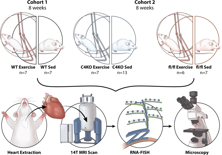
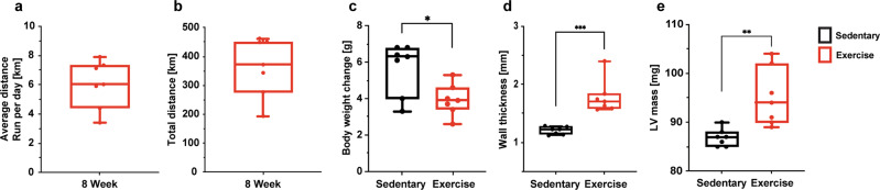
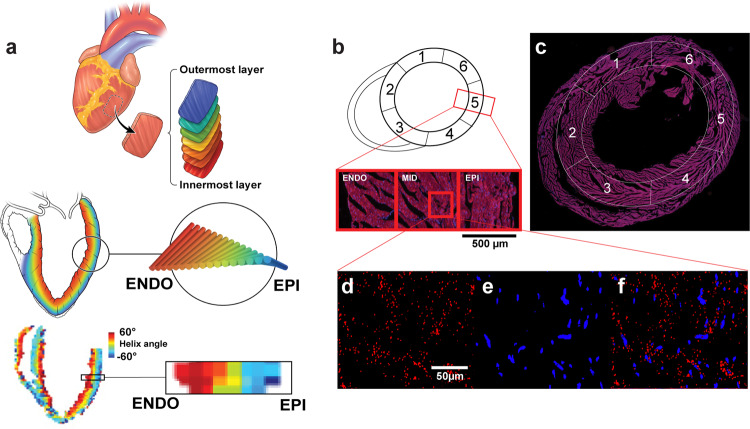
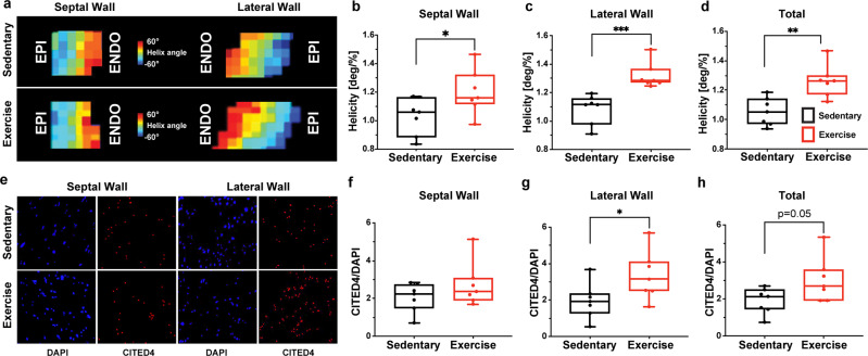
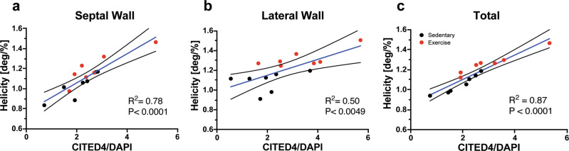
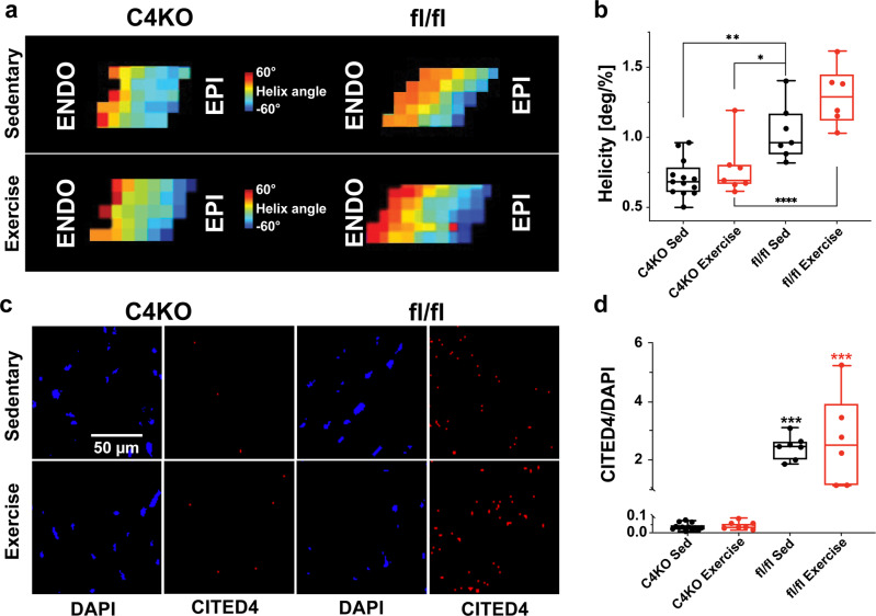
Both exercise-induced molecular mechanisms and physiological cardiac remodeling have been previously studied on a whole heart level. However, the regional microstructural tissue effects of these molecular mechanisms in the heart have yet to be spatially linked and further elucidated. We show in exercised mice that the expression of CITED4, a transcriptional co-regulator necessary for cardioprotection, is regionally heterogenous in the heart with preferential significant increases in the lateral wall compared with sedentary mice. Concordantly in this same region, the heart's local microstructural tissue helicity is also selectively increased in exercised mice. Quantification of CITED4 expression and microstructural tissue helicity reveals a significant correlation across both sedentary and exercise mouse cohorts. Furthermore, genetic deletion of CITED4 in the heart prohibits regional exercise-induced microstructural helicity remodeling. Taken together, CITED4 expression is necessary for exercise-induced regional remodeling of the heart's microstructural helicity revealing how a key molecular regulator of cardiac remodeling manifests into downstream local tissue-level changes.


Conflict of interest statement

The authors declare no competing interests.

Figures

Fig. 1
Fig. 1. Characterization of the experimental set-up.
Cohort 1 consisted of wild-type sedentary (n = 7) and wild-type exercise (n = 7) mice. Cohort 2 included cardiomyocyte-specific CITED4 knock out (C4KO) exercise (n = 7), C4KO sedentary (n = 13), fl/fl exercise (n = 6) and fl/fl sedentary (n = 7) mice. These groups were housed for 8 weeks, and the exercise group had free access to a running wheel. The hearts were perfused and excised after the 8-week holding period. The hearts were imaged using 14 T magnetic resonance imaging (MRI) followed by RNA-FISH of a whole axial slice of the left ventricle. These slides were imaged and analyzed with a fluorescent light microscope.
Fig. 2
Fig. 2. Wheel running promotes significant changes in body weight, heart mass, and thickness.
a The average distance cohort 1 ran per day in kilometers (km). b The average total km cohort 2 ran through 8-weeks. c The body weight changes between the sedentary and exercise groups of cohort 1. d The difference in wall thickness between the sedentary and exercise groups of cohort 1. e The difference in left ventricular (LV) mass between the exercise and sedentary groups. Unpaired two-tailed t-test *P < 0.05, **P < 0.01, ***P < 0.001. Data are presented min to max. Within each box, horizontal black and red lines denote median values; boxes extend from the 25th to the 75th percentile of each group’s distribution of values.
Fig. 3
Fig. 3. Helix angle analysis and RNA-FISH imaging and quantification of exercise and sedentary mouse hearts.
a Helix angles change from the endocardium to epicardium between sedentary and exercised mice. b A visual representation of the location of the six AHA sections and a zoomed in images of the complete cross-section across each transmural layer. c Complete image of sectioned mouse tissue with AHA sections overlayed. d CITED4 signal isolated within mid-myocardium. e DAPI signal isolated and magnified in the mid-myocardium. f CITED4 and DAPI signal imaged from mid-myocardium overlayed.
Fig. 4
Fig. 4. Wheel running in mice promotes physiological remodeling in the heart in coordination with spatial upregulations of CITED4.
a Representative DT-MRI images of helicity changes from the endocardium to epicardium between sedentary and exercised mice in cohort 1. bd Helicity differences within the septum, lateral wall and averaged across all AHA sections. e Magnified representative images of DAPI and CITED4 within the septal and lateral wall regions within exercised and sedentary mice. fh CITED4 signal in sedentary and exercised mice within the septum, lateral wall and averaged across all AHA sections. Unpaired two-tailed t-test. *P < 0.05, **P < 0.01, ***P < 0.001. Data are presented min to max. Within each box, horizontal black and red lines denote median values; boxes extend from the 25th to the 75th percentile of each group’s distribution of values.
Fig. 5
Fig. 5. Regional helicity changes are significantly correlated with CITED4/DAPI expression.
ac Simple linear regressions of CITED4/DAPI signal and helicity within the septum, lateral wall and averaged across all AHA sections. Black dots represent sedentary animals and red dots represent exercised animals. The blue line represents the total simple linear regression of both groups. *P < 0.05, **P < 0.01, ***P < 0.001. Data presented with mean and 95% confidence interval.
Fig. 6
Fig. 6. Cardiomyocyte-specific knock out of CITED4 impacts regional tissue helicity changes.
a Helix angle changes differ significantly between groups. b An analysis of helicity between all groups of cohort 2. c Images of DAPI and CITED4 staining of representative mice from cohort 2. d Quantification of CITED4/DAPI using RNA-FISH with cohort 2. Two-way ANOVA used for multivariant analyses *P < 0.05, **P < 0.01, ***P < 0.001. Data are presented min to max. Within each box, horizontal black and red lines denote median values; boxes extend from the 25th to the 75th percentile of each group’s distribution of values.

References

    1. Vujic A, et al. Exercise induces new cardiomyocyte generation in the adult mammalian heart. Nat. Commun. 2018;9:1–9. doi: 10.1038/s41467-018-04083-1. - DOI - PMC - PubMed
    1. Bezzerides VJ, et al. CITED4 induces physiologic hypertrophy and promotes functional recovery after ischemic injury. JCI Insight. 2016;1:1–14. doi: 10.1172/jci.insight.85904. - DOI - PMC - PubMed
    1. Bernardo BC, Weeks KL, Pretorius L, McMullen JR. Molecular distinction between physiological and pathological cardiac hypertrophy: Experimental findings and therapeutic strategies. Pharmacol. Ther. 2010;128:191–227. doi: 10.1016/j.pharmthera.2010.04.005. - DOI - PubMed
    1. Liu X, et al. MiR-222 is necessary for exercise-induced cardiac growth and protects against pathological cardiac remodeling. Cell Metab. 2015;21:584–595. doi: 10.1016/j.cmet.2015.02.014. - DOI - PMC - PubMed
    1. DeMaria AN, Neumann A, Lee G, Fowler W, Mason DT. Alterations in ventricular mass and performance induced by exercise training in man evaluated by echocardiography. Circulation. 1978;57:237–244. doi: 10.1161/01.CIR.57.2.237. - DOI - PubMed

Publication types

Substances