. 2022 Jul 4;15(1):59.
doi: 10.1186/s13041-022-00944-5.

Increased spine PIP3 is sequestered from dendritic shafts

Affiliations

Increased spine PIP3 is sequestered from dendritic shafts

Yoshibumi Ueda et al. Mol Brain. .

Abstract

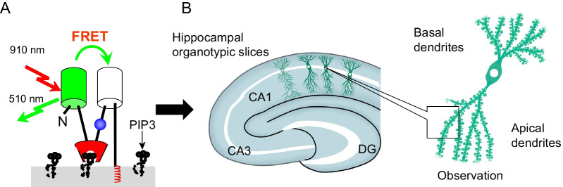
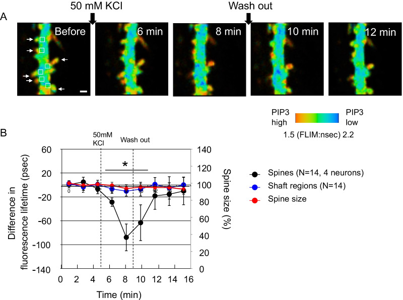
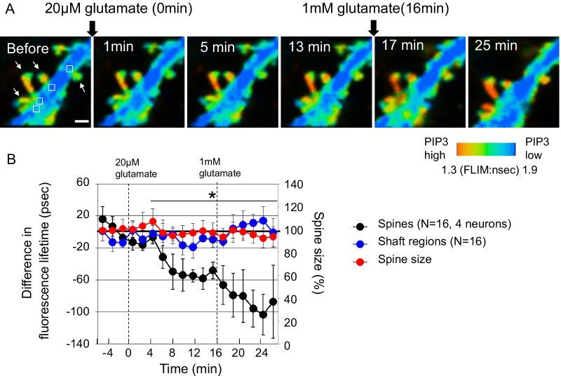
Phosphatidylinositol 3,4,5-trisphosphate (PIP3) is a lipid second messenger that is crucial for the synaptic plasticity underlying learning and memory in pyramidal neurons in the brain. Our previous study uncovered PIP3 enrichment in the dendritic spines of hippocampal pyramidal neurons in the static state using a fluorescence lifetime-based PIP3 probe. However, the extent to which PIP3 enrichment is preserved in different states has not been fully investigated. Here, we revealed that PIP3 accumulation in dendritic spines is strictly controlled even in an active state in which PIP3 is increased by glutamate stimulation and high potassium-induced membrane depolarization. Time-course PIP3 analysis clarified the gradual PIP3 accumulation in dendritic spines over days during neuronal development. Collectively, these results deepen our understanding of PIP3 dynamics in dendritic spines, and the dysregulation of the PIP3 gradient between dendritic spines and shafts could cause neuronal diseases and mental disorders, such as autism spectrum disorder.

Keywords: Dendritic spine; Fluorescence lifetime; Fluorescence resonance energy transfer; Hippocampus; Phosphatidylinositol 3,4,5-trisphosphate; Two-photon microscopy.


Conflict of interest statement

The authors declare that they have no competing interests.

Figures

Fig. 1
Fig. 1
Experimental procedure to observe PIP3 dynamics with FLIMPA3 in hippocampal organotypic slices. A Principle of the use of FLIMPA3 to observe PIP3 dynamics. PIP3 production induces a conformational change in FLIMPA3 through the binding of the PH domain to PIP3, leading to an increase in FRET between monomeric GFP (mGFP) and sREARCh. B FLIMPA3 was expressed in CA1 pyramidal neuronal cells in hippocampal organotypic slices of rat brains. Then, fluorescence lifetime imaging was performed on dendritic spines and primary and secondary dendritic shafts by two-photon microscopy. DG indicates dentate gyrus
Fig. 2
Fig. 2
PIP3 is increased in dendritic spines but not dendritic shafts in response to high potassium-induced depolarization. A Fluorescence lifetime imaging of FLIMPA3. A color gradient was used to represent PIP3 levels, with a reddish color indicating a shorter fluorescence lifetime and higher PIP3 levels. White arrows indicate dendritic spines. The fluorescence lifetime values of dendritic shafts corresponding to dendritic spines were analyzed from the regions indicated by white rectangles in the image before KCl stimulation. White bar = 1 µm. B Time-course analysis of the fluorescence lifetime of dendritic spines and shafts. Asterisks denote a statistically significant difference in the fluorescence lifetime difference between dendritic spines and shafts (p < 0.05)
Fig. 3
Fig. 3
Spatiotemporal PIP3 dynamics at dendritic spines and shafts in response to glutamate administration. A Fluorescence lifetime imaging of FLIMPA3 during glutamate administration. White arrows indicate dendritic spines. The fluorescence lifetime values of dendritic shafts corresponding to dendritic spines were analyzed from the regions indicated by white rectangles in the image before glutamate stimulation. White bar = 1 µm. B Time course of fluorescence lifetime changes in FLIMPA3 in dendritic spines and dendritic shafts. Asterisks denote a statistically significant difference in the fluorescence lifetime difference between dendritic spines and shafts (p < 0.05)
Fig. 4
Fig. 4
The change in PIP3 levels in dendritic spines and shafts during neurodevelopment. A Representative images for each time point during neuronal development. The fluorescence lifetime values of dendritic shafts corresponding to dendritic spines were analyzed from the regions indicated by white rectangles in the image for 8 days. FLIMPA3 was transfected one day before observation in every experiment. White bar = 1 µm. B The change in the difference of fluorescence lifetime between dendritic spines and shafts during neuronal development. A total of 110 dendritic spines from 17 neurons were examined. The fluorescence lifetime of the dendritic spines from one neuron was averaged. The values were obtained by subtracting the fluorescence lifetime values of dendritic spines from the fluorescence lifetime values of the corresponding dendritic shafts. A correlation coefficient was calculated by least squares method. C The correlation between days in vitro and spine size. The number of spines same with B was analyzed

References

    1. Kakumoto T, Nakata T. Optogenetic control of PIP3: PIP3 is sufficient to induce the actin-based active part of growth cones and is regulated via endocytosis. PLoS ONE. 2013;8:e70861. doi: 10.1371/journal.pone.0070861. - DOI - PMC - PubMed
    1. Jiang H, Guo W, Liang X, Rao Y. Both the establishment and the maintenance of neuronal polarity require active mechanisms: critical roles of GSK-3beta and its upstream regulators. Cell. 2005;120:123–135. - PubMed
    1. Garrido JJ, Simon D, Varea O, Wandosell F. GSK3 alpha and GSK3 beta are necessary for axon formation. FEBS Lett. 2007;581:1579–1586. doi: 10.1016/j.febslet.2007.03.018. - DOI - PubMed
    1. Jaworski J, Spangler S, Seeburg DP, Hoogenraad CC, Sheng M. Control of dendritic arborization by the phosphoinositide-3'-kinase-Akt-mammalian target of rapamycin pathway. J Neurosci. 2005;25:11300–11312. doi: 10.1523/JNEUROSCI.2270-05.2005. - DOI - PMC - PubMed
    1. Shi SH, Jan LY, Jan YN. Hippocampal neuronal polarity specified by spatially localized mPar3/mPar6 and PI 3-kinase activity. Cell. 2003;112:63–75. doi: 10.1016/S0092-8674(02)01249-7. - DOI - PubMed

Publication types