

Kindlin-2 loss in condylar chondrocytes causes spontaneous osteoarthritic lesions in the temporomandibular joint in mice
- PMID: 35788130
- PMCID: PMC9253313
- DOI: 10.1038/s41368-022-00185-1
Kindlin-2 loss in condylar chondrocytes causes spontaneous osteoarthritic lesions in the temporomandibular joint in mice
Abstract
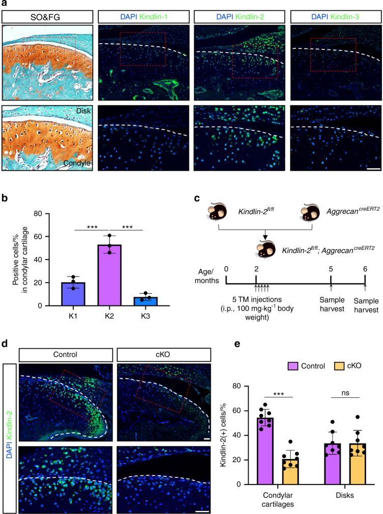
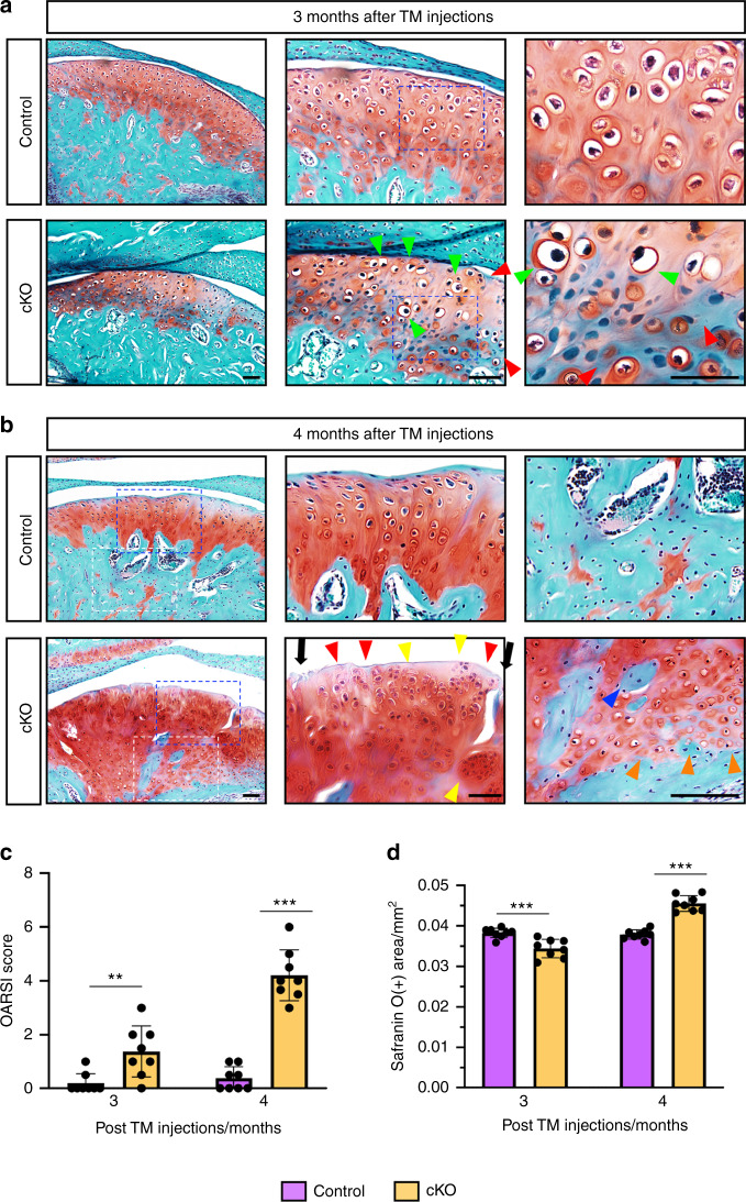
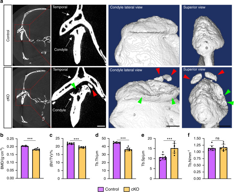
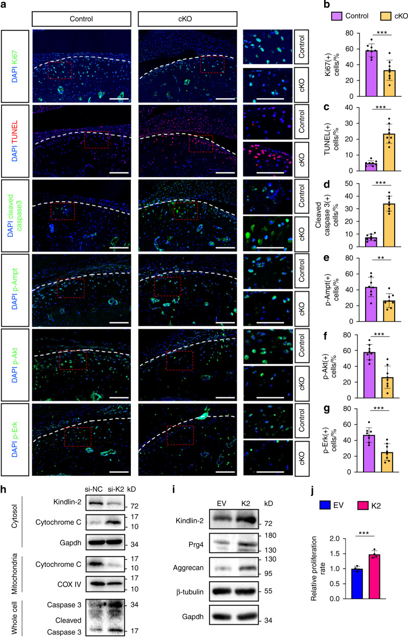
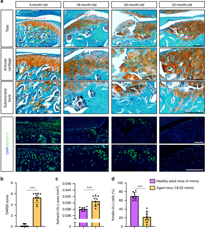
The progressive destruction of condylar cartilage is a hallmark of the temporomandibular joint (TMJ) osteoarthritis (OA); however, its mechanism is incompletely understood. Here, we show that Kindlin-2, a key focal adhesion protein, is strongly detected in cells of mandibular condylar cartilage in mice. We find that genetic ablation of Kindlin-2 in aggrecan-expressing condylar chondrocytes induces multiple spontaneous osteoarthritic lesions, including progressive cartilage loss and deformation, surface fissures, and ectopic cartilage and bone formation in TMJ. Kindlin-2 loss significantly downregulates the expression of aggrecan, Col2a1 and Proteoglycan 4 (Prg4), all anabolic extracellular matrix proteins, and promotes catabolic metabolism in TMJ cartilage by inducing expression of Runx2 and Mmp13 in condylar chondrocytes. Kindlin-2 loss decreases TMJ chondrocyte proliferation in condylar cartilages. Furthermore, Kindlin-2 loss promotes the release of cytochrome c as well as caspase 3 activation, and accelerates chondrocyte apoptosis in vitro and TMJ. Collectively, these findings reveal a crucial role of Kindlin-2 in condylar chondrocytes to maintain TMJ homeostasis.
© 2022. The Author(s).
Conflict of interest statement
The authors declare no competing interests.
Figures

Similar articles
-
Deletion of Axin1 in condylar chondrocytes leads to osteoarthritis-like phenotype in temporomandibular joint via activation of β-catenin and FGF signaling.J Cell Physiol. 2019 Feb;234(2):1720-1729. doi: 10.1002/jcp.27043. Epub 2018 Aug 2. J Cell Physiol. 2019. PMID: 30070692 Free PMC article.
-
Deletion of Bmal1 in aggrecan-expressing cells leads to mouse temporomandibular joint osteoarthritis.J Bone Miner Metab. 2024 Sep;42(5):529-537. doi: 10.1007/s00774-024-01524-4. Epub 2024 Jul 9. J Bone Miner Metab. 2024. PMID: 38981876
-
Loss of Fgfr1 in chondrocytes inhibits osteoarthritis by promoting autophagic activity in temporomandibular joint.J Biol Chem. 2018 Jun 8;293(23):8761-8774. doi: 10.1074/jbc.RA118.002293. Epub 2018 Apr 24. J Biol Chem. 2018. PMID: 29691281 Free PMC article.
-
Pathological mechanism of chondrocytes and the surrounding environment during osteoarthritis of temporomandibular joint.J Cell Mol Med. 2021 Jun;25(11):4902-4911. doi: 10.1111/jcmm.16514. Epub 2021 May 5. J Cell Mol Med. 2021. PMID: 33949768 Free PMC article. Review.
-
Temporomandibular Joint Osteoarthritis: Pathogenic Mechanisms Involving the Cartilage and Subchondral Bone, and Potential Therapeutic Strategies for Joint Regeneration.Int J Mol Sci. 2022 Dec 22;24(1):171. doi: 10.3390/ijms24010171. Int J Mol Sci. 2022. PMID: 36613615 Free PMC article. Review.
Cited by
-
Resatorvid alleviates experimental inflammatory TMJOA by restraining chondrocyte pyroptosis and synovial inflammation.Arthritis Res Ther. 2023 Nov 29;25(1):230. doi: 10.1186/s13075-023-03214-4. Arthritis Res Ther. 2023. PMID: 38031141 Free PMC article.
-
Targeting miR-29 mitigates skeletal senescence and bolsters therapeutic potential of mesenchymal stromal cells.Cell Rep Med. 2024 Aug 20;5(8):101665. doi: 10.1016/j.xcrm.2024.101665. Cell Rep Med. 2024. PMID: 39168101 Free PMC article.
-
A loss of primary cilia by a reduction in mTOR signaling correlates with age-related deteriorations in condylar cartilage.Geroscience. 2024 Dec;46(6):5995-6007. doi: 10.1007/s11357-024-01143-x. Epub 2024 Mar 25. Geroscience. 2024. PMID: 38526843 Free PMC article.
-
FLRT2 mediates chondrogenesis of nasal septal cartilage and mandibular condyle cartilage.Open Med (Wars). 2024 Mar 22;19(1):20240902. doi: 10.1515/med-2024-0902. eCollection 2024. Open Med (Wars). 2024. PMID: 38584835 Free PMC article.
-
Pip5k1γ promotes anabolism of nucleus pulposus cells and intervertebral disc homeostasis by activating CaMKII-Ampk pathway in aged mice.Aging Cell. 2024 Sep;23(9):e14237. doi: 10.1111/acel.14237. Epub 2024 Jun 5. Aging Cell. 2024. PMID: 38840443 Free PMC article.
References
-
- Monasterio, G. et al. in Temporomandibular Joint Pathology—Current Approaches and Understanding. (IntechOpen, London, 2018) 10.5772/intechopen.72496.
Publication types
MeSH terms
Substances
LinkOut - more resources
Full Text Sources
Medical
Molecular Biology Databases
Research Materials
Miscellaneous
