. 2022 Sep 12;81(10):1400-1408.
doi: 10.1136/annrheumdis-2022-222229.

Keratinocyte-derived S100A9 modulates neutrophil infiltration and affects psoriasis-like skin and joint disease

Affiliations

Keratinocyte-derived S100A9 modulates neutrophil infiltration and affects psoriasis-like skin and joint disease

Liliana F Mellor et al. Ann Rheum Dis. .

Abstract

Objectives: S100A9, an alarmin that can form calprotectin (CP) heterodimers with S100A8, is mainly produced by keratinocytes and innate immune cells. The contribution of keratinocyte-derived S100A9 to psoriasis (Ps) and psoriatic arthritis (PsA) was evaluated using mouse models, and the potential usefulness of S100A9 as a Ps/PsA biomarker was assessed in patient samples.

Methods: Conditional S100A9 mice were crossed with DKO* mice, an established psoriasis-like mouse model based on inducible epidermal deletion of c-Jun and JunB to achieve additional epidermal deletion of S100A9 (TKO* mice). Psoriatic skin and joint disease were evaluated in DKO* and TKO* by histology, microCT, RNA and proteomic analyses. Furthermore, S100A9 expression was analysed in skin, serum and synovial fluid samples of patients with Ps and PsA.

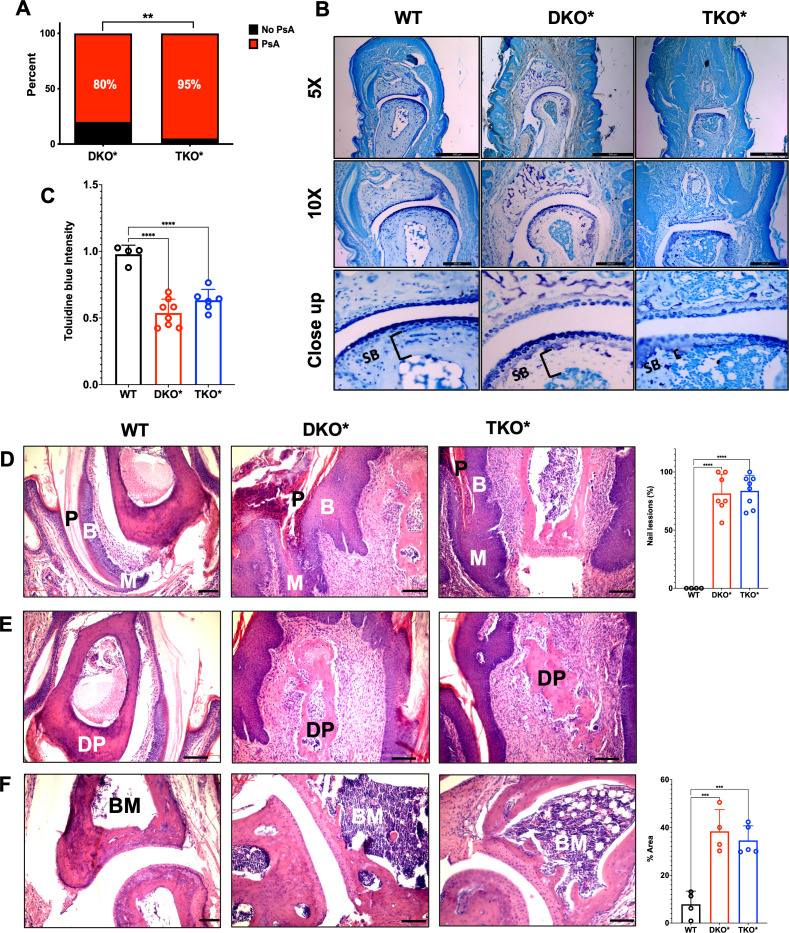
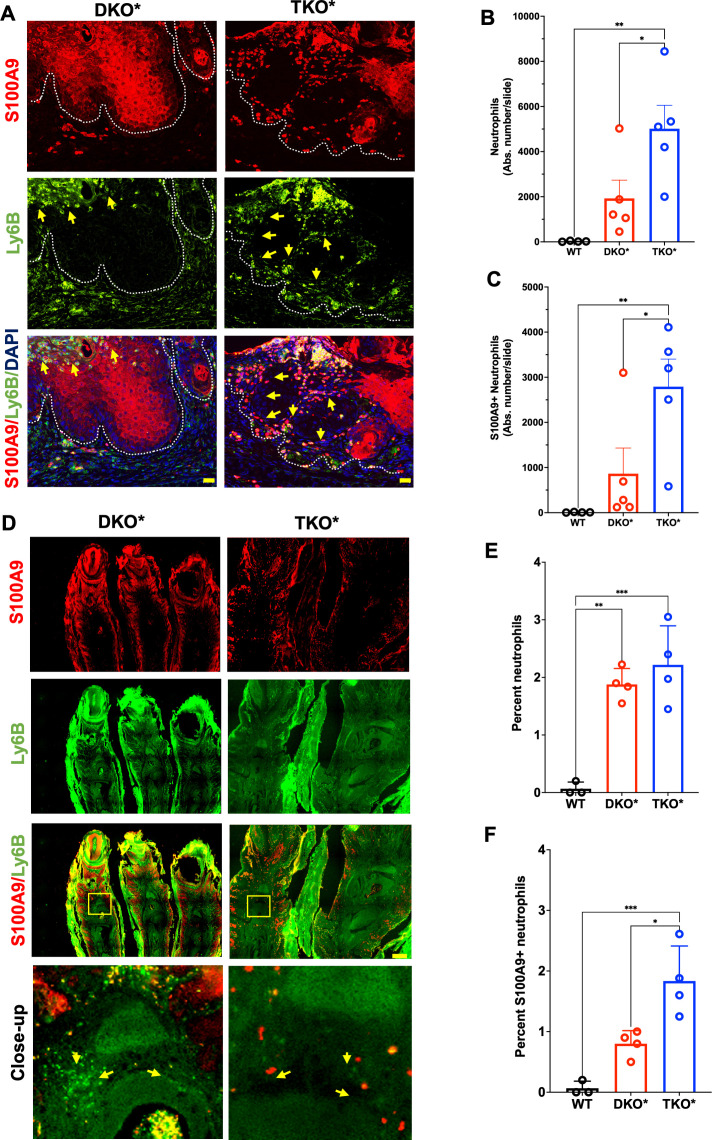
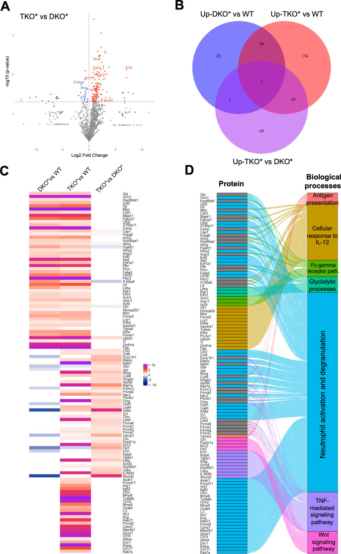
Results: Compared with DKO* littermates, TKO* mice displayed enhanced skin disease severity, PsA incidence and neutrophil infiltration. Altered epidermal expression of selective pro-inflammatory genes and pathways, increased epidermal phosphorylation of STAT3 and higher circulating TNFα were observed in TKO* mice. In humans, synovial S100A9 levels were higher than the respective serum levels. Importantly, patients with PsA had significantly higher serum concentrations of S100A9, CP, VEGF, IL-6 and TNFα compared with patients with only Ps, but only S100A9 and CP could efficiently discriminate healthy individuals, patients with Ps and patients with PsA.

Conclusions: Keratinocyte-derived S100A9 plays a regulatory role in psoriatic skin and joint disease. In humans, S100A9/CP is a promising marker that could help in identifying patients with Ps at risk of developing PsA.

Keywords: cytokines; inflammation; psoriatic arthritis.


Conflict of interest statement

Competing interests: None declared.

Figures

Figure 1
Figure 1
Characterisation of psoriasis-like mouse model with inducible epidermal deletion of S100A9. (A) Immunofluorescence images of the ear skin of mice with inducible dual epidermal deletion of c-Jun and junB (DKO*), inducible triple epidermal deletion of c-Jun, JunB and S100A9 (TKO*) and DKO* mice with total deletion of S100A9 (DKO*-S100A9−/−) 23 days after first tamoxifen (TAM) injection (red: S100A9, green: JunB, scale bar=50 µm). (B) Quantification of S100A9-positive epidermal cells in the ear of DKO*, TKO* and DKO*-S100A9−/− mice (n=4–6). (C) Skin disease severity scoring in DKO* and TKO* mice. (D) Weight of control wild-type (WT), DKO* and TKO* with moderate/severe skin phenotype 23 days after first TAM injection (n=18–26). (E–J) S100A9 (E), S100A8 (F), calprotectin (G), IL-17A (H), IL-6 (I) and TNFα (J) concentrations in the sera of WT, DKO* and TKO* mice with moderate-severe psoriasis-like phenotype (n=4–13).
Figure 2
Figure 2
Psoriatic-arthritic-like (PsA) phenotype in mice with severe psoriasis-like disease. (A) Prevalence of psoriatic arthritis (PsA) in mice with inducible dual epidermal deletion of c-Jun and junB (DKO*) and triple epidermal deletion of c-Jun, JunB and S100A9 (TKO*) with severe psoriasis-like disease (n=20 per group). (B) Toluidine blue staining of the distal interphalangeal (DIP) joint of control wild-type (WT), DKO* and TKO* mice with PsA (scale bar=200 µm). (C) Quantification of toluidine blue staining intensity of articular cartilage in WT, DKO* and TKO* mice (WT n=4; DKO* n=8; TKO* n=6; each point represents the median of several joints measured per sample). (D) H&E-stained histological images showing psoriatic nail involvement with changes in the nail plate (P), nail matrix (M) and nail bed (B) of DKO* and TKO* mice and quantification of nail lesions (scale bar=100 µm). (E) H&E histological images of the distal phalanx (DP) showing enthesitis with high immune infiltration in the areas around the bone (scale bar=100 µm). (F) H&E histological images showing osteitis of the bone marrow (BM) of the distal phalanx (DP) in DKO* and TKO* mice (BM=bone marrow) with quantification of per cent area of bone marrow covered by inflammation (scale bar=100 µm).
Figure 3
Figure 3
Neutrophil infiltration in mice with severe psoriasis-like disease. (A) S100A9 (red) and Ly6B (neutrophils; green) immunostaining of lesional ears in mice with inducible dual epidermal deletion of c-Jun and junB (DKO*) and triple epidermal deletion of c-Jun, JunB and S100A9 (TKO*) (dotted lines represent the basal membrane, yellow arrows point to Ly6B/S100A9-double positive cells, scale bar=20 µm). (B–C) Confocal microscopy–based quantification of absolute number of neutrophils (Ly6B-positive) (B) and S100A9-positive neutrophils (C) in the whole ear sections of DKO* and TKO* mice and wild-type (WT) littermates (n=4–5 mice). (D) S100A9 (red) and Ly6B (green) immunostaining of lesional psoriatic arthritis (PsA)–like paws (scale bar=200 µm). Yellow arrows point to infiltrating cells. (E–F) Confocal microscopy–based quantification of neutrophil (Ly6B-positive) (E) and S100A9-positive neutrophil (F) in the distal interphalangeal joints of DKO* and TKO* mice and WT littermates (average of 3–5 regions per paw, n=4 mice).
Figure 4
Figure 4
Proteomic analyses in whole ear extracts of DKO* and TKO* mice. (A) Volcano plot showing upregulated (red) and downregulated (blue) proteins (n=3–5 per condition; p<0.05); proteins in grey are below statistical significance. (B) Venn diagram depicting statistically significant upregulated proteins in (I) mice with inducible dual epidermal deletion of c-Jun and junB (DKO*) compared towild-type (WT) controls, (II) mice with triple epidermal deletion of c-Jun, JunB and S100A9 (TKO*) compared toWT mice and (III) TKO* compared to DKO* mice (n=3–5 per condition; p<0.05). (C) Heat map of significantly upregulated proteins (left) and Alluvial plot (right) of enriched biological processes associated to each protein.
Figure 5
Figure 5
Gene expression in FACS-sorted neutrophils and keratinocytes qRT-PCR analysis in (A–F) neutrophils and in (G–L) keratinocytes from the ears of mice with inducible dual epidermal deletion of c-Jun and junB (DKO*) and triple epidermal deletion of c-Jun, JunB and S100A9 (TKO*) with severe psoriasis-like phenotype (n=4–9). (M) pSTAT3 immunohistochemistry in the ears of DKO* and TKO* mice with severe psoriasis-like phenotype (scale bar=100 µm). (N) Quantification of positive pSTAT3 cells in the epidermis (n=3–4, 4–5 fields per slide).
Figure 6
Figure 6
Analyses in psoriatic patient samples. (A) S100A8 and S100A9 (red) immunofluorescence staining and quantification in human healthy skin and psoriatic skin (n=5, scale bar=100 µm). (B) S100A8, S100A9, S100A8/A9 (Calprotectin), Interleukin (IL)−17, Tumour necrosis factor alpha (TNF-α) and Interleukin (IL)−6 levels in the serum and synovial fluid of patients with psoriatic arthritis (PsA; n=8, ns=non-significant). (C) S100A8, S100A9, S100A8/A9 (Calprotectin), IL-17, TNF-α and IL-6 serum levels of healthy controls (HC), patients with psoriasis (Ps) or patients with PsA (each group n=24).

Comment in

References

    1. Wagner EF, Schonthaler HB, Guinea-Viniegra J, et al. . Psoriasis: what we have learned from mouse models. Nat Rev Rheumatol 2010;6:704–14. 10.1038/nrrheum.2010.157 - DOI - PubMed
    1. Oliveira MdeFSPde, Rocha BdeO, Duarte GV. Psoriasis: classical and emerging comorbidities. An Bras Dermatol 2015;90:9–20. 10.1590/abd1806-4841.20153038 - DOI - PMC - PubMed
    1. Slobodin G, Rosner I, Rozenbaum M, et al. . Psoriatic arthropathy: where now? Isr Med Assoc J 2009;11:430–4. - PubMed
    1. Gottlieb A, Korman NJ, Gordon KB, et al. . Guidelines of care for the management of psoriasis and psoriatic arthritis: section 2. psoriatic arthritis: overview and guidelines of care for treatment with an emphasis on the biologics. J Am Acad Dermatol 2008;58:851–64. 10.1016/j.jaad.2008.02.040 - DOI - PubMed
    1. Mease PJ. Psoriatic arthritis - update on pathophysiology, assessment, and management. Bull NYU Hosp Jt Dis 2010;68:191–8. - PubMed