. 2022 Jul 5;18(7):e1010665.
doi: 10.1371/journal.ppat.1010665. eCollection 2022 Jul.

A new adenine nucleotide transporter located in the ER is essential for maintaining the growth of Toxoplasma gondii

Affiliations

A new adenine nucleotide transporter located in the ER is essential for maintaining the growth of Toxoplasma gondii

Senyang Li et al. PLoS Pathog. .

Erratum in

Abstract

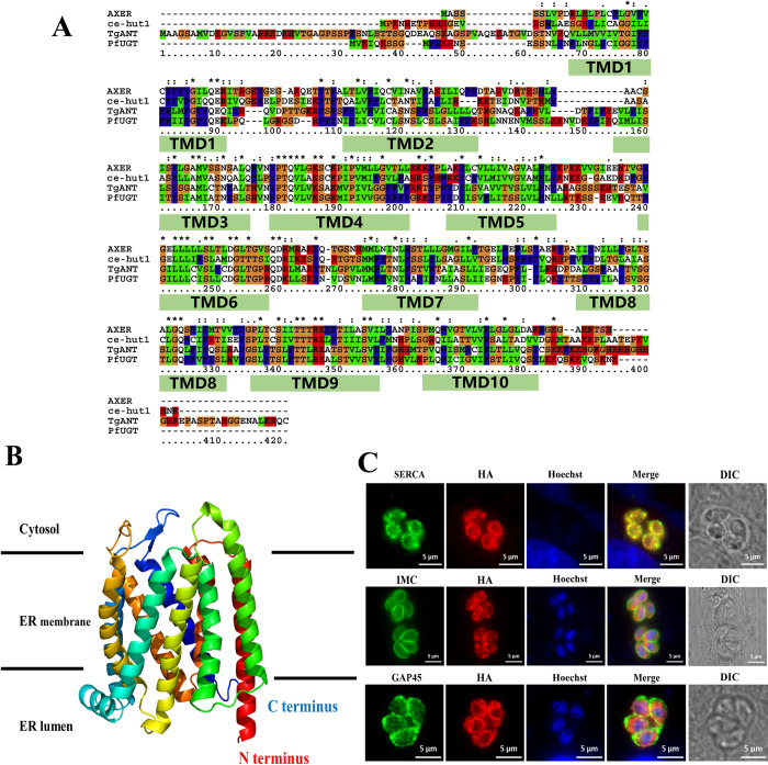
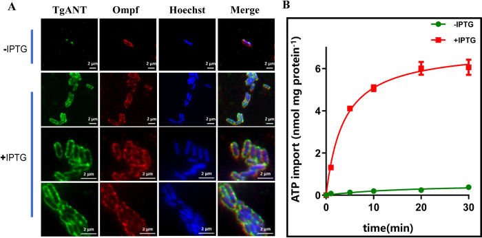
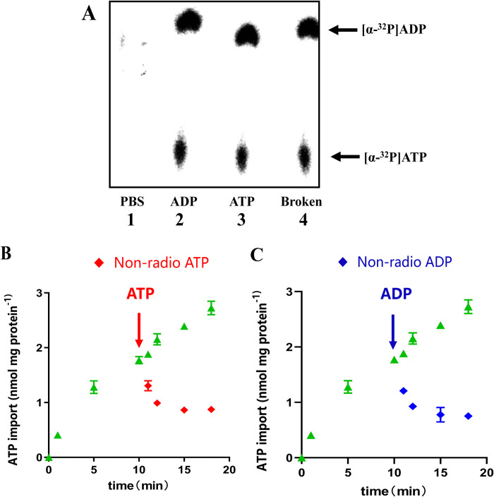
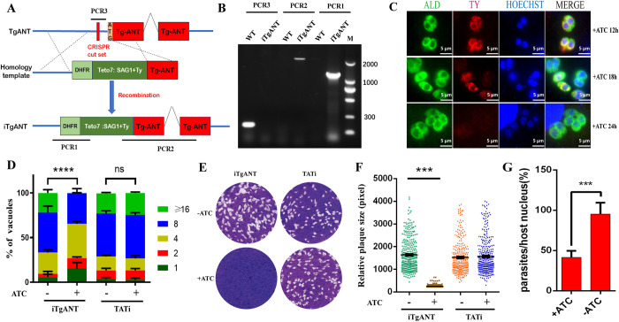
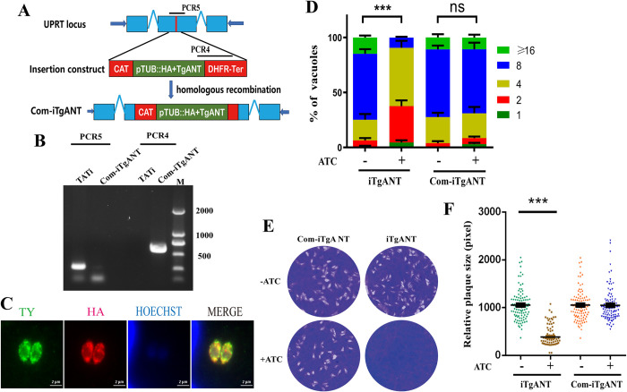
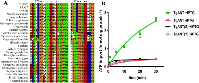
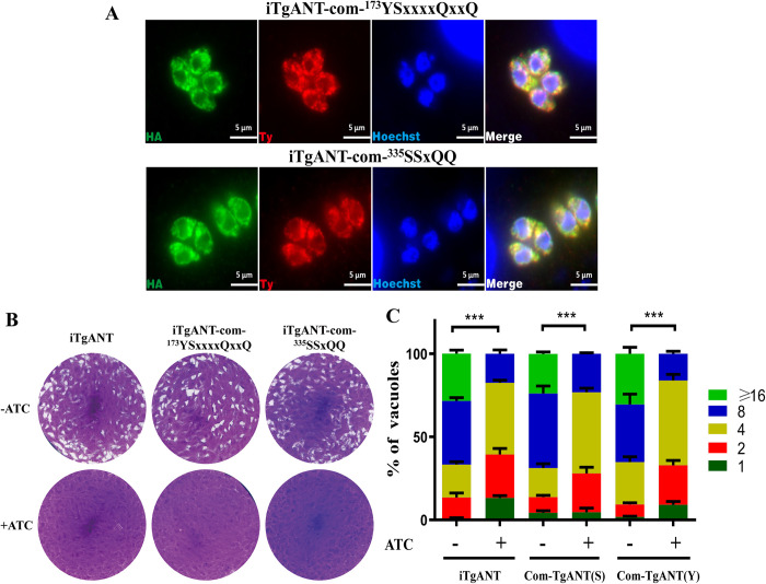
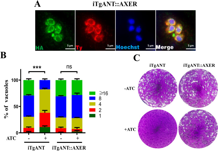
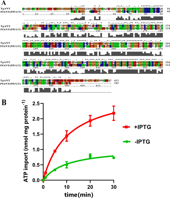
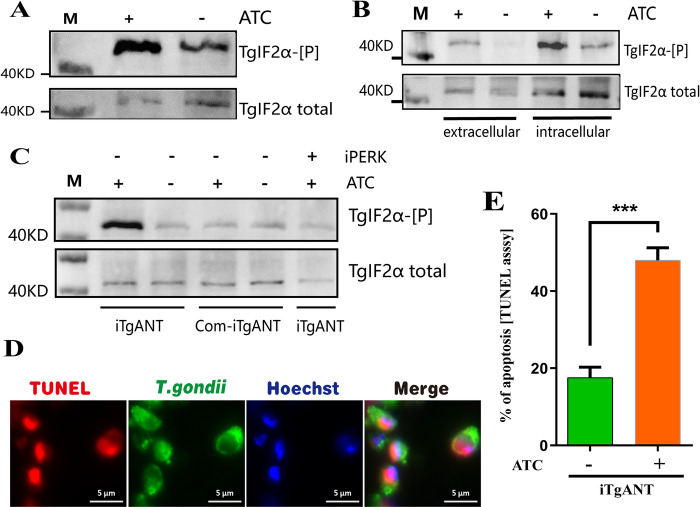
The lumen of the endoplasmic reticulum (ER) is the subcellular site where secretory protein folding, glycosylation and sulfation of membrane-bound proteins, proteoglycans, and lipids occur. The protein folding and degradation in the lumen of the ER require high levels of energy in the form of ATP. Biochemical and genetic approaches show that ATP must first be translocated across ER membrane by particular transporters before serving as substrates and energy sources in the lumenal reactions. Here we describe an ATP/ADP transporter residing in the ER membranes of T.gondii. Immunofluorescence (IFA) assay in transgenic TgANT1-HA tag revealed that TgANT1 is a protein specifically expressed in the ER. In vitro assays, functional integration of TgANT in the cytoplasmic membrane of intact E. coli cells reveals high specificity for an ATP/ADP antiport. The depletion of TgANT leads to fatal growth defects in T.gondii, including a significant slowdown in replication, no visible plaque formation, and reduced ability to invade. We also found that the amino acid mutations in two domains of TgANT lead to the complete loss of its function. Since these two domains are conserved in multiple species, they may share the same transport mechanism. Our results indicate that TgANT is the only ATP/ADP transporter in the ER of T. gondii, and the lack of ATP in the ER is the cause of the death of T. gondii.


Conflict of interest statement

The authors have declared that no competing interests exist.

Figures

Fig 1
Fig 1. Putative structure and intracellular localization of TgANT.
(A) Protein sequences were from NCBI and shown in Gene ID code for Homo sapiens (AXER, Gene ID: 10237), Caenorhabditis elegans (hut-1, Gene ID: 176690), plasmodium (PF3D7-1113300, Gene ID: 810688). The sequences were aligned using ClustalX and TMHMM servers. (B) The hypothetical structural model of TgANT was predicted by the Phyre2 server. The nitrogen terminus and carboxy terminus face the ER lumen, the double lysine motif (-KKQC) is located near the C-terminal end of TgANT. (C) TgANT-HA parasites were subjected to immunostaining using antibodies against the HA tag along with SERCA (sarco/endoplasmic reticulum Ca2+-ATPase, ER protein), The result shows that TgANT was complete co-localization with the ER-resident protein SERCA. But not co-localization with IMC (inner membrane complex, inner membrane protein) and GAP45 (gliding-associated protein 45, plasma membrane protein) of T. gondii. Scale bars: 5 μm.
Fig 2
Fig 2. Heterologously expressed TgANT is an ATP and ADP carrier.
(A) The E. coil harboring pet16b-TgANT-HA were subjected IFA using antibodies against the HA tag, Ompf (outer membrane porin F) and Hoechst. The result shows that TgANT was expressed in the membrane of E. coil. Scale bars: 2 μm. (B) Time course for [α-32P] ATP uptake into intact E. coli cells. IPTG-induced E. coli cells harboring the plasmid encoding TgANT were incubated with 25 nM [α-32P] ATP for the indicated time intervals. Noninduced E. coli cells with the plasmid encoding TgANT were used as control. Means ± s.e.m., three independent assays.
Fig 3
Fig 3. Back exchange experiments with E. coli cells.
(A) E. coli cells expressing TgANT were preloaded with 50nM radioactive [α-32P] ATP for 5 min, followed by carried out back exchange experiment with 5 μM non-labeled ATP, ADP, or PBS for 3 min and terminated by rapid centrifugation. Then, 2 μL sample of the supernatant were loaded onto the TLC PEI cellulose plate. The supernatant of the broken E. coli cells was used to detect the metabolic activity (lane 4). (B) and (C) E. coli cells harboring the plasmid encoding TgANT were incubated with 50 nM [α-32P] ATP for 10 min. Then, efflux was induced by the addition of fifty-fold unlabeled ATP (B) or ADP (C) for 1min, 2min, 5min and 8min. Means ± s.e.m., three independent assays.
Fig 4
Fig 4. Conditional knockdown of TgANT results in severe growth defects.
(A) and (B) schematic showing generation of the iTgANT strains via CRISPR-Cas9–assisted gene engineering, which was done by replacing the endogenous TgANT promoter with a tetracycline regulatable promoter (SAG1::TetO7) in the TATi line (A). PCR1–PCR3 indicates screening of clonal mutant (B). (C) The Ty epitope at the N-terminal of TgANT was detected by IFA (red) at the appointed time after the 0.5 μg/ml ATc treatment. The results showed that TgANT was completely depletion after 24 hours of drug treatment. The fructose-1,6-bisphosphate aldolase (ALD) of T. gondii as a marker. (D) Intracellular replication assay comparing parasite proliferation under indicated conditions. TATi and iTgANT strains were pretreated with 0.5 μg/ml ATc for 40 h or left untreated. Subsequently they were allowed to infect HFF monolayers for 40–60 min, and uninvaded parasites were washed with PBS. Whereafter, invaded parasites were cultured with 0.5 μg/ml ATc or left untreated for 24 h to determine the number of parasites in each parasitophorous vacuole (PV). ***p ≤ 0.001, two-way ANOVA, three independent repeated, a representative one is shown here. (E) and (F) Parasites were grown ±0.5 μg/ml ATc for 7 days to form plaques on HFF monolayers (E). plaque size presented as relative area (pixel size calculated by Photoshop) from three independent assays (F). ***p ≤ 0.001, Student’s t-test, three independent assays. (G) Host cell invasion efficiencies determined by a two-color staining protocol that distinguishes intracellular from extracellular parasites. **p ≤ 0.01, Student’s t-test, three independent repeated, a representative one is shown here.
Fig 5
Fig 5. TgANT complementation fully restored the growth defects of TgANT depletion mutants.
(A) and (B) schematic showing generation of the iTgANT strains via CRISPR-Cas9–assisted gene engineering(A). PCR1–PCR3 indicates screening of clonal mutant (B). (C) IFA confirmed that the ectopic TgANT (red) was successfully expressed and localized to the ER. (D) Intracellular replication assay comparing parasite proliferation under indicated conditions. TATi and Com-iTgANT strains were pretreated with 0.5 μg/ml ATc for 40 h or left untreated. Subsequently, they were allowed to infect HFF monolayers for 40–60 min, and uninvaded parasites were washed with PBS. Whereafter, invaded parasites were cultured with 0.5 μg/ml ATc or left untreated for 24 h to determine the number of parasites in each parasitophorous vacuole (PV). ***p ≤ 0.001, two-way ANOVA, three independent repeated, a representative one is shown here. (E) and (F) Plaque assays comparing the growth of TgANT depletion strain and TgANT complementation strain. Parasites were grown ±0.5 μg/ml ATc for 7 days to form plaques on HFF monolayers (E). plaque size presented as relative area (pixel size calculated by Photoshop) from three independent assays (F) ***p ≤ 0.001, Student’s t-test, three independent repeated.
Fig 6
Fig 6. Conserved protein domain prediction and the absorb experiment of E. coli cells harboring mutant plasmid.
(A) Conserved protein domain of TgANT were searched in NCBI. These two conserved domains are conserved in many species, such as mollusks, fungal, drosophila willistoni. (B) IPTG-induced E. coli cells harboring the plasmid encoding mutant cDNA of TgANT (pet16b-TgANT-173YSxxxxQxxQ, Gray hollow dots, and pet16b-TgANT-335SSxQQ, Black dot) were incubated with 25 nM [α-32P] ATP for the indicated time intervals. IPTG-induced or Noninduced E. coli cells harboring the plasmid encoding wild-type TgANT were used as control. Means ± s.e.m., data of three independent repeated.
Fig 7
Fig 7. The phenotypic experiment of TgANT compensation mutant strains.
(A) IFA confirmed the correct integration and expression in ER of Com-TgANT-173YSxxxxQxxQ strain and Com-TgANT-335SSxQQ strain.HA denote the mutant CDS of TgANT, and Ty denotes the TgANT in situ. (B) plaques experiment of iTgANT strains and mutant compensation strains, parasites were grown ±0.5 μg/ml ATc for 7 days to form plaques on HFF monolayers. (C) iTgANT strains and mutant compensation strains (Com-TgANT(Y) refer to Com-TgANT-173YSxxxxQxxQ strain and Com-TgANT(S) refer to Com-TgANT-335SSxQQ) were pretreated with 0.5 μg/ml ATc for 40 h or left untreated. Subsequently they were allowed to infect HFF monolayers for 40–60 min, and uninvaded parasites were washed with PBS. Whereafter, invaded parasites were cultured with 0.5 μg/ml ATc or left untreated for 24 h to determine the number of parasites in each parasitophorous vacuole (PV). ***p ≤ 0.001, two-way ANOVA, three independent repeated, a representative one is shown here.
Fig 8
Fig 8. The Phenotypic experiment of heterologous supplementation strains.
(A) IFA confirmed the correct integration and expression in ER of iTgANT::AXER strain. (B) and (C) Intracellular replication assay (B) and plaque assay (C) comparing the growth of TgANT depletion strain and AXER complementation strain. ***p ≤ 0.001, two-way ANOVA, three independent repeated, a representative one is shown here.
Fig 9
Fig 9. Homology analysis and [α-32P] ATP absorb experiment.
(A) The sequences of TgANT and PfUNT were aligned using ClustalX. (B) Kinetics of [α-32P] ATP uptake into intact E. coli cells. IPTG-induced E. coli cells harboring the plasmid encoding PfANT were incubated with 25 nM [α-32P] ATP for the indicated time intervals. Noninduced E. coli cells with the plasmid encoding PfANT were used as control. Means ± s.e.m., three independent assays.
Fig 10
Fig 10. ER stress and apoptosis detection.
(A) iTgANT strain were treated with 0.5 μg/ml ATc for 36h or left untreated, subsequently collected the parasites. They were subjected to Western blot analyses using mouse anti-TgIF2α-P and rabbit anti-TgIF2α. (B) Intracellular parasites were treated with 0.5 μg/ml ATc for 36h or left untreated, subsequently collected the extracellular and intracellular parasites. They were subjected to Western blot analyses using mouse anti-TgIF2α-P and rabbit anti-TgIF2α. (C) iTgANT strain and Com-iTgANT were treated with 0.5 μg/ml ATc for 36h or left untreated, subsequently collected the parasites. Besides, the iTgANT strain was treated with ATc for 33h and then add the iPERK for 3h to verify the phosphorylation of TgIF2α is caused by ER stress. All the samples were subjected to Western blot analyses using mouse anti-TgIF2α-P and rabbit anti-TgIF2α. (D) Morphology of iTgANT strain (green, GRA7) and TUNEL labeling in situ (red) following treatment with ATc for 36h. (E) Mean percentages ± SEM of TUNEL-positive T. gondii, results are from three independent experiments, ***P≤0.001, Student’s t-test.

Similar articles

Cited by

References

    1. Schwarz DS, Blower MD. The endoplasmic reticulum: structure, function and response to cellular signaling. Cellular and molecular life sciences: CMLS. 2016;73(1):79–94. Epub 2015/10/05. doi: 10.1007/s00018-015-2052-6 ; PubMed Central PMCID: PMC4700099. - DOI - PMC - PubMed
    1. Li H, Musayev FN, Yang J, Su J, Liu Q, Wang W, et al.. A novel and unique ATP hydrolysis to AMP by a human Hsp70 BiP. Protein science: a publication of the Protein Society. 2021. Epub 2021/12/24. doi: 10.1002/pro.4267 . - DOI - PMC - PubMed
    1. Braakman I, Hebert DN. Protein folding in the endoplasmic reticulum. Cold Spring Harbor perspectives in biology. 2013;5(5):a013201. Epub 2013/05/03. doi: 10.1101/cshperspect.a013201 ; PubMed Central PMCID: PMC3632058. - DOI - PMC - PubMed
    1. Carmeille R, Lomoriello PS, Devarakonda PM, Kellermeier JA, Heaslip AT. Actin and an unconventional myosin motor, TgMyoF, control the organization and dynamics of the endomembrane network in Toxoplasma gondii. Plos Pathogens. 2021;17(2). ARTN e1008787 doi: 10.1371/journal.ppat.1008787 WOS:000616583700001. - DOI - PMC - PubMed
    1. Faust O, Abayev-Avraham M, Wentink AS, Maurer M, Nillegoda NB, London N, et al.. HSP40 proteins use class-specific regulation to drive HSP70 functional diversity. Nature. 2020;587(7834):489–+. doi: 10.1038/s41586-020-2906-4 WOS:000588617900010. - DOI - PubMed

MeSH terms