. 2022 Sep;37(9):1750-1760.
doi: 10.1002/jbmr.4644. Epub 2022 Jul 22.

Skeletal Effects of Inducible ERα Deletion in Osteocytes in Adult Mice

Affiliations

Skeletal Effects of Inducible ERα Deletion in Osteocytes in Adult Mice

Madison L Doolittle et al. J Bone Miner Res. 2022 Sep.

Abstract

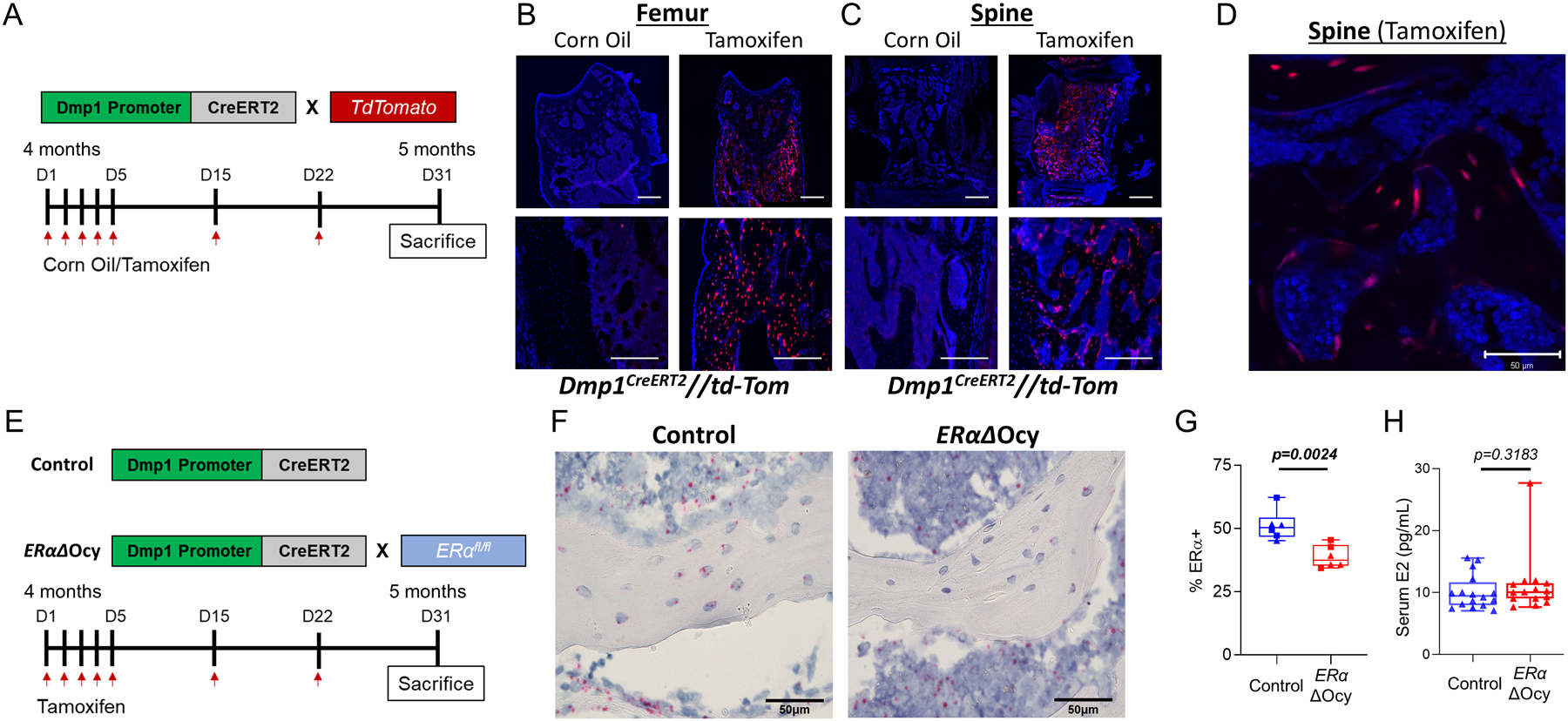
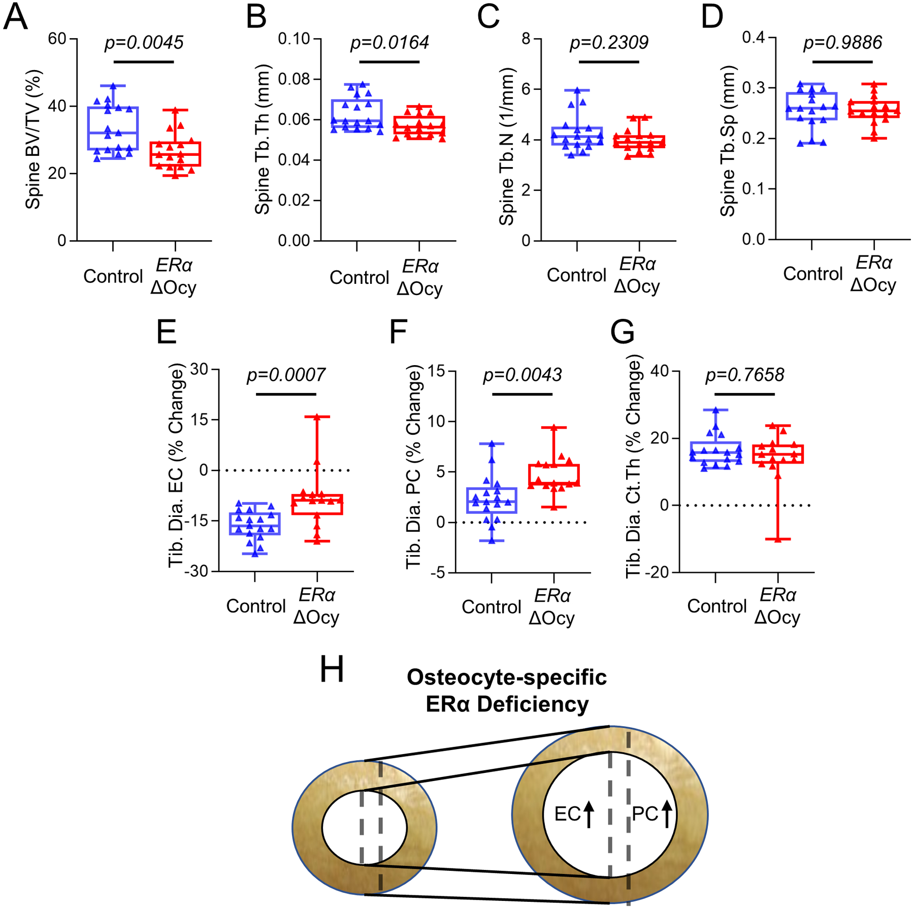
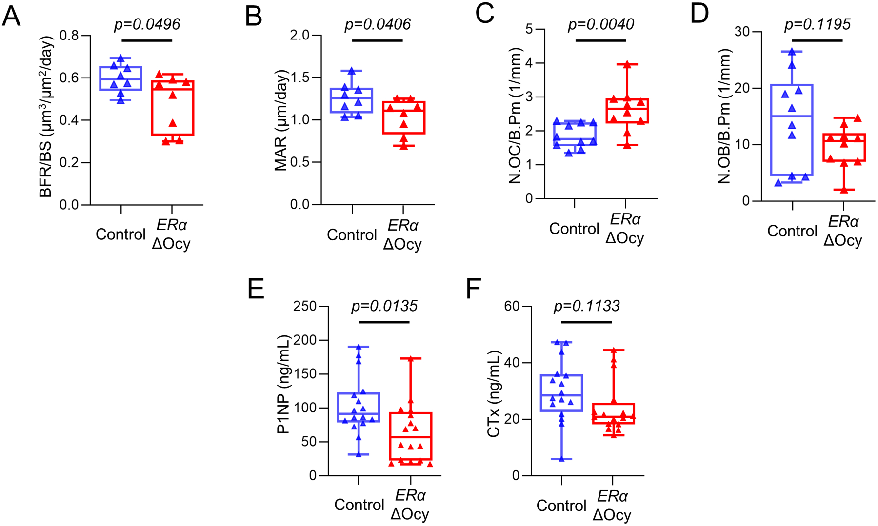
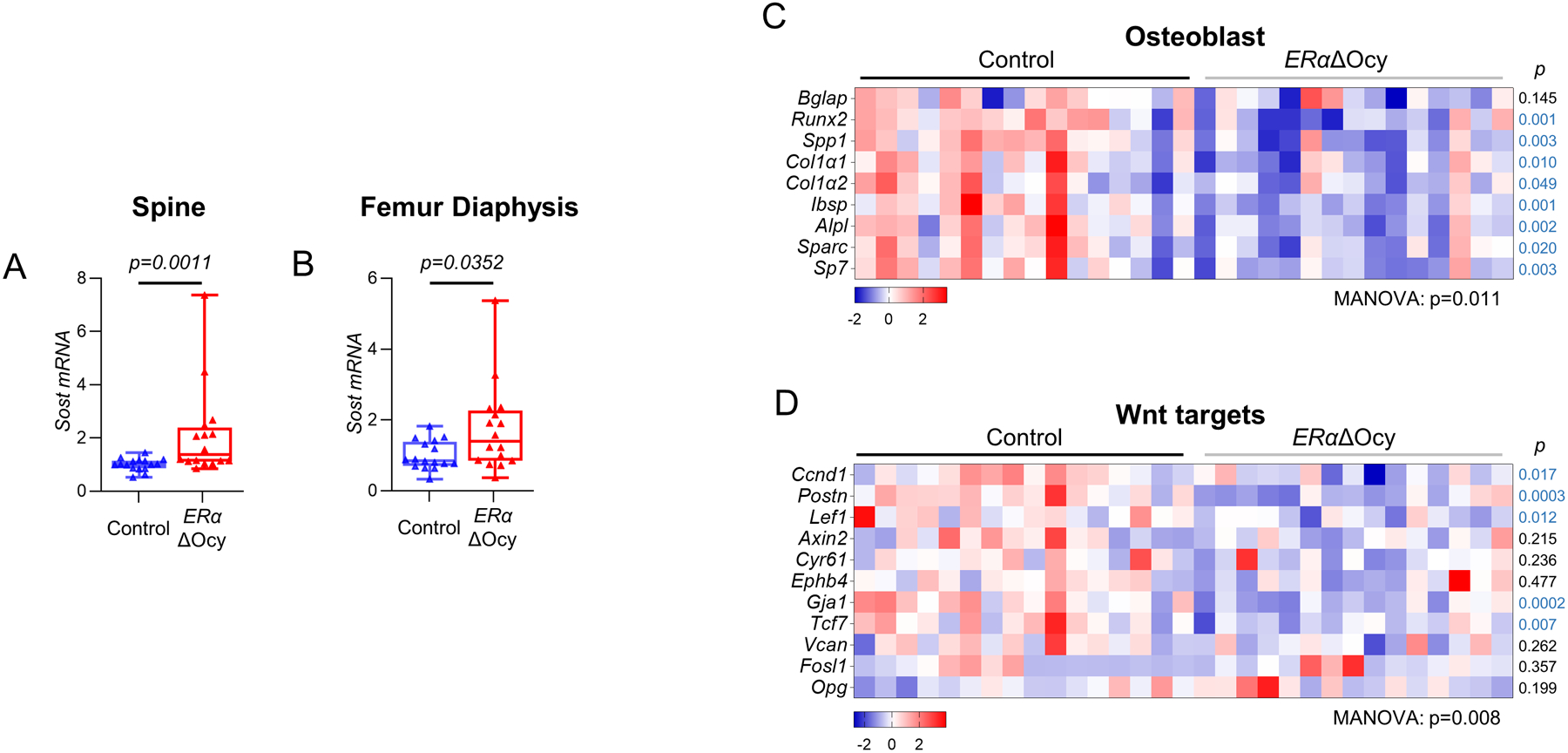
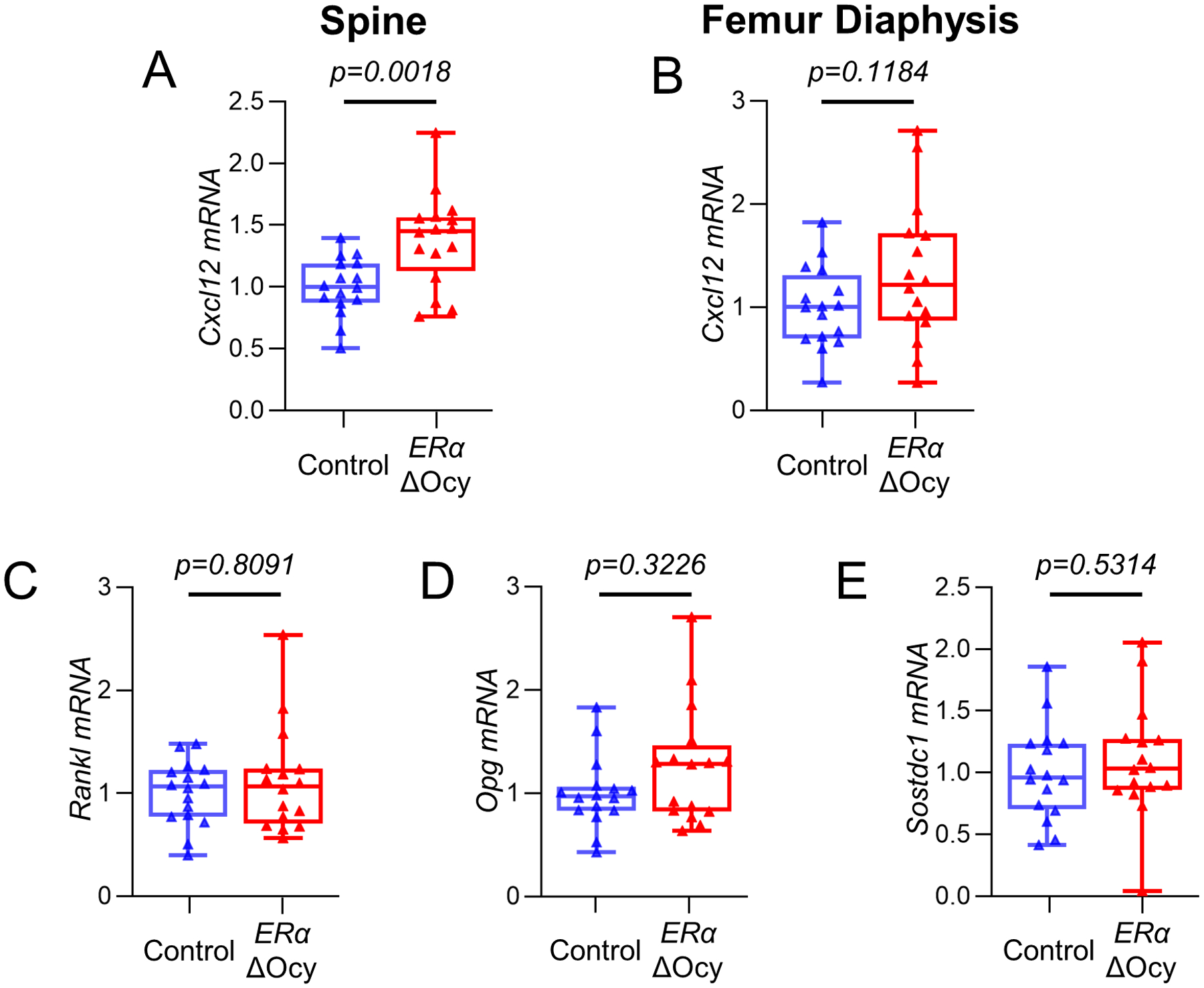
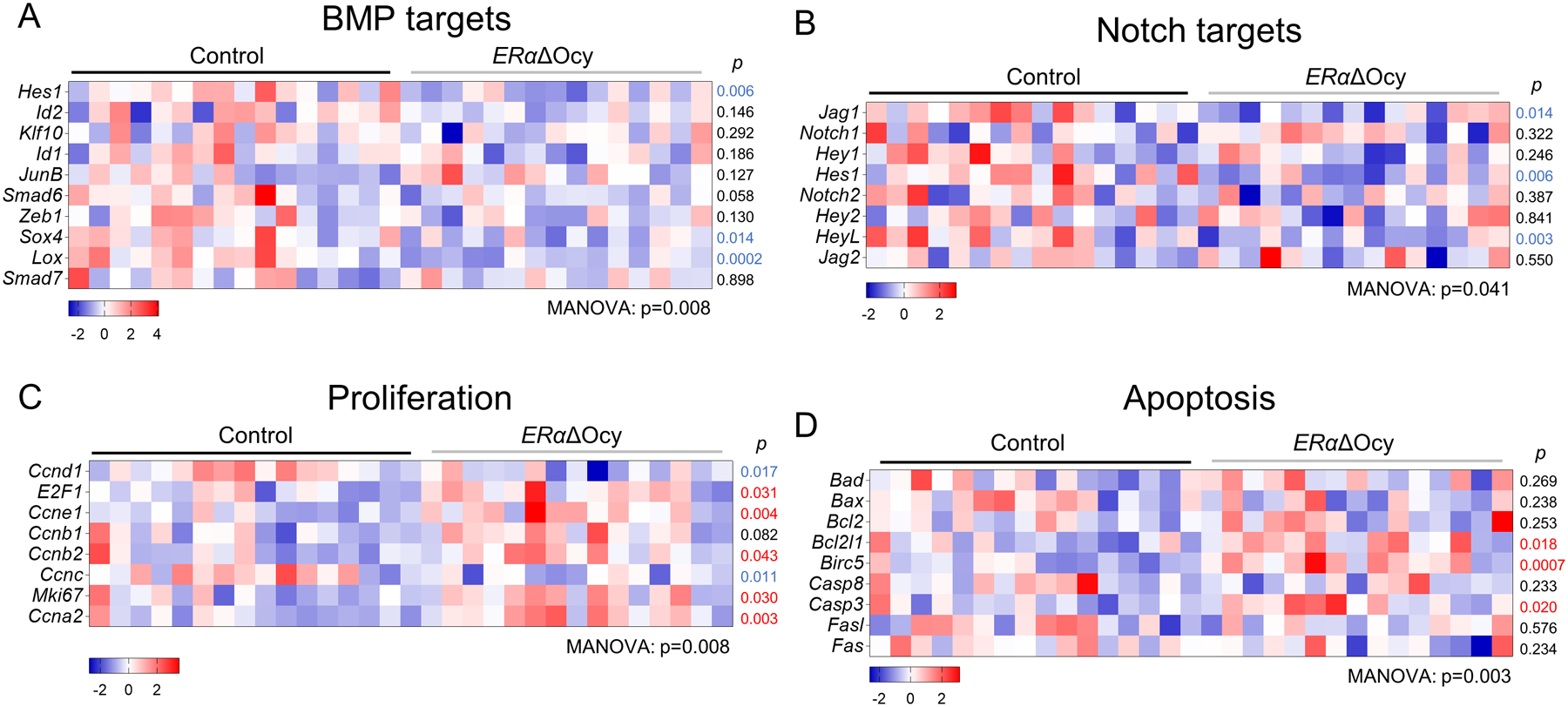
Estrogen is known to regulate bone metabolism in both women and men, but substantial gaps remain in our knowledge of estrogen and estrogen receptor alpha (ERα) regulation of adult bone metabolism. Studies using global ERα-knockout mice were confounded by high circulating sex-steroid levels, and osteocyte/osteoblast-specific ERα deletion may be confounded by ERα effects on growth versus the adult skeleton. Thus, we developed mice expressing the tamoxifen-inducible CreERT2 in osteocytes using the 8-kilobase (kb) Dmp1 promoter (Dmp1CreERT2 ). These mice were crossed with ERαfl//fl mice to create ERαΔOcy mice, permitting inducible osteocyte-specific ERα deletion in adulthood. After intermittent tamoxifen treatment of adult 4-month-old mice for 1 month, female, but not male, ERαΔOcy mice exhibited reduced spine bone volume fraction (BV/TV (-20.1%, p = 0.004) accompanied by decreased trabecular bone formation rate (-18.9%, p = 0.0496) and serum P1NP levels (-38.9%, p = 0.014). Periosteal (+65.6%, p = 0.004) and endocortical (+64.1%, p = 0.003) expansion were higher in ERαΔOcy mice compared to control (Dmp1CreERT2 ) mice at the tibial diaphysis, reflecting the known effects of estrogen to inhibit periosteal apposition and promote endocortical formation. Increases in Sost (2.1-fold, p = 0.001) messenger RNA (mRNA) levels were observed in trabecular bone at the spine in ERαΔOcy mice, consistent with previous reports that estrogen deficiency is associated with increased circulating sclerostin as well as bone SOST mRNA levels in humans. Further, the biological consequences of increased Sost expression were reflected in significant overall downregulation in panels of osteoblast and Wnt target genes in osteocyte-enriched bones from ERαΔOcy mice. These findings thus establish that osteocytic ERα is critical for estrogen action in female, but not male, adult bone metabolism. Moreover, the reduction in bone formation accompanied by increased Sost, decreased osteoblast, and decreased Wnt target gene expression in ERαΔOcy mice provides a direct link in vivo between ERα and Wnt signaling. © 2022 American Society for Bone and Mineral Research (ASBMR).

Keywords: ESTROGENS AND SERMs; GENETIC ANIMAL MODELS; OSTEOCYTES; OSTEOPOROSIS; SEX STEROIDS.


Figures

Figure 1.
Figure 1.
Dmp1-CreERT2 targets osteocytes. (A) Experimental outline for reporter induction in Dmp1CreERT2 x TdTomato mice. (B) Representative images of induced TdTomato expression in osteocytes in the femur and (C, D) spine of corn oil- versus tamoxifen-treated Dmp1CreERT2 x TdTomato mice at low (10X, 20X, C) and high (40X, D) magnification. Black areas are bone, blue areas show extensive DAPI staining in the bone marrow. Scale bars = 10μm for 10X, 20X and 50μm for 40X. ;(E) Experimental outline for inducible deletion of ERα in osteocytes. (F) Images of RNAscope for Esr1 mRNA (encoding ERα) performed on spine bone sections from tamoxifen-treated Dmp1CreERT2 (Control) or ERαΔOcy mice. Scale bars = 50μm (G) Quantification of percentage of osteocytes positive for ERα observed from RNAscope (n = 6 per group [3 males (squares), 3 females (triangles)]). (H) Measurement of serum estradiol (E2) from cardiac blood (n = 15–16 per group). Boxes represent median and interquartile range and whiskers represent minimum and maximum values. Datasets were analyzed by unpaired t-test (G) or Mann-Whitney test (H).
Figure 2.
Figure 2.
Deletion of osteocyte ERα in adult female mice leads to reduced bone mass and expansion of endosteal and periosteal surfaces. (A-D) Micro-CT analyses of the lumbar spine of tamoxifen-treated Control or ERαΔOcy mice: (A) bone volume fraction (BV/TV), (B) trabecular thickness (Tb.Th), (C) trabecular number (Tb.N), and (D) trabecular spacing (Tb.Sp). (E-H) Longitudinal micro-CT analyses of cortical bone at the tibial diaphysis (Tib. Dia.) showing percent (%) change between baseline and endpoint. (E) Endocortical circumference (EC), (F) periosteal circumference (PC), and (G) cortical thickness (Ct.Th).. (H) Diagram of changes in tibial cortical bone with osteocyte-specific ERα knockout. n=16–17 mice per group. Boxes represent median and interquartile range and whiskers represent minimum and maximum values. Datasets analyzed by unpaired t-test (A, D, F) or Mann-Whitney test (B, C, E, G).
Figure 3.
Figure 3.
ERα deletion in osteocytes leads to reduced bone formation and increased osteoclast numbers. (A) Measurement of bone formation rate per bone surface (BFR/BS) and (B) mineral apposition rates (MAR) in the lumbar spines of tamoxifen-treated Control or ERαΔOcy mice using double-label dynamic histomorphometry. (C) Counted number of osteoclasts (N.OC/B.Pm) and (D) osteoblasts (N.OB/B.Pm) normalized to bone perimeter using static histomorphometry. (E) PINP and (F) CTx levels measured from endpoint cardiac serum. n=8–16 per group. Boxes represent median and interquartile range and whiskers represent minimum and maximum values. Datasets analyzed by unpaired t-test.
Figure 4.
Figure 4.
ERαΔOcy mice display increased Sost expression and a reduction in bone anabolic pathways. (A) mRNA expression of Sost in Dmp1CreERT2 and ERαΔOcy mice at the spine and (B) femur diaphysis by qRT-PCR. (C) Heatmap of mRNA expression changes in osteoblast and (D) Wnt signaling pathways from spines of Control and ERαΔOcy mice by qRT-PCR. n=16–20 mice per group. Boxes represent median and interquartile range and whiskers represent minimum and maximum values. Datasets analyzed by Mann-Whitney test (A, B) or MANOVA (C, D) with pathway-level p-values listed below each heatmap and individual gene p-values beside each row (blue font p-value indicates downregulated gene in the ERαΔOcy mice).
Figure 5.
Figure 5.
Cxcl12, but not other markers of bone turnover are altered in ERαΔOcy mice. (A) mRNA expression of Cxcl12 at the spine and (B) femur diaphysis of Control and ERαΔOcy mice by qRT-PCR. (C) Rankl, (D) Opg, and (E) Sostdc1 mRNA expression by qRT-PCR at the spine. Boxes represent median and interquartile range and whiskers represent minimum and maximum values. Datasets analyzed by unpaired t-test (A, B, E) or Mann-Whitney test (C, D).
Figure 6.
Figure 6.
Osteocyte ERαΔOcy deletion suppresses osteogenic pathways, while promoting proliferation and apoptosis gene expression. (A-D) Heatmaps of mRNA expression changes in (A) BMP signaling, (B) Notch signaling, (C) proliferation, and (D) apoptosis pathways from spines of tamoxifen-treated Control and ERαΔOcy mice by qRT-PCR. Datasets analyzed by MANOVA with pathway-level significance-values listed below each heatmap and individual gene p-values beside each row. Blue font p-value represent significantly downregulated expression in ERαΔOcy mice, while red font p-values represent upregulated expression in ERαΔOcy mice.

References

    1. Khosla S, Melton LJ 3rd, Riggs BL. The unitary model for estrogen deficiency and the pathogenesis of osteoporosis: is a revision needed? J Bone Miner Res. Mar 2011;26(3):441–51. Epub 2010/10/12. - PMC - PubMed
    1. Khosla S, Monroe DG. Regulation of Bone Metabolism by Sex Steroids. Cold Spring Harb Perspect Med. Jan 2 2018;8(1). Epub 2017/07/16. - PMC - PubMed
    1. Nicks KM, Fujita K, Fraser D, McGregor U, Drake MT, McGee-Lawrence ME, et al. Deletion of Estrogen Receptor Beta in Osteoprogenitor Cells Increases Trabecular but Not Cortical Bone Mass in Female Mice. J Bone Miner Res. Mar 2016;31(3):606–14. Epub 2015/09/30. - PMC - PubMed
    1. Sims NA, Dupont S, Krust A, Clement-Lacroix P, Minet D, Resche-Rigon M, et al. Deletion of estrogen receptors reveals a regulatory role for estrogen receptors-beta in bone remodeling in females but not in males. Bone. Jan 2002;30(1):18–25. Epub 2002/01/17. - PubMed
    1. Almeida M, Iyer S, Martin-Millan M, Bartell SM, Han L, Ambrogini E, et al. Estrogen receptor-alpha signaling in osteoblast progenitors stimulates cortical bone accrual. J Clin Invest. Jan 2013;123(1):394–404. Epub 2012/12/12. - PMC - PubMed

Publication types