. 2022 Aug 19;25(8):104685.
doi: 10.1016/j.isci.2022.104685. Epub 2022 Jun 30.

Immunomodulatory LncRNA on antisense strand of ICAM-1 augments SARS-CoV-2 infection-associated airway mucoinflammatory phenotype

Affiliations

Immunomodulatory LncRNA on antisense strand of ICAM-1 augments SARS-CoV-2 infection-associated airway mucoinflammatory phenotype

Dinesh Devadoss et al. iScience. .

Abstract

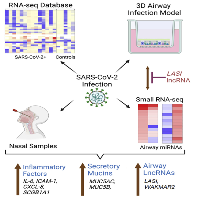
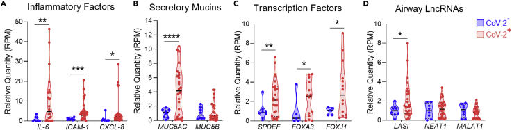
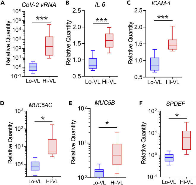
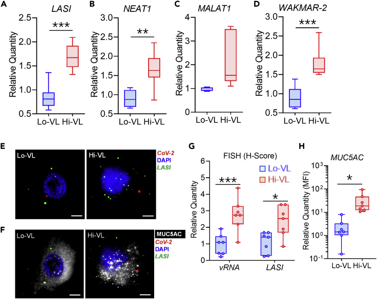
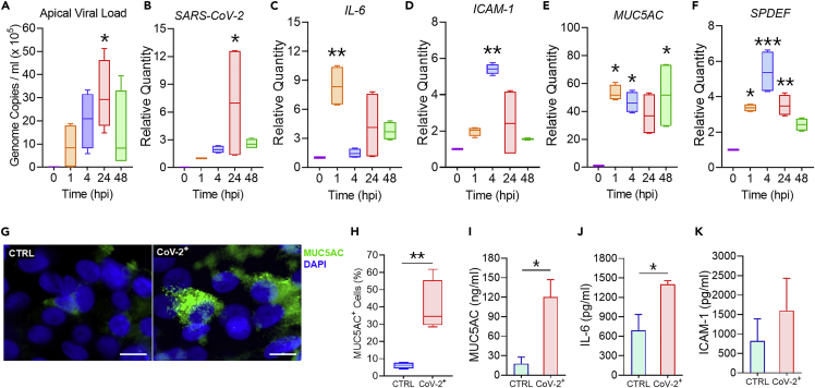
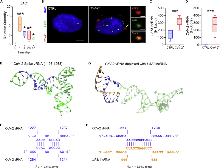
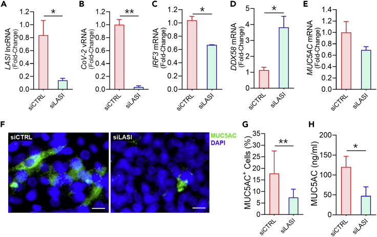
Noncoding RNAs are important regulators of mucoinflammatory response, but little is known about the contribution of airway long noncoding RNAs (lncRNAs) in COVID-19. RNA-seq analysis showed a more than 4-fold increased expression of IL-6, ICAM-1, CXCL-8, and SCGB1A1 inflammatory factors; MUC5AC and MUC5B mucins; and SPDEF, FOXA3, and FOXJ1 transcription factors in COVID-19 patient nasal samples compared with uninfected controls. A lncRNA on antisense strand to ICAM-1 or LASI was induced 2-fold in COVID-19 patients, and its expression was directly correlated with viral loads. A SARS-CoV-2-infected 3D-airway model largely recapitulated these clinical findings. RNA microscopy and molecular modeling indicated a possible interaction between viral RNA and LASI lncRNA. Notably, blocking LASI lncRNA reduced the SARS-CoV-2 replication and suppressed MUC5AC mucin levels and associated inflammation, and select LASI-dependent miRNAs (e.g., let-7b-5p and miR-200a-5p) were implicated. Thus, LASI lncRNA represents an essential facilitator of SARS-CoV-2 infection and associated airway mucoinflammatory response.

Keywords: Immunology; Molecular biology; Molecular mechanism of gene regulation; Virology.


Conflict of interest statement

Dr. Hitendra S. Chand and Dr. Madhavan Nair are coinventor on a US utility patent #10,851,376 for long noncoding RNAs in pulmonary airway inflammation. The authors have no competing financial interests to declare.

Figures

None
Graphical abstract
Figure 1
Figure 1
Expression levels of transcripts encoding inflammatory factors, airway secretory mucins, associated transcription factors, and select lncRNAs in the RNA-seq database of nasopharyngeal swabs of SARS-CoV-2 positive individuals (A–D) Relative expression of (A) airway inflammatory factors IL-6, ICAM-1, and CXCL-8 mRNAs; (B) airway mucins MUC5AC and MUC5B mRNAs; (C) transcription factors SPDEF, FOXA3, and FOXJ1 mRNAs; and (D) lncRNAs LASI, NEAT1, and MALAT1 in SARS-CoV-2 positive (CoV-2+) individuals compared with SARS-CoV-2 negative (CoV-2) control individuals. The expression levels were calculated from the transcript reads per million (RPM) RNA-seq data (Table S1) of nasopharyngeal swabs from CoV-2+ subjects (n = 26) compared with CoV-2 control subjects (n = 8) as reported recently (Cheemarla et al., 2021). (Data shown as violin plots with individual data points, medians, and quartiles; ∗p < 0.05; ∗∗p < 0.01; ∗∗∗p < 0.001; ∗∗∗∗p < 0.0001 by Student’s t test). Similar increased expression of IFIH1 and SCGB1A1 mRNAs was observed in CoV-2+ subjects (Figure S1).
Figure 2
Figure 2
COVID-19 positive individuals with high nasopharyngeal viral load show increased mucoinflammatory phenotype compared with low viral load individuals (A–F) Total RNA from nasopharyngeal swab samples of COVID-19 positive individuals (n = 20) was analyzed. Based on the SARS-CoV-2 nucleocapsid viral RNA expression, the individuals with high viral load (Hi-VL) had average CT values of 25.9 ± 1.3 (n = 10), whereas those with low viral load (Lo-VL) had average CT values of 35.5 ± 0.7 (n = 10). Relative mRNA expression of (A) SARS-CoV-2 viral RNA (CoV-2 vRNA); innate inflammatory factors, (B) IL-6, and (C) ICAM-1; airway mucins (D) MUC5AC and (E) MUC5B; and (F) mucin regulatory transcriptional factor SPDEF in Hi-VL compared with Lo-VL patient swab samples. (Data shown as box and whisker plots with minimum to maximum range; n = 10/gp; ∗p < 0.05; ∗∗p < 0.01; ∗∗∗p < 0.001 by Student’s t test). Increased expression of MUC4 and SCGB1A1 mRNAs was also observed in Hi-VL subjects with no changes in select viral entry facilitating host factor mRNAs and IFN1B mRNA expression (Figure S2) and select host mucoinflammatory genes and lncRNAs strongly correlated with CoV-2 viral load (Figure S3).
Figure 3
Figure 3
COVID-19 positive individuals with high viral load show increased immunomodulatory lncRNAs and the secretory mucin MUC5AC expression (A–D) Relative expression of immunomodulatory lncRNAs, (A) LASI, (B) NEAT1, (C) MALAT1, and (D) WAKMAR2 in Hi-VL compared with Lo-VL individuals’ swab samples. (E) The dual-FISH analysis detected colocalization of SARS-CoV-2 viral RNA (vRNA) and LASI lncRNA in nasal swab samples. Representative micrographs from Lo-VL and Hi-VL swab samples display detection of SARS-CoV-2 nucleocapsid (CoV-2) vRNA (green) and LASI lncRNA (red) along with DAPI-stained nuclei (blue). (F) Nasal swab cells showing immunoreactive MUC5AC expression (shown in white) in the dual-FISH labeled cells; scale bar: 2 μm. (G) H-score quantitation of vRNA and LASI lncRNA in COVID-19-positive individuals. (H) Relative quantitation of mean fluorescence intensity (MFI) of MUC5AC expression in Lo-VL and Hi-VL swab samples. (n = 10/gp for data in A, B, C, & D; and n = 7/gp for data in G & H with 10 cells analyzed per sample; ∗p < 0.05; ∗∗p < 0.01; ∗∗∗p < 0.001 by Student’s t test). There was no change in MUC5B expression levels in Lo-VL and Hi-VL samples (Figure S4).
Figure 4
Figure 4
SARS-CoV-2 infection of human respiratory epithelial cells induces robust mucoinflammatory response in a 3D airway tissue model (A–F) Respiratory airway epithelial cells differentiated on air-liquid interface were infected with one MOI of SARS-CoV-2 clinical isolate (USA-WA1/2020 isolate) and analyzed at 0, 1, 4, 24, and 48 h postinfection (hpi). Viral loads were determined in (A) the apical washes and (B) the total cellular RNA. Relative expression levels of the inflammatory factors, (C) IL-6, and (D) ICAM-1 mRNA; and (E) airway mucin MUC5AC; and (F) SPDEF transcriptional factor in the total cellular RNA was analyzed by qRT-PCR. (n = 4/gp; ∗p < 0.05; ∗∗p < 0.01; ∗∗∗p < 0.001 by ANOVA). (G) Representative micrographs of uninfected control (CTRL) and SARS-CoV-2-infected (CoV-2+) cells showing MUC5AC (shown in green) immunoreactivity along with the DAPI stained nuclei (shown in blue); scale bar: 5 μm. (H) Percentage of MUC5AC+ cells within each treatment group. (I–K) Secreted protein levels of (I) MUC5AC mucin in apical washes and (J) IL-6 and (K) ICAM-1 in culture media supernatants as determined by specific ELISA assays (n = 4/gp; ∗p < 0.05; ∗∗p < 0.01; by Student’s t test). There was a significant suppression of viral entry host factors with elevated expression of other mucin genes and SCGB1A1 mRNA following CoV-2 infection (Figure S5).
Figure 5
Figure 5
SARS-CoV-2 infection induces LASI lncRNA expression in human respiratory epithelial cells that potentially show direct interaction with CoV-2 spike RNA (A) Relative expression levels of LASI lncRNA in SARS-CoV-2-infected cells at 0, 1, 4, 24, and 48 hpi. (n = 4/gp; ∗p < 0.05; ∗∗p < 0.01; ∗∗∗p < 0.001 by ANOVA). (B) Colocalization of SARS-CoV-2 vRNA and LASI transcripts in CoV-2-infected (CoV-2+) cells as determined by dual-FISH staining and the structured-illumination imaging analysis. Representative micrographs of dual-FISH-stained cells showing SARS-CoV-2 N1 vRNA (in red) and LASI lncRNAs (in green) along with DAPI-stained nuclei (in blue); scale bar: 2 μm. (C and D) H-score quantitation of (C) CoV-2 vRNA and (D) LASI lncRNAs per cell in CoV-2+ and control cells. (n = 9–10 cells/gp; ∗∗p < 0.01; ∗∗∗p < 0.001 by Student’s t test). (E) Modeled 3D structure of SARS-CoV-2 spike vRNA nucleotide sequence from 1198 to 1268, and the LASI lncRNA interacting region (1227-1237) is highlighted in blue. (F) The intra-sequence base-pairing of spike nucleotides forms the hairpin stem structure. (G) Modeled 3D structure of the CoV-2 Spike vRNA duplexed with LASI lncRNA sequence 646-635 (highlighted in orange) at the end of 100 ns simulation (see Video S1 at online supplemental data). (H) Inter-sequence base-pairing of CoV-2 vRNA with LASI lncRNA sequence (shown in orange). There was no significant change in interferon-related gene expression; however, expression of other immunomodulatory lncRNAs were differentially regulated following CoV-2 infection (Figure S6). LASI-interacting sequence is conserved in Spike viral RNAs of CoV-2 Delta and Omicron variants (Figure S7).
Figure 6
Figure 6
Blocking LASI lncRNA expression reduces the SARS-CoV-2 viral load and suppresses the MUC5AC mucin expression (A–E) Relative expression of (A) LASI lncRNA and (B) SARS-CoV-2 vRNA in cells transfected with siRNA targeting LASI (siLASI) compared with control siRNA (siCTRL)-transfected cells and infected with one MOI of SARS-CoV-2. Relative expression of (C) IRF3, (D) DDX58, and (E) MUC5AC mRNA following SARS-CoV-2 infection in siLASI-transfected cells compared with siCTRL cells. (n = 4/gp from two independent experiments; ∗p < 0.05; ∗∗p < 0.01; by Student’s t test). (F) The micrographs of SARS-CoV-2-infected cells following siCTRL and siLASI transfection show MUC5AC (green) immunoreactivity and DAPI-stained nuclei (blue), scale bar: 5 μm. (G) Percentage of MUC5AC+ cells within each treatment group (n = 10/gp; ∗∗p < 0.001 by Student’s t test). (H) MUC5AC mucin protein levels in the apical wash of CoV-2-infected cells and transfected with siLASI or siCTRL as determined by ELISA assay. (n = 4/gp from two independent experiments; ∗p < 0.05 by Student’s t test). Expression of MUC2 and MUC4 mucin mRNAs were also suppressed in siLASI cells with no change in expression of IL-6, ICAM-1, and other lncRNAs (Figure S8).
Figure 7
Figure 7
Airway epithelial miRNAs are modulated by SARS-CoV-2 infection and regulated by LASI lncRNA Relative expression levels of miRNAs in siLASI-treated cells following 48 h SARS-CoV-2 infection compared with control-infected cells as analyzed by small RNA-seq analysis. (A) Heatmap of select miRNAs upregulated by CoV-2 infection and suppressed in siLASI-treated cells (∗miR-4488 was upregulated >200-fold in infected control cells); see Table S2 for the comprehensive list. (B) Expression levels of miRNAs that are downregulated by CoV-2 infection but induced in siLASI-treated cells. (C–E) Relative quantitation of miRNAs: (C) miR-4488, (D) let-7b-5p, and (E) miR-150-5p that were upregulated by SARS-CoV-2 infection in siCTRL cells but suppressed in siLASI-transfected cells. (F–H) Relative expression of miRNAs: (F) miR-6510-3p, (G) miR-200a-5p, and (H) miR-197-3p that were downregulated by SARS-CoV-2 infection in siCTRL cells but induced in siLASI-transfected cells. (∗p < 0.05; ∗∗p < 0.001 by Student’s t test). Expression levels of select miRNAs were also elevated in Hi-VL nasopharyngeal swab samples of our study cohort (Figure S9).

Update of

Similar articles

Cited by

References

    1. Abdelmoaty M.M., Yeapuri P., Machhi J., Olson K.E., Shahjin F., Kumar V., Zhou Y., Liang J., Pandey K., Acharya A., et al. Defining the innate immune responses for SARS-CoV-2-human macrophage interactions. Front. Immunol. 2021;12:741502. doi: 10.3389/fimmu.2021.741502. - DOI - PMC - PubMed
    1. Acharya A., Pandey K., Thurman M., Klug E., Trivedi J., Sharma K., Lorson C.L., Singh K., Byrareddy S.N. Discovery and evaluation of entry inhibitors for SARS-CoV-2 and its emerging variants. J. Virol. 2021;95:e0143721. doi: 10.1128/jvi.01437-21. - DOI - PMC - PubMed
    1. Abu-Izneid T., AlHajri N., Ibrahim A.M., Javed M.N., Salem K.M., Pottoo F.H., Kamal M.A. Micro-RNAs in the regulation of immune response against SARS CoV-2 and other viral infections. J. Adv. Res. 2021;30:133–145. doi: 10.1016/j.jare.2020.11.013. - DOI - PMC - PubMed
    1. Agirre X., Meydan C., Jiang Y., Garate L., Doane A.S., Li Z., Verma A., Paiva B., Martín-Subero J.I., Elemento O., et al. Long non-coding RNAs discriminate the stages and gene regulatory states of human humoral immune response. Nat. Commun. 2019;10:821. doi: 10.1038/s41467-019-08679-z. - DOI - PMC - PubMed
    1. Akula S.M., Bolin P., Cook P.P. Cellular miR-150-5p may have a crucial role to play in the biology of SARS-CoV-2 infection by regulating nsp10 gene. RNA Biol. 2022;19:1–11. doi: 10.1080/15476286.2021.2010959. - DOI - PMC - PubMed

LinkOut - more resources