. 2022 Oct;31(10):1638-1640.
doi: 10.1111/exd.14640. Epub 2022 Jul 25.

Familial primary cutaneous amyloidosis: Caspase activation may be involved in amyloid formation

Affiliations

Familial primary cutaneous amyloidosis: Caspase activation may be involved in amyloid formation

Javier Antoñanzas et al. Exp Dermatol. 2022 Oct.
No abstract available

Keywords: amyloidosis; apoptosis; caspases.


Conflict of interest statement

The authors declare no conflict of interest.

Figures
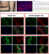
FIGURE 1
FIGURE 1
Clinical phenotype as well as the histological, immunohistochemical and molecular findings. (A) Hypopigmented and hyperpigmented macules in the proband with FPLCA. (B) Amyloid deposits with Congo Red staining (×100). (C) OSMR heterozygous missense sequence 1891G>C p.(Val631Leu) as detected by the Sanger method. (D) Immunofluorescence studies in proband and normal skin. (D‐Fig1a) Presence of caspases in the dermis within the amyloid deposits. No OSMR is expressed in the amyloid material. Colocalized expression of caspases and OSMR is not found. (D‐Fig1b) Presence of keratins of high molecular weight (34βE12) in the dermis, coinciding with the amyloid deposits. No OSMR expression is found in the amyloid material. (D‐Fig1c) Absence of caspase expression in the dermis of normal skin. Deposits of OSMR are found in keratinocytes of the epidermis. (D‐Fig1d) No expression of high molecular weight keratins (34βE12) in the dermis of the skin of a healthy daughter. Deposits of OSMR in keratinocytes of the epidermis. Caspase‐3 and Keratin (green), OSMR (red), DAPI Nuclear staining (blue). Scale Bar = 50 μm.

References

    1. Tanaka A, Arita K, Lai‐Cheong JE, Palisson F, Hide M, McGrath JA. New insight into mechanisms of pruritus from molecular studies on familial primary localized cutaneous amyloidosis. Br J Dermatol. 2009;161(6):1217‐1224. - PubMed
    1. Tanaka A, Lai‐Cheong JE, van den Akker PC, et al. The molecular skin pathology of familial primary localized cutaneous amyloidosis. Exp Dermatol. 2010;19(5):416‐423. - PubMed
    1. Chen SH, Benveniste EN. Oncostatin M: a pleiotropic cytokine in the central nervous system. Cytokine Growth Factor Rev. 2004;15(5):379‐391. - PubMed
    1. Heinrich PC, Behrmann I, Haan S, et al. Principles of interleukin (IL)‐6‐type cytokine signalling and its regulation. Biochem J. 2003;374(1):1‐20. - PMC - PubMed
    1. Bando T, Morikawa Y, Komori T, Senba E. Complete overlap of interleukin‐31 receptor a and oncostatin M receptor β in the adult dorsal root ganglia with distinct developmental expression patterns. Neuroscience. 2006;142(4):1263‐1271. - PubMed

Supplementary concepts