. 2022 Sep;50(9):1151-1160.
doi: 10.1124/dmd.122.000918. Epub 2022 Jul 5.

The COVID-19 Oral Drug Molnupiravir Is a CES2 Substrate: Potential Drug-Drug Interactions and Impact of CES2 Genetic Polymorphism In Vitro

Affiliations

The COVID-19 Oral Drug Molnupiravir Is a CES2 Substrate: Potential Drug-Drug Interactions and Impact of CES2 Genetic Polymorphism In Vitro

Yue Shen et al. Drug Metab Dispos. 2022 Sep.

Abstract

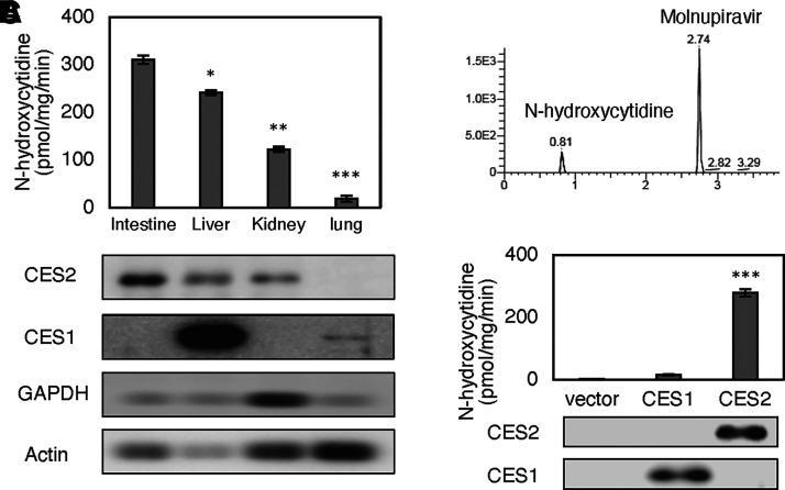
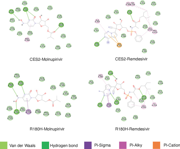
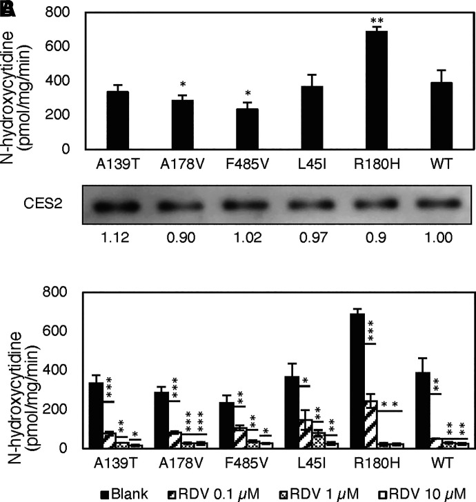
Molnupiravir is one of the two coronavirus disease 2019 (COVID-19) oral drugs that were recently granted the emergency use authorization by the Food and Drug Administration (FDA). Molnupiravir is an ester and requires hydrolysis to exert antiviral activity. Carboxylesterases constitute a class of hydrolases with high catalytic efficiency. Humans express two major carboxylesterases (CES1 and CES2) that differ in substrate specificity. Based on the structural characteristics of molnupiravir, this study was performed to test the hypothesis that molnupiravir is preferably hydrolyzed by CES2. Several complementary approaches were used to test this hypothesis. As many as 24 individual human liver samples were tested and the hydrolysis of molnupiravir was significantly correlated with the level of CES2 but not CES1. Microsomes from the intestine, kidney, and liver, but not lung, all rapidly hydrolyzed molnupiravir and the magnitude of hydrolysis was related closely to the level of CES2 expression among these organs. Importantly, recombinant CES2 but not CES1 hydrolyzed molnupiravir, collectively establishing that molnupiravir is a CES2-selective substrate. In addition, several CES2 polymorphic variants (e.g., R180H) differed from the wild-type CES2 in the hydrolysis of molnupiravir. Molecular docking revealed that wild-type CES2 and its variant R180H used different sets of amino acids to interact with molnupiravir. Furthermore, molnupiravir hydrolysis was significantly inhibited by remdesivir, the first COVID-19 drug granted the full approval by the FDA. The results presented raise the possibility that CES2 expression and genetic variation may impact therapeutic efficacy in clinical situations and warrants further investigation. SIGNIFICANCE STATEMENT: COVID-19 remains a global health crisis, and molnupiravir is one of the two recently approved oral COVID-19 therapeutics. In this study, we have shown that molnupiravir is hydrolytically activated by CES2, a major hydrolase whose activity is impacted by genetic polymorphic variants, disease mediators, and many potentially coadministered medicines. These results presented raise the possibility that CES2 expression and genetic variation may impact therapeutic efficacy in clinical situations and warrants further investigation.


Figures

Fig. 1.
Fig. 1.
Structure and elution profile of molnupiravir and its hydrolytic metabolite N-hydroxycytidine. (A) Structure of molnupiravir. (B) Structure of N-hydroxycytidine. (C) Elution profile of molnupiravir. (D) Elution profile of N-hydroxycytidine. Representative chromatograms of molnupiravir (0.6 ng) and N-hydroxycytidine (0.18 ng) eluted with a gradient mobile phase constituting ammonium acetate (1 mM, pH 4,3) in water and acetonitrile as described in the text.
Fig. 2.
Fig. 2.
Hydrolysis of molnupiravir by individual human liver samples and correlation analysis of the hydrolysis with the level of CES1 or CES2. (A) Molnupiravir hydrolysis by individual liver samples. Liver S9 fractions (2 µg) were incubated with molnupiravir at a final concentration of 1 µM for 40 minutes and the formation of the hydrolytic metabolite N-hydroxycytidine was analyzed by LC-MS/MS. For Western blotting, S9 fractions (3 µg) were subjected to SDS-polyacrylamide gel electrophoresis, electrophoretically transferred to nitrocellulose membrane, and detected by the chemiluminescent detection system for CES1, CES2, or GAPDH. (B) Correlation between molnupiravir hydrolysis and the level of CES2 or CES1. The intensity of immunostaining was captured and quantified by ChemiDoc Imaging system. The immunostaining intensity of CES1 or CES2 was plotted against the relative hydrolysis of molnupiravir. The correlation coefficient and the corresponding P values were calculated. An arrow sign indicates potential outliers. (C) Correlation between CES1 and CES2 expression. Statistical significance of correlation at P < 0.01.
Fig. 3.
Fig. 3.
Hydrolysis of molnupiravir by organ-specific microsomes and cell lysates containing recombinant CES1 or CES2. (A) Molnupiravir hydrolysis by organ-specific microsomes. Pooled microsomes (2 µg) from the intestine (n = 6), liver (n = 20), kidney (n = 20) or lung (n = 10) were incubated with molnupiravir at a final concentration of 1 µM for 40 minutes and the formation of the hydrolytic metabolite N-hydroxycytidine was analyzed by LC-MS/MS. For Western blotting, microsomes (1.2 µg) were subjected to SDS-polyacrylamide gel electrophoresis, electrophoretically transferred to nitrocellulose membrane, and detected by the chemiluminescent detection system for CES1, CES2, GAPDH, or β-actin. Single asterisk for statistical significance at P < 0.05, double at P < 0.01, and triple at P < 0.001 based on the comparison from the hydrolysis by intestinal microsomes. (B) Representative chromatogram of molnupiravir hydrolysis by pooled microsomes from the intestine (MPV: molnupiravir, NHC: N-hydroxycytidine). (C) Molnupiravir hydrolysis by cell lysates containing recombinant CES1 or CES2. Cells (293T) were transfected by CES1, CES2, or the corresponding vector. Cell lysates (0.1 µg) were tested for the hydrolysis of molnupiravir hydrolysis. Western blotting confirmed the expression of CES1 and CES2 upon transfection. Triple asterisks for statistical significance at P < 0.001 from vector control.
Fig. 4.
Fig. 4.
Remdesivir-inhibition of molnupiravir hydrolysis in microsomes. (A) Selective inhibition of CES2 by remdesivir. Pooled microsomes (2 µg) from the intestine (n = 6), liver (n = 20), and kidney (n = 20) were incubated with remdesivir (RDV at 0, 1, or 10 µM) for 2 hours and then subjected to native gel electrophoresis. The gel was then stained for esterase activity by 4-methylumbelliferyl acetate. Images were captured by ChemiDoc Imaging system. (B) Inhibition of molnupiravir hydrolysis. Pooled microsomes (2 µg) were incubated with remdesivir (RDV) for 2 hours and then with 1 µM molnupiravir for 40 minutes. The hydrolysis of molnupiravir was monitored for the production of the formation of N-hydroxycytidine was analyzed by LC-MS/MS. Single asterisk for statistical significance at P < 0.05, double at P < 0.01, and triple at P < 0.001 for the comparison indicated by a line.
Fig. 5.
Fig. 5.
Remdesivir-inhibition of molnupiravir hydrolysis in microsomes. (A) Intracellular inhibition of CES2 by remdesivir (RDV) with 4-methylumbelliferylacetate as the substrate. Human primary hepatocytes suspension (106) was incubated with RDV at various concentrations (0, 1, or 10 µM) for 2 hours and then centrifuged at 1000 g and washed extensively. The hepatocytes were then lysed and analyzed for CES2 inhibition by native gel electrophoresis. (B) Intracellular inhibition of molnupiravir hydrolysis by RDV with molnupiravir as the substrate. Human primary hepatocytes suspension (106) was incubated with RDV at various concentrations (0, 1, or 10 µM) for 2 hours and washed as above. The pellets were resuspended and then incubated with molnupiravir at 1 µM for 40 minutes. The formation of N-hydroxycytidine by LC-MS/MS. Single asterisk denotes statistical significance at P < 0.05, double at P < 0.01, and triple at P < 0.001 for the comparison indicated by a line.
Fig. 7.
Fig. 7.
Molecular docking of molnupiravir, remdesivir, and sofosbuvir. The molecular docking was performed by Autodock Vina. The ligands were sourced from PubChem as three-dimensional spatial data files. The wild-type CES2 was downloaded from the PDB and Swiss Model websites and cleaned to remove the ligand and any other unwanted molecules, such as water using the Chimera. The CES2 mutants were generated in the Chimera with the rotamer function. These files were then prepared for simulation by Autodock Tools. The center for the Autodock Vina simulation was the center of CES2 or its mutants. The search box was set to 126 × 126 × 126 with an exhaustiveness of 100.
Fig. 6.
Fig. 6.
Hydrolysis of molnupiravir by CES2 polymorphic variants and inhibited hydrolysis by remdesivir. (A) Hydrolysis of molnupiravir by CES2 polymorphic variants. Cells (293T) were transfected by the wild-type CES2 or mutant. Cell lysates (0.5 µg) were tested for the hydrolysis of molnupiravir hydrolysis. The same amount of lysates was analyzed by Western blotting for the expression. The hydrolysis was normalized based on the intensity of the immunostaining. Single asterisk denotes statistical significance at P < 0.05 and double at P < 0.01 for the comparison with the wild-type CES2. (B) Remdesivir-inhibited hydrolysis of molnupiravir by CES2 polymorphic variants. Lysates (0.5 µg) from transfected cells were incubated with remdesivir (RDV) at 0, 0.1, 1, and 10 µM for 2 hours and then with 1 µM molnupiravir for 40 minutes. The hydrolysis of molnupiravir was monitored for the production of the formation of N-hydroxycytidine was analyzed by LC-MS/MS. Single asterisk denotes statistical significance at P < 0.05, double at P < 0.01, and triple at P < 0.001 for the comparison indicated by a line.

References

    1. Asif Z, Chen Z, Stranges S, Zhao X, Sadiq R, Olea-Popelka F, Peng C, Haghighat F, Yu T (2022) Dynamics of SARS-CoV-2 spreading under the influence of environmental factors and strategies to tackle the pandemic: A systematic review. Sustain Cities Soc 81:103840. - PMC - PubMed
    1. Ayele AG, Enyew EF, Kifle ZD (2021) Roles of existing drug and drug targets for COVID-19 management. Metabol Open 11:100103. - PMC - PubMed
    1. Baba S, Yumoto R, Kawami M, Takano M (2021) Functional expression of equilibrative and concentrative nucleoside transporters in alveolar epithelial cells. Pharmazie 76:416–421. - PubMed
    1. Bienert S, Waterhouse A, de Beer TA, Tauriello G, Studer G, Bordoli L, Schwede T (2017) The SWISS-MODEL repository-new features and functionality. Nucleic Acids Res 45 :D313–D319. - PMC - PubMed
    1. Bojkova D, Widera M, Ciesek S, Wass MN, Michaelis M, Cinatl J (2022) Reduced interferon antagonism but similar drug sensitivity in omicron variant compared to delta variant of SARS-CoV-2 isolates. Cell Res 32:319–321. - PMC - PubMed

Publication types