. 2022 Dec;5(4):350-361.
doi: 10.1002/ame2.12225. Epub 2022 Jul 6.

Development of a humanized HLA-A30 transgenic mouse model

Affiliations

Development of a humanized HLA-A30 transgenic mouse model

Meng-Min Zhu et al. Animal Model Exp Med. 2022 Dec.

Abstract

Background: There are remarkable genetic differences between animal major histocompatibility complex (MHC) systems and the human leukocyte antigen (HLA) system. HLA transgenic humanized mouse model systems offer a much better method to study the HLA-A-related principal mechanisms for vaccine development and HLA-A-restricted responses against infection in human.

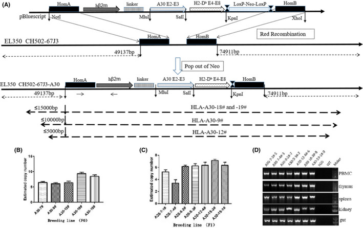
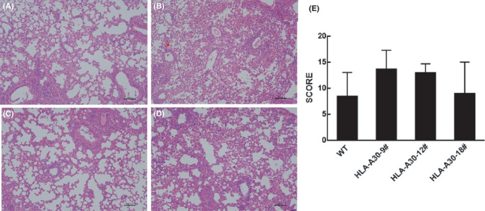
Methods: A recombinant gene encoding the chimeric HLA-A30 monochain was constructed. This HHD molecule contains the following: α1-α2 domains of HLA-A30, α3 and cytoplasmic domains of H-2Db , linked at its N-terminus to the C-terminus of human β2m by a 15-amino-acid peptide linker. The recombinant gene encoding the chimeric HLA-A30 monochain cassette was introduced into bacterial artificial chromosome (BAC) CH502-67J3 containing the HLA-A01 gene locus by Red-mediated homologous recombination. Modified BAC CH502-67J3 was microinjected into the pronuclei of wild-type mouse oocytes. This humanized mouse model was further used to assess the immune responses against influenza A virus (H1N1) pdm09 clinically isolated from human patients. Immune cell population, cytokine production, and histopathology in the lung were analyzed.

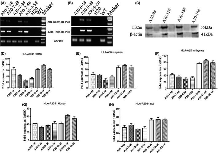
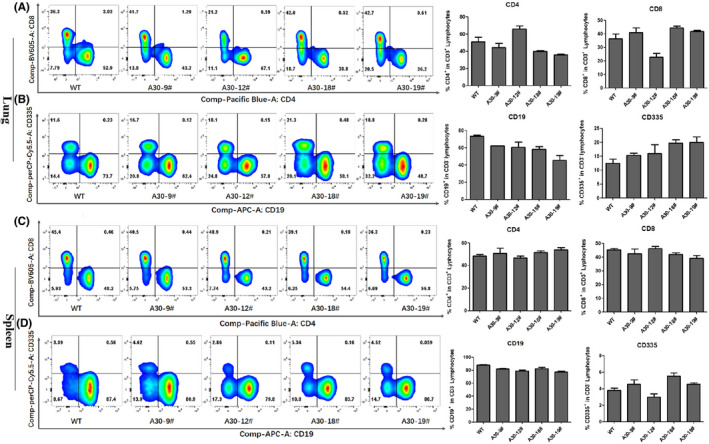
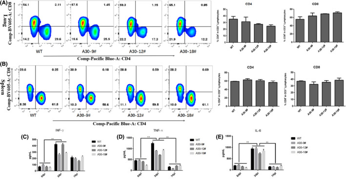
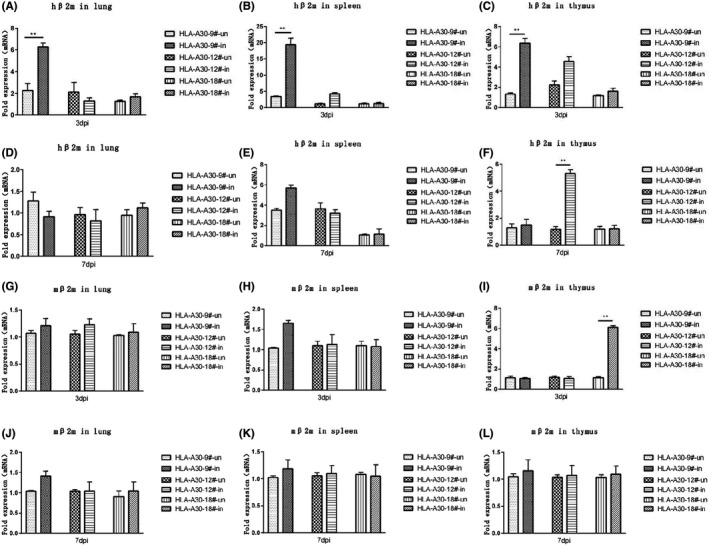
Results: We describe a novel human β2m-HLA-A30 (α1α2)-H-2Db (α3 transmembrane cytoplasmic) (HHD) monochain transgenic mouse strain, which contains the intact HLA-A01 gene locus including 49 kb 5'-UTR and 74 kb 3'-UTR of HLA-A01*01. Five transgenic lines integrated into the large genomic region of HLA-A gene locus were obtained, and the robust expression of exogenous transgene was detected in various tissues from A30-18# and A30-19# lines encompassing the intact flanking sequences. Flow cytometry revealed that the introduction of a large genomic region in HLA-A gene locus can influence the immune cell constitution in humanized mice. Pdm09 infection caused a similar immune response among HLA-A30 Tg humanized mice and wild-type mice, and induced the rapid increase of cytokines, including IFN-γ, TNF-α, and IL-6, in both HLA-A30 humanized Tg mice and wild-type mice. The expression of HLA-A30 transgene was dramatically promoted in tissues from A30-9# line at 3 days post-infection (dpi).

Conclusions: We established a promising preclinical research animal model of HLA-A30 Tg humanized mouse, which could accelerate the identification of novel HLA-A30-restricted epitopes and vaccine development, and support the study of HLA-A-restricted responses against infection in humans.

Keywords: HLA-A30; humanized mouse; immunology; major histocompatibility complex (MHC).


Conflict of interest statement

The authors declare that they have no conflicts of interest relevant to this work. We declare that we do not have any commercial or associative interest that represents a conflict of interest in connection with the work submitted.

Xiao‐hui Zhou is an Editorial Board member of AMEM and a co‐author of this article. To minimize bias, he was excluded from all editorial decision‐making related to the acceptance of this article for publication.

Figures

FIGURE 1
FIGURE 1
Derivation of HLA‐A30 humanized transgenic mice. (A) Map of HLA‐A30 transgene constructs and strategy for manipulating BAC clone CH502‐67 J3 through Lambda Red‐mediated recombineering. The code sequence of HLA‐A01*01 gene is replaced with the recombinant gene encoding a human β2m‐HLA‐A30 (α1α2)‐H‐2Db (α3 transmembrane cytoplasmic) (HHD) monochains. Several transgenic lines integrated with HLA‐A gene locus are shown on the dotted line. (B) BAC copy number estimated by quantitative PCR in 5 F0 transgenic mice. (C) BAC copy number in several F1 transgenic lines estimated by quantitative PCR. (D) Expression of chimeric gene analyzed in PBMC, kidney, gut, thymus, and spleen by RT‐PCR with the specifically designed primer. WT, wild type. Marker is 5 Kb DNA marker
FIGURE 2
FIGURE 2
Measurement of HLA‐A30 expression levels in several naive humanized mice lines. The full‐length transcripts of chimeric gene are amplified by RT‐PCR with the specifically designed primer in lung from 7# transgenic lines (A) and 9# transgenic lines (B). (C) Immunoblotting was performed using thymus tissue to measure the expression of the chimeric protein in several humanized mice. A band at ~55 kDa for HLA‐A30 is shown. The annotated tissues were used to investigate the mRNA levels of the HLA‐A30 by qPCR: (D) PBMC, (E) spleen, (F) thymus, (G) kidney, and (H) gut. Data are shown as mean ± SD of 3 individual mice. WT, wild type
FIGURE 3
FIGURE 3
Immunofluorescence staining revealing the expression of hβ2m in HLA‐A30 humanized mice. (A) hβ2m expression in thymus from HLA‐A30‐9#. (B) No hβ2m expression in thymus from WT mice. WT, wild type. Scale bar, 25 μm
FIGURE 4
FIGURE 4
Changes in the ratios of different immune cell subsets analyzed by flow cytometry in lung and spleen from naive HLA‐A30 humanized mice. One representative FACS plot is shown, and the percentages indicate the proportions of CD4+ and CD8+ T cells and CD19+ and CD335+ cells in lung and spleen (n = 4). (A) Percentage of CD4+ and CD8+ T cells in lung. (B) Percentage of CD19+ and CD335+ cells in lung. (C) Percentage of CD4+ and CD8+ T cells in spleen. (D) Percentage of CD19+ and CD335+ cells in spleen. WT, wild type. The data are presented as mean ± SD
FIGURE 5
FIGURE 5
Immune responses in lung and spleen from wild‐type and HLA‐A30 humanized mice at 7 dpi after pdm09 infection. Wild‐type and HLA‐A30 humanized mice were infected intranasally with 9.85 × 106 TCID50 A/Shanghai/37 T/2009(H1N1) (pdm09) virus. One representative FACS plot is shown, and the percentages indicate the proportions of CD4+ and CD8+ T cells in lung (n = 4). (A) Percentage of CD4+ and CD8+ T cells in lung at 7 dpi after pdm09 infection. (B) Percentage of CD4+ and CD8+ T cells in spleen at 7 dpi after pdm09 infection. The pulmonary homogenates were used to measure the levels of inflammatory cytokines by ELISA assay at indicated times after infection (C–E). WT, wild type. Data are shown as mean ± SD of 3 individual mice
FIGURE 6
FIGURE 6
Measurement of hβ2m levels in lung, spleen, and thymus of humanized mice infected with pdm09 virus. HLA‐A30 humanized mice were infected intranasally with 9.85 × 106 TCID50 A/Shanghai/37 T/2009(H1N1) (pdm09) virus. The indicated tissue homogenates were used to measure the expression levels of the hβ2m molecules (A–F) and the mβ2m molecules (G–L) by qPCR at indicated times after infection. WT, wild type. Data are shown as mean ± SD of 3 individual mice. ** p < .01
FIGURE 7
FIGURE 7
Histopathological examination of lung tissue. (A–D) H&E‐stained slides from HLA‐A30 humanized mice and wild‐type mice infected with 9.85 × 106 TCID50 A/Shanghai/37 T/2009(H1N1) (pdm09) virus and killed at 7 dpi were examined for pathological changes (original magnification ×10; scale bars, 100 μm). (E) The histology scores (see Section 2) for the infected group are shown for day 7 after infection

Similar articles

Cited by

References

    1. Cresswell P, Ackerman AL, Giodini A, Peaper DR, Wearsch PA. Mechanisms of MHC class I‐restricted antigen processing and cross‐presentation. Immunol Rev. 2005;207:145‐157. - PubMed
    1. Kelly A, Trowsdale J. Genetics of antigen processing and presentation. Immunogenetics. 2019;71(3):161‐170. - PMC - PubMed
    1. Viret C, Janeway CA Jr. MHC and T cell development. Rev Immunogenet. 1999;1(1):91‐104. - PubMed
    1. Boucherma R, Kridane‐Miledi H, Bouziat R, et al. HLA‐A*01:03, HLA‐A*24:02, HLA‐B*08:01, HLA‐B*27:05, HLA‐B*35:01, HLA‐B*44:02, and HLA‐C*07:01 monochain transgenic/H‐2 class I null mice: novel versatile preclinical models of human T cell responses. J Immunol. 2013;191(2):583‐593. - PMC - PubMed
    1. Ru Z, Xiao W, Pajot A, et al. Development of a humanized HLA‐A2.1/DP4 transgenic mouse model and the use of this model to map HLA‐DP4‐restricted epitopes of HBV envelope protein. PLoS One. 2012;7(3):e32247. - PMC - PubMed

Publication types