Review
. 2023 Apr;163(4):933-941.
doi: 10.1016/j.chest.2022.06.036. Epub 2022 Jul 2.

Evaluation of Dyspnea and Exercise Intolerance After Acute Pulmonary Embolism

Affiliations
Review

Evaluation of Dyspnea and Exercise Intolerance After Acute Pulmonary Embolism

Timothy A Morris et al. Chest. 2023 Apr.

Abstract

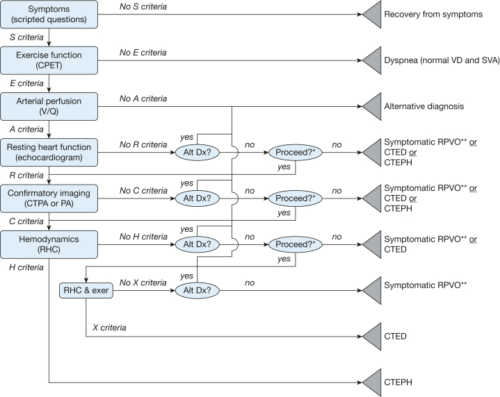
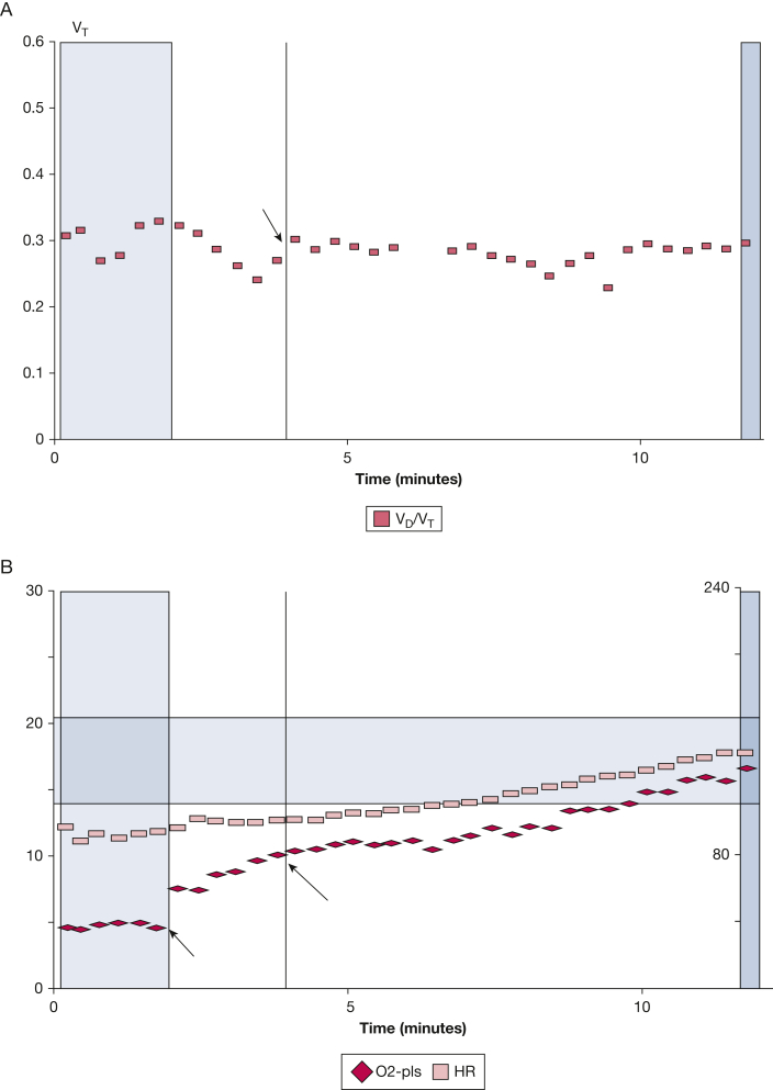
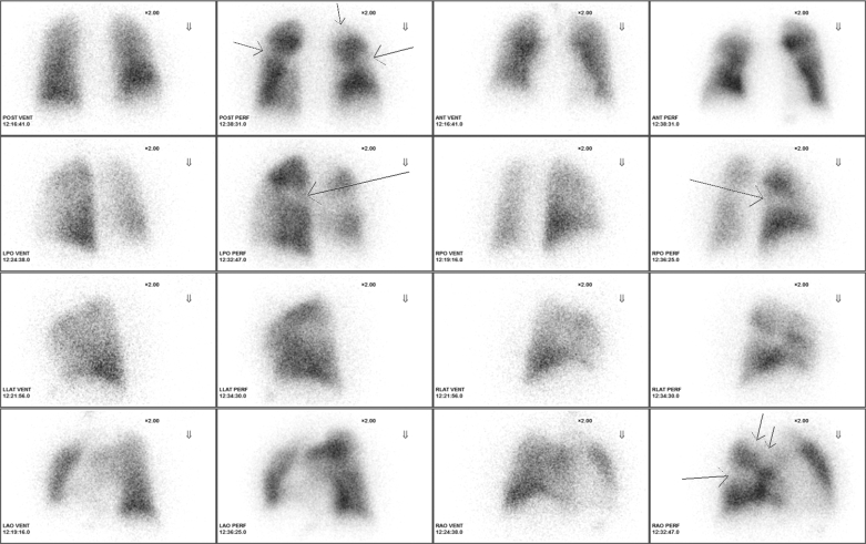
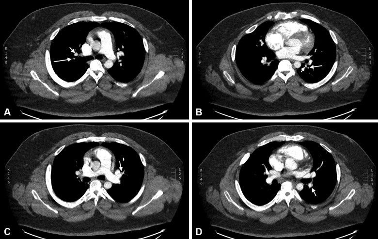
Long-term dyspnea and exercise intolerance are common clinical problems after acute pulmonary embolism. Unfortunately, no single test can distinguish among the range of potential pathologic outcomes after pulmonary embolism. We illustrate a stepwise approach to post-pulmonary embolism evaluation that uses a hierarchic series of clinically validated diagnostic tests. The algorithm is represented by the acronym SEARCH, which stands for Symptom screening, Exercise testing, Arterial perfusion, Resting echocardiography, Confirmatory chest imaging, and Hemodynamics measured by right heart catheterization. We illustrate the algorithm with a patient whom we saw in our pulmonary embolism follow-up clinic. Patients are asked at least 6 months after pulmonary embolism whether they have returned to their baseline level of respiratory comfort and exercise tolerance. Patients with dyspnea and exercise intolerance undergo noninvasive cardiopulmonary exercise testing to identify elevated ventilatory dead space ratios, decreased stroke volume augmentation with exercise, and other physiologic abnormalities during exertion. Ventilation-perfusion scanning is performed on those patients with exercise-related physiologic findings to confirm the presence of residual pulmonary arterial obstruction or to suggest alternative diagnoses. Resting echocardiography may provide evidence of pulmonary hypertension; confirmatory imaging with pulmonary angiography or CT angiography may disclose findings characteristic of chronic pulmonary artery obstruction. Finally, right heart catheterization is performed to confirm chronic thromboembolic pulmonary hypertension; if resting pulmonary hemodynamics are normal, then invasive cardiopulmonary exercise testing may disclose exercise-induced defects.

Keywords: dyspnea; exercise intolerance; pulmonary embolism.


Figures

Figure 1
Figure 1
SEARCH (symptom screen, exercise function, arterial perfusion, resting heart function, confirmatory imaging, and hemodynamics) algorithm decision tree. The SEARCH algorithm grades potential outcomes after pulmonary embolism into distinct, nonoverlapping diagnostic categories. Rectangles represent criteria-driven nodes that reflect dichotomous objective test results. The ovals represent subjective clinical decision nodes. The triangles represent endpoint nodes that reflect the specific differential diagnoses warranted from the clinical data. Alt Dx = alternative diagnosis; CPET = cardiopulmonary exercise testing; CTED = chronic thromboembolic disease (pulmonary hypertension with exercise); CTEPH = chronic thromboembolic pulmonary hypertension; CTPA = CT pulmonary arteriogram; exer = exercise; PA = pulmonary arteriogram; SVA = stroke volume augmentation during exercise; symptomatic RPVO = symptomatic residual pulmonary vascular occlusion; VD = physiologic dead space ventilation; X Criteria = pattern of findings during exercise right heart catheterization. ∗The choice to proceed will depend on the clinical importance of distinguishing among the remaining diagnostic possibilities (eg, symptomatic residual pulmonary vascular occlusion vs chronic thromboembolic disease (pulmonary hypertension with exercise vs chronic thromboembolic pulmonary hypertension) in the patient being evaluated. ∗∗Symptomatic residual pulmonary vascular occlusion is subtyped as chronic thromboembolic disease with increased physiologic dead space ventilation, chronic thromboembolic disease with decreased stroke volume augmentation, chronic thromboembolic disease with increased physiologic dead space ventilation and decreased stroke volume augmentation, or chronic thromboembolic disease with unspecified physiologic effect. S, E, A, R, C, and H criteria represent prespecified test results, as described in the text.
Figure 2
Figure 2
A-B, Elevated ventilatory dead space ratio and decreased stroke volume augmentation at anaerobic threshold and during cardiopulmonary exercise testing. Noninvasive cardiopulmonary exercise testing disclosed physiologic defects that corresponded to the patient’s dyspnea and exercise tolerance. A, Ventilatory dead space ratio (squares) at anaerobic threshold (arrow) is elevated (0.29; predicted = 0.20) and remains elevated for the remainder of exercise. B, Stroke volume augmentation at anaerobic threshold, which is estimated by the ratio of the rate of oxygen consumption/heart rate (diamonds) at anaerobic threshold (9.7 mL oxygen/beat; long arrow) to the rate of oxygen consumption/heart rate at rest (5.2 mL oxygen/beat; short arrow) multiplied by 0.55, is decreased (102%; predicted 140%). HR = heart rate; O2-Pls = rate of oxygen consumption/heart rate; VD/VT = dead space proportion of tidal volume; VT = tidal volume.
Figure 3
Figure 3
SPECT perfusion scan. Pleural-based segmental perfusion defects (arrows) are present in the superior segment of the left lower lobe, the superior segment of the right lower lobe, and the anterior and the apical segments of the right upper lobe. There were no matching ventilation defects. ANT = anterior view; LAO = left anterior oblique; LLAT = left lateral; LPO = left posterior oblique; PERF = perfusion scan; POST = posterior view; RAO = right anterior oblique; RLAT = right lateral; RPO = right posterior oblique; SPECT = single-phton emission CT; VENT = ventilation scan.
Figure 4
Figure 4
A-D, Confirmatory chest imaging. Contrast to enhance the pulmonary arteries disclosed multiple regions smaller-then-normal pulmonary artery caliber. A, Apical segment of the right upper lobe. The pulmonary artery (long arrow) is smaller in caliber than the corresponding vein (short arrow). B, Superior segment of the left lower lobe. The pulmonary artery (long arrow) is smaller in caliber than the corresponding vein (short arrow). C, Superior branch of the lingular pulmonary artery. An eccentric filling defect (arrow) is apparent on the anterior wall of the artery. D, Descending branch of the left pulmonary artery. A web-like filling defect (arrow) is present on the lateral aspect of the artery.

References

    1. Klok F.A., Tijmensen J.E., Haeck M.L., van Kralingen K.W., Huisman M.V. Persistent dyspnea complaints at long-term follow-up after an episode of acute pulmonary embolism: results of a questionnaire. Eur J Intern Med. 2008;19(8):625–629. - PubMed
    1. Klok F.A., van Kralingen K.W., van Dijk A.P., et al. Quality of life in long-term survivors of acute pulmonary embolism. Chest. 2010;138(6):1432–1440. - PubMed
    1. van Es J., den Exter P.L., Kaptein A.A., et al. Quality of life after pulmonary embolism as assessed with SF-36 and PEmb-QoL. Thromb Res. 2013;132(5):500–505. - PubMed
    1. Tavoly M., Utne K.K., Jelsness-Jorgensen L.P., et al. Health-related quality of life after pulmonary embolism: a cross-sectional study. BMJ Open. 2016;6(11) - PMC - PubMed
    1. Fernandes T.M., Alotaibi M., Strozza D.M., et al. Dyspnea postpulmonary embolism from physiological dead space proportion and stroke volume defects during exercise. Chest. 2020;157(4):936–944. - PubMed