. 2022 Oct 15:260:119434.
doi: 10.1016/j.neuroimage.2022.119434. Epub 2022 Jul 2.

Psilocybin induces spatially constrained alterations in thalamic functional organizaton and connectivity

Affiliations

Psilocybin induces spatially constrained alterations in thalamic functional organizaton and connectivity

Andrew Gaddis et al. Neuroimage. .

Erratum in

Abstract

Background: Classic psychedelics, such as psilocybin and LSD, and other serotonin 2A receptor (5-HT2AR) agonists evoke acute alterations in perception and cognition. Altered thalamocortical connectivity has been hypothesized to underlie these effects, which is supported by some functional MRI (fMRI) studies. These studies have treated the thalamus as a unitary structure, despite known differential 5-HT2AR expression and functional specificity of different intrathalamic nuclei. Independent Component Analysis (ICA) has been previously used to identify reliable group-level functional subdivisions of the thalamus from resting-state fMRI (rsfMRI) data. We build on these efforts with a novel data-maximizing ICA-based approach to examine psilocybin-induced changes in intrathalamic functional organization and thalamocortical connectivity in individual participants.

Methods: Baseline rsfMRI data (n=38) from healthy individuals with a long-term meditation practice was utilized to generate a statistical template of thalamic functional subdivisions. This template was then applied in a novel ICA-based analysis of the acute effects of psilocybin on intra- and extra-thalamic functional organization and connectivity in follow-up scans from a subset of the same individuals (n=18). We examined correlations with subjective reports of drug effect and compared with a previously reported analytic approach (treating the thalamus as a single functional unit).

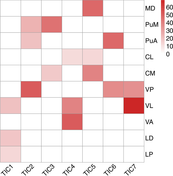
Results: Several intrathalamic components showed significant psilocybin-induced alterations in spatial organization, with effects of psilocybin largely localized to the mediodorsal and pulvinar nuclei. The magnitude of changes in individual participants correlated with reported subjective effects. These components demonstrated predominant decreases in thalamocortical connectivity, largely with visual and default mode networks. Analysis in which the thalamus is treated as a singular unitary structure showed an overall numerical increase in thalamocortical connectivity, consistent with previous literature using this approach, but this increase did not reach statistical significance.

Conclusions: We utilized a novel analytic approach to discover psilocybin-induced changes in intra- and extra-thalamic functional organization and connectivity of intrathalamic nuclei and cortical networks known to express the 5-HT2AR. These changes were not observed using whole-thalamus analyses, suggesting that psilocybin may cause widespread but modest increases in thalamocortical connectivity that are offset by strong focal decreases in functionally relevant intrathalamic nuclei.

Keywords: Functional MRI; Functional connectivity; Independent component analysis; Psilocybin; Psychedelics; Resting state; Thalamocortical connectivity; Thalamus.


Figures

Fig. 1.
Fig. 1.
Visual display of thalamic parcellation derived from spatially constrained group Independent Component Analysis (ICA) applied to the Phase I data, comparing two voxel-wise assignment strategies. Each panel plots Thalamic Independent Component (TIC) assignment for all visible intra-thalamic voxels on an axial slice of the CH-best template from MRIcron. Lighter shades represent a more inclusive winner-take-all assignment strategy, in which every thalamic voxel is assigned to the component with the highest loading at baseline. Darker shades represent only voxels with robust component loading at baseline (z ≥ 2). Components are labeled in two shades (one for each assignment strategy) of each of the following colors: TIC01- brown, TIC02- green, TIC03- blue, TIC04- orange, TIC05- red, TIC06- pink, TIC07- purple. The visualization shows that components have larger and more weakly contributing peripheral areas that encompass smaller cores with robust contribution. For each component, both areas are clearly spatially contiguous and bilaterally symmetrical.
Fig. 2.
Fig. 2.
Percent overlap between histologically identified thalamic nuclei (Morel et al., 1997) and thalamic components thresholded at z ≥ 2. Only those nuclei with ≥10% overlap for any TIC are displayed. MD = mediodorsal; PuM = medial pulvinar; PuA = anterior pulvinar; CL = central lateral; CM = central medial; VP = ventral posterior; VL = ventral lateral; VA = ventral anterior; LD = lateral dorsal; LP = lateral posterior.
Fig. 3.
Fig. 3.
Overall engagement (number of significant voxels) for each Thalamic Independent Component (TIC01 to TIC07) in each drug condition. Each point represents a single participant and session for each IC; box-plots represent the distribution across participants. Non-parametric Wilcoxon two-sided paired t-test p-values for the comparison of voxel counts between placebo and psilocybin sessions are displayed at the top right-hand corner of each panel, corrected at FDR = 0.05. Engagement within only voxels demonstrating robust component loading at baseline (z ≥ 2) was also significantly decreased for TIC03 and TIC05, consistent with the total voxel count results displayed in Fig. 2, with additional components TIC02 and TIC07 also showing significantly decreased engagement during the psilocybin vs. placebo session. These results are displayed in the supplementary material (Fig. S1).
Fig. 4.
Fig. 4.
Left - Percent overlap between histologically identified Morel thalamic nuclei and thresholded thalamic component probabilistic effect maps showing significant decreases in engagement during psilocybin vs. placebo. Only those nuclei with ≥10% overlap are displayed. MD = mediodorsal; PuM = medial pulvinar; PuA = anterior pulvinar; CL = central lateral; VP = ventral posterior; VL = ventral lateral.
Fig. 5.
Fig. 5.
Spearman rank correlations between change in engagement voxel counts and change in subjective effects from placebo to psilocybin. Correlations with r2 ≥0.20 (meeting criteria for a very large association) are indicated with an asterisk∗. Negative correlations represent larger subjective effects of psilocybin associated with larger decreases in TIC engagement; positive correlations represent smaller subjective effects of psilocybin associated with larger decreases in TIC engagement.
Fig. 6.
Fig. 6.
Changes in functional connectivity between placebo (PLA) and psilocybin (PSI) sessions (FDR = 0.05). Red lines between networks indicate increased connectivity on psilocybin (PLA < PSI); blue lines indicate decreased connectivity on psilocybin (PLA > PSI). Axial or sagittal slices outside of the circumference of the connectogram depict components included in the connectogram, and color codes on the circumference of the connectogram denote the overall network sets to which each component belongs. Purple: Thalamic Independent Components (TIC01 to TIC07). Pink: visual network (IC-08, IC-09, and IC-10). Red: auditory network (IC-14). Yellow: cerebellar network (IC-12). Chartreuse: Default Mode Network (“DMN”, IC-11). Spring Green: Executive Control Network (“Executive”, IC-15). Cyan: Right/Left Frontoparietal Networks (“FPN”, IC-16 and IC-17). Dark Blue: Somatosensory network (“SMN”, IC-13). Results using a correction level of FDR = 0.01 are shown in the Supplement (Fig. S2).
Fig. 7.
Fig. 7.
Changes in the strength of significant edges (FDR = 0.05) are associated with changes in subjective experience from placebo to psilocybin (shown using Spearman rank correlations). Correlations with r2 ≥ 0.20 are highlighted with an asterisk∗. Negative correlations represent larger subjective effects of psilocybin associated with decreased connectivity; positive correlations represent larger subjective effects of psilocybin associated with increased connectivity. IC01–07: Thalamic Independent Components (“TIC”s), IC08–10: Visual Network, IC11: Default Mode Network (“DMN”), IC12: Cerebellar Network, IC13: Somatosensory Network (“SMN”), IC14: Auditory Network, IC15: Executive Control Network, IC16–17: Right/Left Frontoparietal Networks (“FPN”).
Fig. 8.
Fig. 8.
Voxelwise connectivity between all thalamic voxels (N = 2268) and both the Default Mode Network (“DMN”, IC11) and Occipital Pole network (IC09). (Panel A) Voxelwise thalamocortical connectivity t-statistics for DMN and Occipital Pole cortical ICs. Blue = placebo > psilocybin (decreased connectivity on psilocybin). Red = placebo < psilocybin (increased connectivity on psilocybin). The green outline shows the area of robustly contributing voxels for each TIC, shown on the corresponding row in panel B. (Panel B) Mean TIC engagement from baseline data thresholded at z ≥ 2. Images are displayed using neurological convention.

References

    1. Avram M, Müller F, Rogg H, Korda A, Andreou C, Holze F, Vizeli P, Ley L, Liechti ME, Borgwardt S, 2022. Characterizing thalamocortical (dys)connectivity following d-amphetamine, LSD, and MDMA administration. Biol. Psychiatry Cogn. Neurosci. Neuroimaging. doi:10.1016/j.bpsc.2022.04.003. - DOI - PubMed
    1. Barre A, Berthoux C, De Bundel D, Valjent E, Bockaert J, Marin P, Bécamel C, 2016. Presynaptic serotonin 2A receptors modulate thalamocortical plasticity and associative learning. Proc. Natl. Acad. Sci. U. S. A. 113, E1382–E1391. doi:10.1073/pnas.1525586113. - DOI - PMC - PubMed
    1. Barrett FS, Carbonaro TM, Hurwitz E, Johnson MW, Griffiths RR, 2018a. Double-blind comparison of the two hallucinogens psilocybin and dextromethorphan: effects on cognition. Psychopharmacology doi:10.1007/s00213-018-4981-x. - DOI - PMC - PubMed
    1. Barrett FS, Johnson MW, Griffiths RR, 2015. Validation of the revised Mystical Experience Questionnaire in experimental sessions with psilocybin. J. Psychopharmacol. 29, 1182–1190. doi:10.1177/0269881115609019. - DOI - PMC - PubMed
    1. Barrett FS, Krimmel SR, Griffiths R, Seminowicz DA, Mathur BN, 2020. Psilocybin acutely alters the functional connectivity of the claustrum with brain net- works that support perception, memory, and attention. Neuroimage 218, 116980. doi:10.1016/j.neuroimage.2020.116980. - DOI - PMC - PubMed

Publication types