Review
. 2022 Aug:54:102389.
doi: 10.1016/j.redox.2022.102389. Epub 2022 Jun 30.

Signal amplification in the KEAP1-NRF2-ARE antioxidant response pathway

Affiliations
Review

Signal amplification in the KEAP1-NRF2-ARE antioxidant response pathway

Shengnan Liu et al. Redox Biol. 2022 Aug.

Abstract

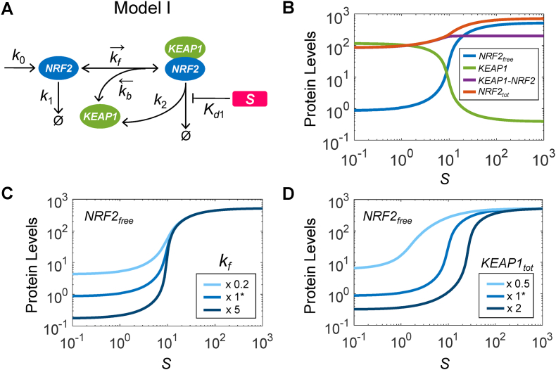
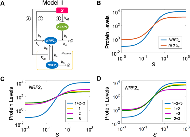
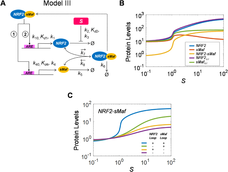
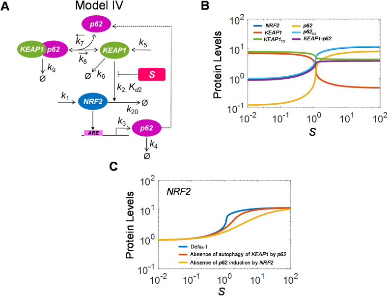
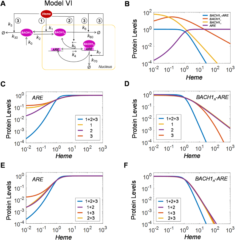
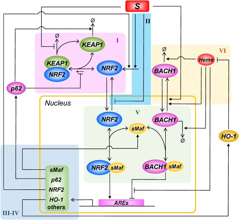
The KEAP1-NRF2-ARE signaling pathway plays a central role in mediating the adaptive cellular stress response to oxidative and electrophilic chemicals. This canonical pathway has been extensively studied and reviewed in the past two decades, but rarely was it looked at from a quantitative signaling perspective. Signal amplification, i.e., ultrasensitivity, is crucially important for robust induction of antioxidant genes to appropriate levels that can adequately counteract the stresses. In this review article, we examined a number of well-known molecular events in the KEAP1-NRF2-ARE pathway from a quantitative perspective with a focus on how signal amplification can be achieved. We illustrated, by using a series of mathematical models, that redox-regulated protein sequestration, stabilization, translation, nuclear trafficking, DNA promoter binding, and transcriptional induction - which are embedded in the molecular network comprising KEAP1, NRF2, sMaf, p62, and BACH1 - may generate highly ultrasensitive NRF2 activation and antioxidant gene induction. The emergence and degree of ultrasensitivity depend on the strengths of protein-protein and protein-DNA interaction and protein abundances. A unique, quantitative understanding of signal amplification in the KEAP1-NRF2-ARE pathway will help to identify sensitive targets for the prevention and therapeutics of oxidative stress-related diseases and develop quantitative adverse outcome pathway models to facilitate the health risk assessment of oxidative chemicals.

Keywords: ARE; KEAP1; NRF2; Oxidative stress; Signal amplification; Ultrasensitivity.


Conflict of interest statement

The authors declare no conflict of interest.

Figures

Fig. 1
Fig. 1
Schematic illustration of the KEAP1-NRF2-ARE pathway embedded in negative feedback and incoherent feedforward circuitry. (A) Biologist's view. (B) Engineer's view.
Fig. 2
Fig. 2
Model I: NRF2 ultrasensitivity arising from saturable NRF2 sequestration and degradation by KEAP1. (A) Structure of Model I, containing constitutive synthesis of NRF2 (k0 step), KEAP1-independent NRF2 degradation (k1), reversible binding of NRF2 and KEAP1 (kf and kb), and KEAP1-dependent NRF2 degradation and concurrent recycling of KEAP1 (k2) which can be inhibited by electrophilic stressor S (Kd1). (B) Simulated steady-state dose-response of various state variables as indicated, with free NRF2 (NRF2free) exhibiting ultrasensitivity. (C) The degree of ultrasensitivity of the NRF2free response is modulated by the binding affinity between NRF2 and KEAP1 as indicated by different kf values. (D) The degree of ultrasensitivity of the NRF2free response is modulated by the total abundance of KEAP1 (KEAP1tot) as indicated. x 1* denotes using the default parameter values, x 0.5 and x 2 denote using half and twice the default values respectively.
Fig. 3
Fig. 3
Model II: Nuclear NRF2 ultrasensitivity arising from multistep signaling. (A) Structure of Model II, containing basal synthesis of cytosolic NRF2 (NRF2c) (k0) and stressor S-induced synthesis due to translation enhancement (k1), KEAP1-independent NRF2c degradation (k3), KEAP1-dependent NRF2c degradation (k2), inhibition of KEAP1 activity by S (Kd2), importation of NRF2c into the nucleus (k4), exportation of nuclear NRF2 (NRF2n) (k5) which is inhibited by S (Kd5), and NRF2n degradation (k6). (B) Simulated steady-state dose-response of NRF2c and NRF2n, with the latter exhibiting a higher degree of ultrasensitivity. (C) Loss of ultrasensitivity of NRF2n when only one of the three steps, (i) NRF2c stabilization, (ii) enhanced NRF2c translation, and (iii) inhibited NRF2n exportation, is present as indicted. (D) Reduced ultrasensitivity of NRF2n when two of the three steps are present as indicted.
Fig. 4
Fig. 4
Model III: Ultrasensitivity arising from coupled NRF2 and sMaf positive autoregulation and heterodimerization. (A) Structure of Model III, containing basal synthesis of NRF2 (k10) and sMaf (k40), NRF2-sMaf dimer-induced synthesis of NRF2 (k1 and Kd1) and sMaf (k4 and Kd4), stressor S-inhibited degradation of NRF2 (k2 and Kd2) and S-independent degradation of NRF2 (k3), degradation of sMaf (k5), association (k7) and dissociation (k8) between NRF2 and sMaf, and degradation of NRF2-sMaf dimer (k6). (B) Simulated steady-state dose-response of NRF2, sMaf, and NRF2-sMaf as indicated, exhibiting strong ultrasensitivity. (C) The degree of ultrasensitivity of NRF2-sMaf is altered when the NRF2 and sMaf autoregulatory loops are both present, only one is present, and both are absent.
Fig. 5
Fig. 5
Model IV: Ultrasensitivity arising from positive autoregulation of NRF2 through p62-mediated sequestration and autophagy of KEAP1. (A) Structure of Model IV, containing basal synthesis of NRF2 (k1), KEAP1-independent NRF2 degradation (k20), KEAP1-dependent NRF2 degradation which can be inhibited by stressor S (k2, Kd2), NRF2-induced synthesis of p62 (k3), basal degradation of p62 (k4), synthesis of KEAP1 (k5), p62-independent degradation of KEAP1 (k6), association (k7) and dissociation (k8) between KEAP1 and p62, and autophagic degradation of KEAP1-p62 complex (k9). (B) Simulated steady-state dose-response of NRF2, KEAP1, and p62 variables as indicated, exhibiting ultrasensitivity. (C) Ultrasensitivity of NRF2 when (i) the p62 autoregulatory loop and p62-mediated KEAP1 sequestration and degradation are intact, (ii) p62-mediated KEAP1 autophagy is absent (by setting k9=k6, and k5=1.27573E-4 so that free KEAP1 remains at the same basal level as in the intact model), and (iii) p62 induction by NRF2 is disabled such that p62 remains at the same basal level as in the intact model.
Fig. 6
Fig. 6
Model V: Release of sequestration of sMaf and ARE by nuclear BACH1 exportation. (A) Structure of Model V, containing stressor S-induced nuclear exportation of BACH1n (k1), nuclear importation of BACH1c (k2), reversible binding between BACH1n and sMaf (k3 and k4), reversible binding between NRF2 and sMaf (k5 and k6), transcriptional activation of an ARE Target gene by NRF2-sMaf and its competitive inhibition by BACH1n-sMaf (k7, Kd7, Ki7), and degradation of the protein product of the Target gene (k8). (B-C) Simulated steady-state dose-response of free sMaf, free BACH1n, NRF2-sMaf, BACH1n-sMaf, and Target gene expression as indicated, when the model operates in titration mode (B) where k3 = 0.1, k4 = 0.01, and total BACH1 = 100, thus BACH1n-sMaf is the dominant form of nuclear BACH1, or in equilibrium mode (C) where k3 = 0.01, k4 = 0.1, and total BACH1 = 10000, thus free BACH1n is the dominant form of nuclear BACH1, respectively. None of the variables exhibit ultrasensitivity.
Fig. 7
Fig. 7
Model VI: Ultrasensitivity arising from multistep inhibition of BACH1 by heme. (A) Structure of Model VI, containing basal synthesis of BACH1 (k0), basal degradation of BACH1 (k30, k60, and k70), Heme-stimulated degradation of BACH1 (k3, k6, and k7), Heme-stimulated nuclear exportation of BACH1n (k1), nuclear importation of BACH1c (k2), Heme-inhibited reversible binding between BACH1n and ARE (k4 and k5). (B) Simulated steady-state dose-response of BACH1c, free nuclear BACH1n, free ARE, and BACH1n-ARE complex. Both free ARE and BACH1n-ARE exhibit ultrasensitivity. (CD) Ultrasensitivity of free ARE and BACH1n-ARE, respectively, is lost or weakened when only one of the three Heme-regulated steps, (i) BACH1n nuclear exportation, (ii) BACH1n binding to ARE, and (iii) BACH1n degradation, is present as indicted. (EF) Ultrasensitivity of free ARE and BACH1-ARE, respectively, is weakened when two of the three steps are present as indicted.
Fig. 8
Fig. 8
A global view of the KEAP1-NRF2-ARE signaling pathway integrating multiple ultrasensitive modules of molecular interactions. The modules, corresponding approximately to models I-VI, are shaded and numerically labeled. S: oxidative or electrophilic species.

References

    1. Nguyen T., Sherratt P.J., Pickett C.B. Regulatory mechanisms controlling gene expression mediated by the antioxidant response element. Annu. Rev. Pharmacol. Toxicol. 2003;43:233–260. - PubMed
    1. Baird L., Yamamoto M. The molecular mechanisms regulating the KEAP1-NRF2 pathway. Mol. Cell Biol. 2020;40(13) - PMC - PubMed
    1. Torrente L., DeNicola G.M. Targeting NRF2 and its downstream processes: opportunities and challenges. Annu. Rev. Pharmacol. Toxicol. 2022;62:279–300. - PubMed
    1. Zhang Q., Pi J., Woods C.G., Andersen M.E. A systems biology perspective on Nrf2-mediated antioxidant response. Toxicol. Appl. Pharmacol. 2010;244(1):84–97. - PMC - PubMed
    1. Zhang Q., Bhattacharya S., Pi J., Clewell R.A., Carmichael P.L., Andersen M.E. Adaptive posttranslational control in cellular stress response pathways and its relationship to toxicity testing and safety assessment. Toxicol. Sci. 2015;147(2):302–316. - PMC - PubMed

Publication types

MeSH terms

Substances