. 2022 Jul;44(7):951-961.
doi: 10.1007/s00276-022-02973-0. Epub 2022 Jul 6.

Anatomical variants of the acromioclavicular joint influence its visibility in the standard MRI protocol in patients aged 18-31 years

Affiliations

Anatomical variants of the acromioclavicular joint influence its visibility in the standard MRI protocol in patients aged 18-31 years

Fredrik Helleberg et al. Surg Radiol Anat. 2022 Jul.

Abstract

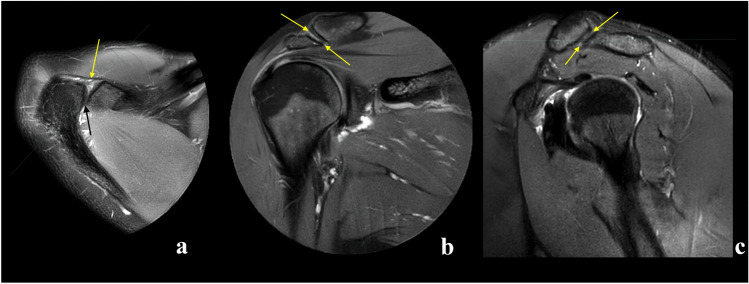
Purpose: Visualization of a structure in orthogonal planes is essential for correct radiological assessment. The aim was to assess the utility of the standard MRI protocol for the shoulder in the assessment of the acromioclavicular joint (ACJ).

Methods: A total of 204 MRI scans of the shoulder were re-reviewed. Visibility of the ACJ in orthogonal planes was assessed, and the type of acromion and the angle between the ACJ and the glenoid cavity were assessed by two observers.

Results: Agreement in the assessment of ACJ visibility was moderate to substantial. The ACJ was visible in the three anatomical views in 48% (confidence interval [CI] 95% = [41-54%]) of the examinations, and no significant difference regarding gender or age was noticed. The mean angle between the ACJ and the glenoid cavity was 41.12 deg. CI95% = (39.72, 42.53) in the axial plane, 33.39 deg. CI95% = (31.33, 35.45) in the coronal plane and 52.49 deg. CI95% = (50.10, 54.86) in the sagittal plane. When the ACJ was visible in the sagittal and axial planes, significant differences were noticed in the remaining planes (p < .05).

Conclusion: Anatomical variations of the ACJ influence its visibility in the standard MRI protocol for examining the shoulder, making this protocol insufficient for ACJ assessment in the examined population.

Keywords: Acromioclavicular joint; Acromion; Anatomy; Magnetic resonance imaging; Shoulder pain.


Conflict of interest statement

The authors declare no competing interests.

Figures

Fig. 1
Fig. 1
The angles between the acromioclavicular joint (yellow) and the glenoid (dashed line) was measured in axial (a and b), coronal (c and d) and sagittal section (e and f). The joint space is visible on all presented sections (af). A and b—proton density (PD)-weighted turbo spin echo (TSE) Spectral Attenuated Inversion Recovery (SPAIR), bf —T2-weighted TSE SPAIR
Fig. 2
Fig. 2
Examples of the visible joint cavity in axial (a), coronal (b) and sagittal view c, respectively (arrows). Three different patients, a a 23-year-old patient with the shoulder pain, b an 18-year-old patient with suspicion of the subacromial impingement, c a 28-year-old patient with suspicion of the supraspinatus tear. A—proton density (PD)-weighted turbo spin echo (TSE) Spectral Attenuated Inversion Recovery (SPAIR), b and c—T2-weighted TSE SPAIR
Fig. 3
Fig. 3
Example of an examination where the joint cavity is not visible in axial plane. The joint space is not visible on the axial plane (a and b) while visible on the sagittal plane (c) and not visible on the coronal plane (d). The localization of the joint is marked by arrows. An 18-year-old patient with suspicion of the subacromial bursitis. A and b—proton density (PD)-weighted turbo spin echo (TSE) Spectral Attenuated Inversion Recovery (SPAIR), c and d—T2-weighted TSE SPAIR
Fig. 4
Fig. 4
Example of an examination where the joint cavity is not visible in coronal plane. The arrows show the localization of the acromioclavicular joint. The joint space is visible on the axial plane (a) and sagittal plane (b). The joint space is not visible on the coronal plane (c and d), localization of the joint is marked by arrows. A 26-year-old patient with suspicion of supraspinatus tear. A—proton density (PD)-weighted turbo spin echo (TSE) Spectral Attenuated Inversion Recovery (SPAIR), b, c and d—T2-weighted TSE SPAIR
Fig. 5
Fig. 5
Example of an examination where the joint cavity is not visible in sagittal view. The arrows show the localization of the acromioclavicular joint. The joint space is visible on the axial plane (a and b) and coronal plane (c). The joint space is not visible on the sagittal plane (d), localization of the joint is marked by arrows. A 30-year-old patient with suspicion of subacromial bursitis. A and b—proton density (PD)-weighted turbo spin echo (TSE) Spectral Attenuated Inversion Recovery (SPAIR), c and d—T2-weighted TSE SPAIR
Fig. 6
Fig. 6
Configuration of the acromioclavicular joint space on the coronal (a and b) and sagittal planes (c and d) when the joint cavity is visible (a and c) or not (b and d) in the axial plane. Larger values of the acromioclavicular joint space angle in the coronal (b) and sagittal (d) planes were noticed when the joint cavity was not visible in the axial plane. Differences in values of angles of the acromioclavicular joint were statistically significant (Table 7). Figure prepared by PS
Fig. 7
Fig. 7
Configuration of the acromioclavicular joint space on the axial (a and b) and sagittal planes (c and d) when the joint cavity is visible (a and c) or not (b and d) in the coronal plane. Larger values of the acromioclavicular joint space angle in the axial (b) plane are noticed when the joint cavity is not visible in the coronal plane. Differences in values of angles of the acromioclavicular joint were not statistically significant (Table 8). Figure prepared by PS
Fig. 8
Fig. 8
Configuration of the acromioclavicular joint space on the axial (a and b) and coronal planes (c and d) when the joint cavity was visible (a and c) or not (b and d) in the sagittal plane. Larger values of the acromioclavicular joint space angle in the axial plane (a) were noticed when the joint cavity was visible in the sagittal plane. Larger values of the acromioclavicular joint space angle in the coronal plane (d) were noticed when the joint cavity was not visible in the sagittal plane. Differences in values of angles of the acromioclavicular joint were statistically significant (Table 9). Figure prepared by PS
Fig. 9
Fig. 9
The plots show the difference in the angles of the shoulder–clavicular joint with respect to the glenoid cavity in the sagittal (A), transverse (B) and frontal (C) planes in groups where the shoulder–clavicular joint is visible or not in the coronal plane (a), the sagittal plane (b) and the axial plane (c). The figure was done in SPSS Statistics software version 22.0

Similar articles

Cited by

References

    1. Alyas F, Curtis M, Speed C, et al. MR imaging appearances of acromioclavicular joint dislocation. Radiographics. 2008;28:463–479. doi: 10.1148/rg.282075714. - DOI - PubMed
    1. Choo HJ, Lee SJ, Kim JH, et al. Can symptomatic acromioclavicular joints be differentiated from asymptomatic acromioclavicular joints on 3-T MR imaging? Eur J Radiol. 2013;82:e184–191. doi: 10.1016/j.ejrad.2012.10.027. - DOI - PubMed
    1. Depalma AF. Surgical anatomy of acromioclavicular and sternoclavicular joints. Surg Clin North Am. 1963;43:1541–1550. doi: 10.1016/s0039-6109(16)37142-0. - DOI - PubMed
    1. Ernberg LA, Potter HG. Radiographic evaluation of the acromioclavicular and sternoclavicular joints. Clin Sports Med. 2003;22:255–275. doi: 10.1016/s0278-5919(03)00006-1. - DOI - PubMed
    1. Fialka C, Krestan CR, Stampfl P, et al. Visualization of intraarticular structures of the acromioclavicular joint in an ex vivo model using a dedicated MRI protocol. AJR Am J Roentgenol. 2005;185:1126–1131. doi: 10.2214/ajr.04.1433. - DOI - PubMed