. 2022 Jul 6;17(7):e0270657.
doi: 10.1371/journal.pone.0270657. eCollection 2022.

Butyrate ameliorates maternal high-fat diet-induced fetal liver cellular apoptosis

Affiliations

Butyrate ameliorates maternal high-fat diet-induced fetal liver cellular apoptosis

Yu-Jyun Huang et al. PLoS One. .

Abstract

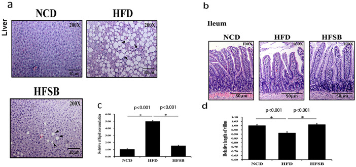
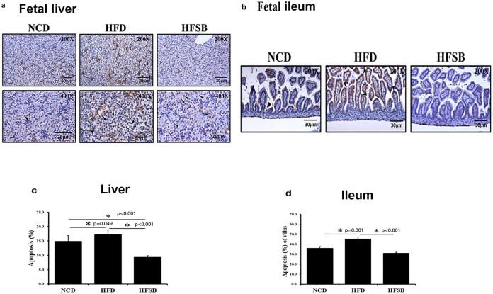
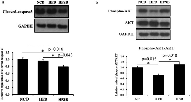
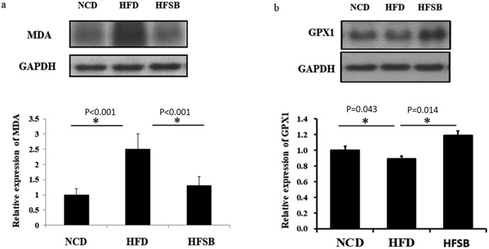
A maternal high-fat diet (HFD) can impact the offspring's development of liver steatosis, with fetal development in utero being a crucial period. Therefore, this study investigated the mechanism and whether butyrate can rescue liver injury caused by maternal HFD in the fetus. Pregnant female Sprague Dawley rats were randomly divided into two groups, prenatal HFD (58% fat) exposure or normal control diet (4.5% fat). The HFD group was fed an HFD 7 weeks before mating and during gestation until sacrifice at gestation 21 days. After confirmation of mating, the other HFD group was supplemented with sodium butyrate (HFSB). The results showed that maternal liver histology showed lipid accumulation with steatosis and shortened ileum villi in HFD, which was ameliorated in the HFSB group (P<0.05). There was increased fetal liver and ileum TUNEL staining and IL-6 expression with increased fetal liver TNF-α and malondialdehyde expression in the HFD group (P<0.05), which decreased in the HFSB group (P<0.05). The fetal liver expression of phospho-AKT/AKT and GPX1 decreased in the HFD group but increased in the HFSB group (P<0.05). In conclusion that oxidative stress with inflammation and apoptosis plays a vital role after maternal HFD in the fetus liver that can be ameliorated with butyrate supplementation.


Conflict of interest statement

The authors have declared that no competing interests exist.

Figures

Fig 1
Fig 1. Histological analysis of maternal liver and ileum.
(a) H&E staining in the liver and (b) ileal tissue. Semi-quantitative analysis of (c) hepatic lipid accumulation and (d) ileal villous length. NCD: normal-chow diet, HFD: high-fat diet, HFSB: high-fat diet with butyrate supplementation during pregnancy (n = 6), * p < 0.05. Black arrowheads: lipid droplets.
Fig 2
Fig 2. Short chain fatty acids profile of maternal rats.
Maternal stool levels of (a) acetic acid, (b) propionic acid, (c) and butyric acid. NCD: normal-chow diet, HFD: high-fat diet, HFSB: high-fat diet with butyrate supplementation during pregnancy (n = 6), * p < 0.05.
Fig 3
Fig 3. Fetal liver and ileum inflammation.
(a) IL-6 staining in fetal liver and (b) fetal ileum. (c, d) Semi-quantitative analysis of IL-6 staining in fetal liver and fetal ileum, (e) western blotting of TNF-α expression in fetal liver, and (f) western blotting of fetal ileum tight junction ZO-1, occludin and claudin-3 expressions. NCD: normal-chow diet, HFD: high-fat diet, HFSB: high-fat diet with butyrate supplementation during pregnancy (n = 6), * p < 0.05. Arrowheads: positive IL-6 staining cells.
Fig 4
Fig 4. Apoptosis in the fetal liver and ileum.
(a) TUNEL staining in the fetus liver and (b) fetus ileum. (c, d) Semi-quantitative analysis of fetus liver and ileum TUNEL stained positive cells. NCD: normal-chow diet, HFD: high-fat diet, HFSB: high-fat diet with butyrate supplementation during pregnancy (n = 6), * p < 0.05. Black arrowheads: positive TUNEL stained cells.
Fig 5
Fig 5. Western blotting of apoptotic protein expression in fetal liver.
(a) cleaved-caspase 3, (b) phosphor-AKT/AKT. NCD: normal-chow diet, HFD: high-fat diet, HFSB: high-fat diet with butyrate supplementation during pregnancy (n = 6), * p < 0.05.
Fig 6
Fig 6. Fetal liver oxidative stress.
(a) western blotting of MDA expression and (b) antioxidative stress GPX1 expression. NCD: normal-chow diet, HFD: high-fat diet, HFSB: high-fat diet with butyrate supplementation during pregnancy (n = 6), * p < 0.05.

References

    1. Haugen AC, Schug TT, Collman G, Heindel JJ: Evolution of DOHaD: the impact of environmental health sciences. J Dev Orig Health Dis 2015, 6(2):55–64. doi: 10.1017/S2040174414000580 - DOI - PMC - PubMed
    1. Kozyrskyj AL, Kalu R, Koleva PT, Bridgman SL: Fetal programming of overweight through the microbiome: boys are disproportionately affected. J Dev Orig Health Dis 2016, 7(1):25–34. doi: 10.1017/S2040174415001269 - DOI - PubMed
    1. Catalano PM, Presley L, Minium J, Hauguel-de Mouzon S: Fetuses of obese mothers develop insulin resistance in utero. Diabetes care 2009, 32(6):1076–1080. doi: 10.2337/dc08-2077 - DOI - PMC - PubMed
    1. Brumbaugh DE, Friedman JE: Developmental origins of nonalcoholic fatty liver disease. Pediatr Res 2014, 75(1–2):140–147. doi: 10.1038/pr.2013.193 - DOI - PMC - PubMed
    1. Parente LB, Aguila MB, Mandarim-de-Lacerda CA: Deleterious effects of high-fat diet on perinatal and postweaning periods in adult rat offspring. Clin Nutr 2008, 27(4):623–634. doi: 10.1016/j.clnu.2008.05.005 - DOI - PubMed

Publication types