. 2022 Sep 6;21(9):1449-1461.
doi: 10.1158/1535-7163.MCT-22-0362.

An Enzymatically Cleavable Tripeptide Linker for Maximizing the Therapeutic Index of Antibody-Drug Conjugates

Affiliations

An Enzymatically Cleavable Tripeptide Linker for Maximizing the Therapeutic Index of Antibody-Drug Conjugates

Summer Y Y Ha et al. Mol Cancer Ther. .

Abstract

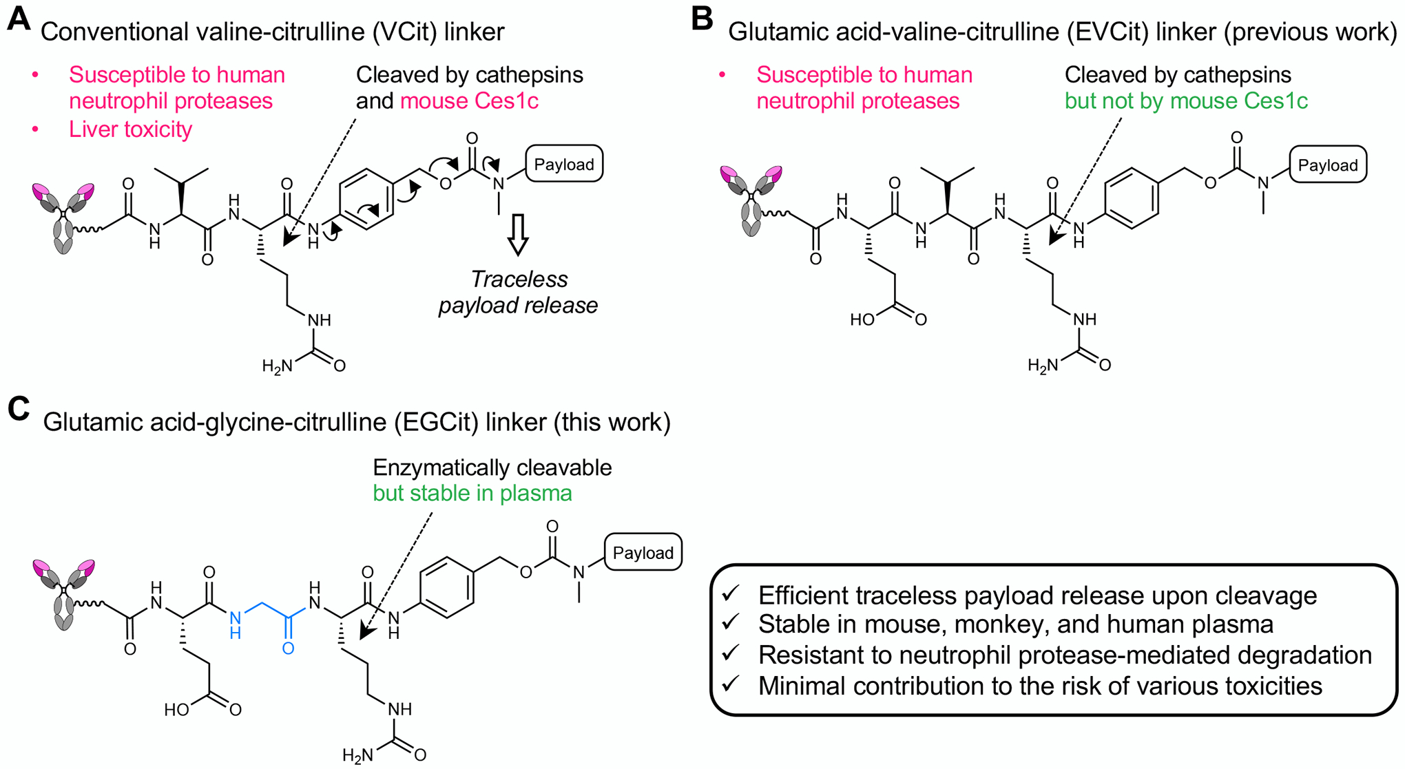
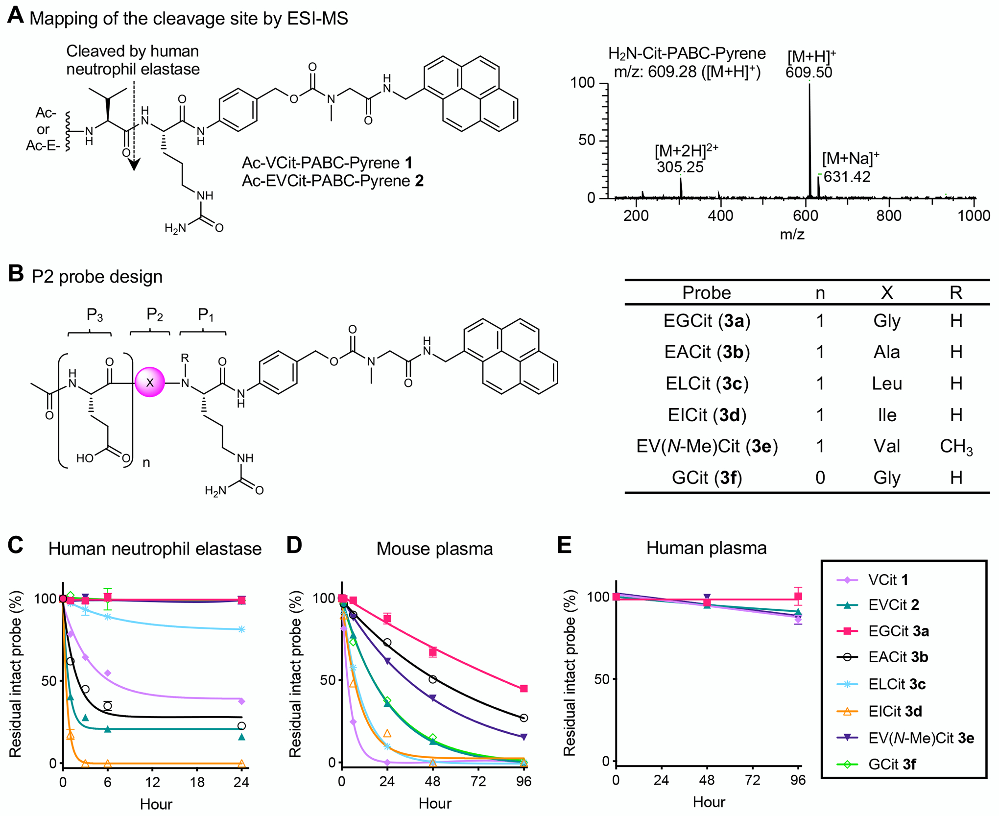
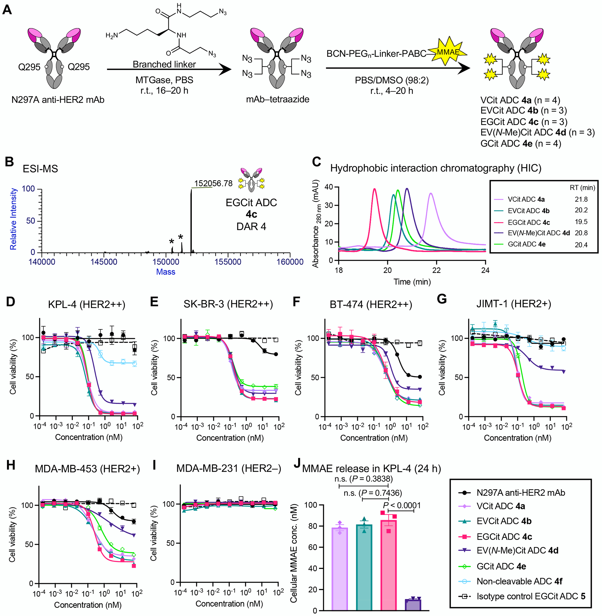
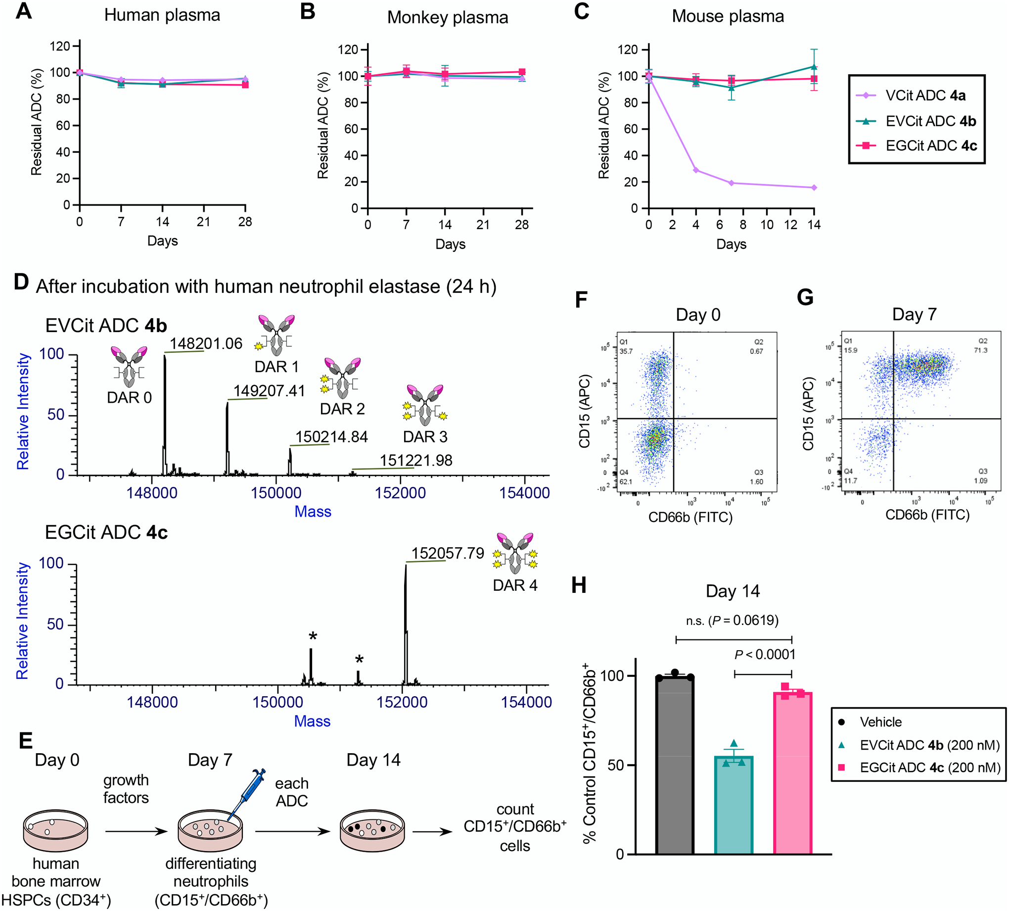
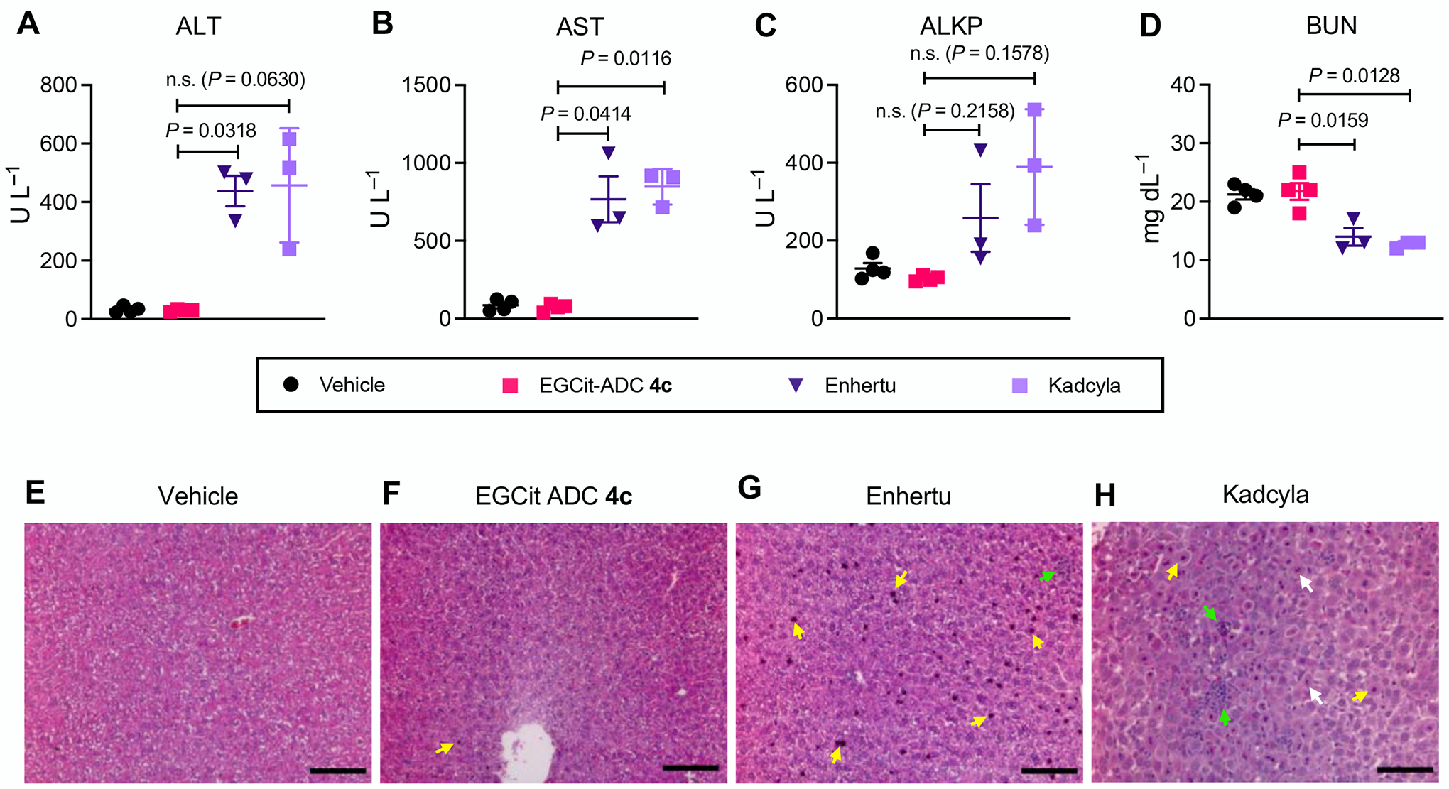
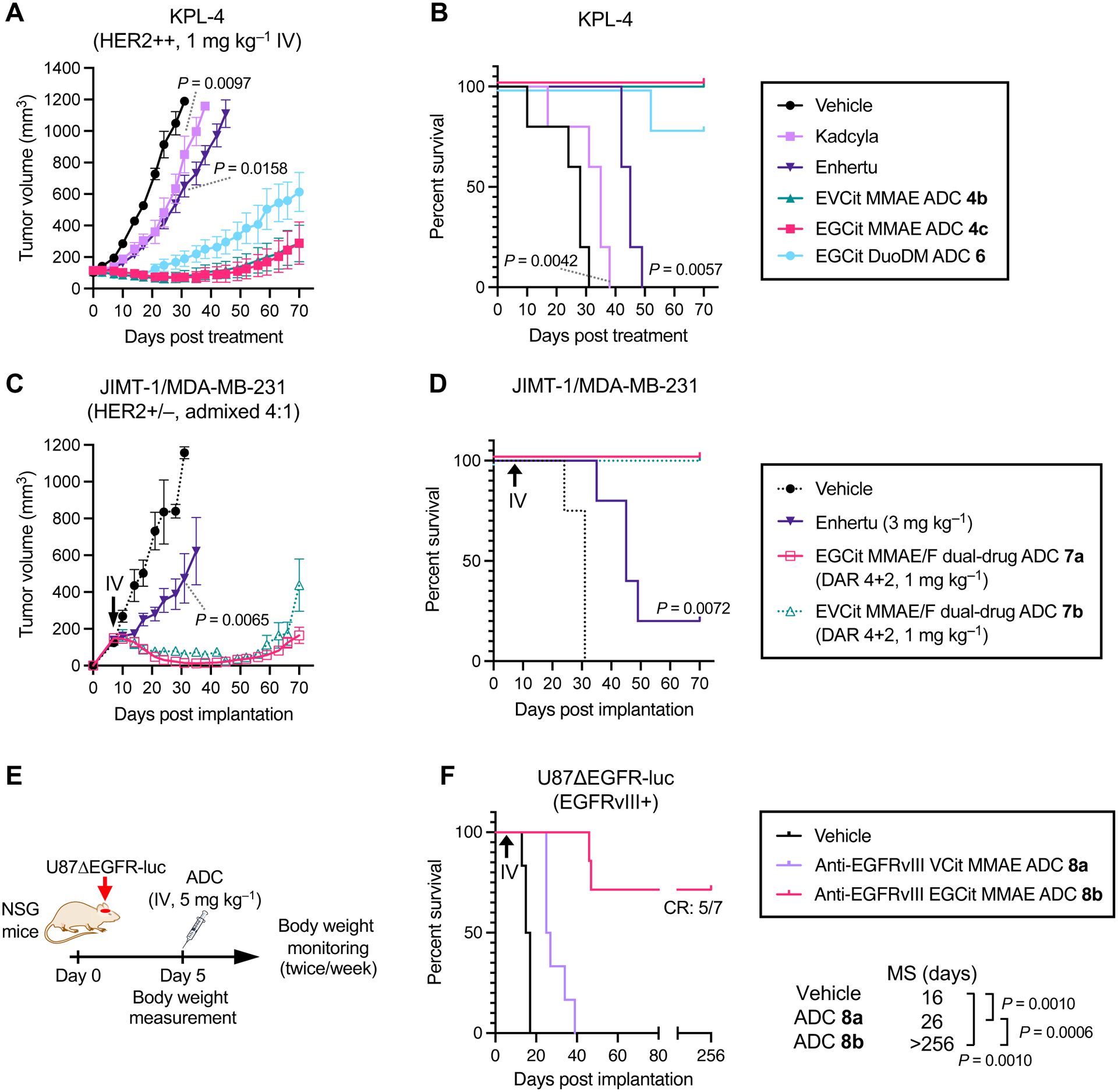
Valine-citrulline is a protease-cleavable linker commonly used in many drug delivery systems, including antibody-drug conjugates (ADC) for cancer therapy. However, its suboptimal in vivo stability can cause various adverse effects such as neutropenia and hepatotoxicity, leading to dose delays or treatment discontinuation. Here, we report that glutamic acid-glycine-citrulline (EGCit) linkers have the potential to solve this clinical issue without compromising the ability of traceless drug release and ADC therapeutic efficacy. We demonstrate that our EGCit ADC resists neutrophil protease-mediated degradation and spares differentiating human neutrophils. Notably, our anti-HER2 ADC shows almost no sign of blood and liver toxicity in healthy mice at 80 mg kg-1. In contrast, at the same dose level, the FDA-approved anti-HER2 ADCs Kadcyla and Enhertu show increased levels of serum alanine aminotransferase and aspartate aminotransferase and morphologic changes in liver tissues. Our EGCit conjugates also exert greater antitumor efficacy in multiple xenograft tumor models compared with Kadcyla and Enhertu. This linker technology could substantially broaden the therapeutic windows of ADCs and other drug delivery agents, providing clinical options with improved efficacy and safety.


Conflict of interest statement

Conflicts of Interest Statement

Y.A., C.M.Y., N.Z., Z.A., and K.T. are named inventors on patent applications relating to the work (PCT/US2018/034363, US-2020-0115326-A1, EU18804968.8-1109/3630189). S.Y.Y.H., Y.A., C.M.Y., N.Z., Z.A., and K.T. are named inventors on a pending patent application relating to the work. All patent applications were filed by the Board of Regents of the University of Texas System. The remaining authors declare no competing interests.

Figures

Fig. 1.
Fig. 1.
The structures and stability profiles of cleavable peptide linkers. A VCit-based ADC linker. VCit linkers are unstable in mouse circulation due to susceptibility to the extracellular carboxylesterase Ces1c. VCit linkers are also labile to human neutrophil elastase-mediated degradation. This instability often triggers premature payload release, leading to poor efficacy in preclinical rodent models and safety concerns including neutropenia and liver toxicity in humans. B EVCit-based ADC linker. We have previously developed EVCit linkers that are stable in human and mouse plasma. However, as shown in this study, this linker is also incapable of withstanding neutrophil elastase-mediated degradation. C EGCit-based ADC linker. This study demonstrates that EGCit linkers resist degradation in circulation and cleavage mediated by human neutrophil proteases while remaining capable of releasing payloads in a traceless manner upon intracellular cleavage.
Fig. 2.
Fig. 2.
Incorporating glycine at the P2 position and glutamic acid at the P3 position increases resistance to undesired degradation leading to premature payload release. A ESI-MS-based mapping of the cleavage site in the presence of human neutrophil elastase. We observed cleavage of the amide bond between valine and citrulline within VCit (1) and EVCit (2) probes. B Structures of small-molecule P2 probes containing EGCit (3a), EACit (3b), ELCit (3c), EICit (3d), EV(N-Me)Cit (3e), or GCit (3f). A pyrene group was used as a surrogate of hydrophobic ADC payloads. CE Stability of probes 1, 2, and 3af in the presence of human neutrophil elastase (C), in undiluted BALB/c mouse plasma (D), and in undiluted human plasma (E) at 37 °C. (1) light purple diamond; (2) green triangle; (3a) magenta square; (3b) black open circle; (3c) cyan asterisk; (3d) orange open triangle; (3e) purple inversed triangle; (3f) light green open diamond. All assays were performed at least three times in technical duplicate, and representative data from the replicates are shown (n = 2). Data are presented as mean values ± SEM. PABC, p-aminobenzyloxycarbonyl.
Fig. 3.
Fig. 3.
EGCit linker increases ADC hydrophilicity and cell killing potency with efficient intracellular payload release. A Construction of ADCs (4ae) by MTGase-mediated branched linker conjugation and following strain-promoted azide–alkyne cycloaddition (yellow spark: MMAE). B Deconvoluted ESI-MS trace of EGCit ADC 4c. Asterisk (*) indicates a fragment ion detected in ESI-MS analysis. See Supplementary Notes for mass traces of the other ADCs. C Overlay of five HIC traces (VCit ADC 4a: light purple; EVCit ADC 4b: green; EGCit ADC 4c: magenta; EV(N-Me)Cit ADC 4d: purple; GCit ADC 4e: light green) under physiological conditions (phosphate buffer, pH 7.4). DI Cell killing potency in the breast cancer cell lines KPL-4 (D), SK-BR-3 (E), BT-474 (F), JIMT-1 (G), MDA-MB-453 (H), and MDA-MB-231 (I). We tested unconjugated N297A anti-HER2 mAb (black circle), VCit ADC 4a (light purple diamond), EVCit ADC 4b (green triangle), EGCit ADC 4c (magenta square), EV(N-Me)Cit 4d (purple inversed triangle), GCit ADC 4e (light green open diamond), non-cleavable ADC 4f (cyan open circle), and isotype control EGCit ADC 5 (black open rectangle with dotted curve). J ESI-MS-based quantification of free MMAE released from ADCs 4ac in KPL-4 cells after incubation at 37 °C for 24 h. All assays were performed in triplicate. Data are presented as mean values ± SEM. For statistical analysis, a one-way ANOVA with a Dunnett’s post hoc test was used (comparison control: EGCit ADC 4c). BCN, bicyclo[6.1.0]nonyne; DAR, drug-to-antibody ratio; MMAE, monomethyl auristatin E; MTGase, microbial transglutaminase; PEG, polyethylene glycol; RT, retention time.
Fig. 4.
Fig. 4.
EGCit ADC is stable in plasma and spares differentiating human neutrophils derived from the bone marrow. AC Stability at 37 °C in undiluted human plasma (A), cynomolgus monkey plasma (B), and BALB/c mouse plasma (C). VCit ADC 4a (light purple diamond), EVCit ADC 4b (green triangle), and EGCit ADC 4c (magenta square) were tested. D ESI-MS traces of ADCs 4b,c after incubation with human neutrophil elastase at 37 °C for 24 h. EGCit ADC 4c did not undergo cleavage, whereas EVCit ADC 4b underwent linker degradation and lost part of its payloads. E Study schedule for differentiation of human bone marrow HSPCs into neutrophils and subsequent treatment with ADCs 4b,c. After 3-day expansion (Day 0), HSPCs were treated with growth factors for 7 days and differentiated into CD15+ and CD66b+ granulocytes/neutrophils. F,G Flow cytometry before (Day 0, F) and after (Day 7, G) differentiation. H The effects of ADCs (vehicle, dark gray; EVCit ADC 4b, green; EGCit ADC 4c, magenta) on the population of human neutrophils relative to those of vehicle control (n = 3). All assays were performed in triplicate. Data are presented as mean values ± SEM. For statistical analysis, a one-way ANOVA with a Dunnett’s post hoc test was used (comparison control: EGCit ADC 4c). HSPCs, hematopoietic stem and progenitor cells.
Fig. 5.
Fig. 5.
The EGCit linker has the potential to minimize antigen-independent hepatotoxicity of ADCs. AD Blood chemistry parameters (ALT (A), AST (B), ALKP (C), and BUN (D)) measured 5 days post ADC injection to 6–8 weeks female CD-1® mice. Mice were injected intravenously with a single dose of vehicle control (n = 4), EGCit ADC 4c (magenta square, n = 4), Enhertu® (purple inversed triangle, n = 3), or Kadcyla® (light purple square, n = 3) at 80 mg kg−1. Data are presented as mean values (bars) ± SEM. For statistical analysis, a Welch’s t-test (two-tailed, unpaired, uneven variance) was used. To control the family-wise error rate in multiple comparisons, crude P values were adjusted by the Holm–Bonferroni method. EH H&E-stained liver sections 5 days post treatment with vehicle (E), EGCit ADC 4c (F), Enhertu® (G), or Kadcyla® (H). Morphological changes are indicated with color arrows (yellow: condensed nuclei; green: necrosis and inflammation; white: fragmented nuclei). Scale bar: 100 μm. This experiment was repeated more than twice independently with similar results. ALKP, alkaline phosphatase; ALT, alanine transaminase; AST, aspartate transaminase; BUN, blood urea nitrogen, H&E, hematoxylin and eosin.
Fig. 6.
Fig. 6.
EGCit ADCs exert improved antitumor effects in various xenograft models compared to conventional ADCs. AD Anti-tumor activity (A, C) and survival benefit (B, D) in orthotopic xenograft mouse models of human breast cancer. KPL-4 model (A, B): a single dose of vehicle control (black circle), Kadcyla® (light purple square), Enhertu® (purple inversed triangle), EVCit-MMAE ADC 4b (green triangle), EGCit-MMAE ADC 4c (magenta square) or EGCit-DuoDM ADC 6 (cyan circle) was intravenously administered at 1 mg kg–1 to tumor-bearing female NSG mice at a mean tumor volume of ~100 mm3 (n = 5 for all groups). JIMT-1/MDA-MB-231 4:1 admixed model (C, D): eight days post implantation (indicated with a black arrow), female NU/J mice were intravenously administered with a single dose of Enhertu® (3 mg kg –1, purple inversed triangle, n = 5) or EGCit-MMAE/F DAR 4+2 dual-drug ADC 7a, (1 mg kg–1, magenta open square, n = 6). Note: The tumor volume and survival curve data of vehicle control (black circle with dotted curve, n = 4) and EVCit dual-drug ADC 7b (1 mg kg–1, green open triangle with dotted curve, n = 5) presented here were previously reported by us(30). Data are presented as mean values ± SEM. E,F study schedule in the U87ΔEGFR-luc orthotopic xenograft model (E) and survival curves after treatment (F). U87ΔEGFR-luc cells were intracranially implanted to male NSG mice. Five days post implantation, mice were intravenously administered with a single dose of vehicle control (black, n = 6), anti-EGFRvIII VCit-MMAE ADC 8a (5 mg kg–1, light purple, n = 6), or anti-EGFRvIII EGCit-MMAE ADC 8b (5 mg kg–1, magenta, n = 7). All animals other than the ones that were found dead or achieved complete remission were killed at the pre-defined humane endpoint, which were counted as deaths. For statistical analysis of the tumor volume data, a Welch’s t-test (two-tailed, unpaired, uneven variance) was used. Kaplan-Meier survival curve statistics were analyzed with a logrank (Mantel–Cox) test. To control the family-wise error rate in multiple comparisons, crude P values were adjusted by the Holm–Bonferroni method. CR, complete remission; DuoDM, duocarmycin DM.

Similar articles

Cited by

References

    1. Drago JZ, Modi S, Chandarlapaty S. Unlocking the potential of antibody–drug conjugates for cancer therapy. Nat Rev Clin Oncol. Nature Publishing Group; 2021;18:327–44. - PMC - PubMed
    1. Khongorzul P, Ling CJ, Khan FU, Ihsan AU, Zhang J. Antibody–Drug Conjugates: A Comprehensive Review. Mol Cancer Res. American Association for Cancer Research; 2020;18:3–19. - PubMed
    1. Lehar SM, Pillow T, Xu M, Staben L, Kajihara KK, Vandlen R, et al. Novel antibody–antibiotic conjugate eliminates intracellular S. aureus. Nature. Nature Publishing Group; 2015;527:323–8. - PubMed
    1. Wang RE, Liu T, Wang Y, Cao Y, Du J, Luo X, et al. An Immunosuppressive Antibody–Drug Conjugate. J Am Chem Soc. American Chemical Society; 2015;137:3229–32. - PMC - PubMed
    1. Esnault C, Schrama D, Houben R, Guyétant S, Desgranges A, Martin C, et al. Antibody-drug conjugates as an emerging therapy in oncodermatology. Cancers (Basel). MDPI AG; 2022;14:778. - PMC - PubMed

Publication types