Review
. 2022 Sep 1;323(3):E281-E289.
doi: 10.1152/ajpendo.00058.2022. Epub 2022 Jul 6.

Sex differences in autonomic responses to stress: implications for cardiometabolic physiology

Affiliations
Review

Sex differences in autonomic responses to stress: implications for cardiometabolic physiology

Carley Dearing et al. Am J Physiol Endocrinol Metab. .

Abstract

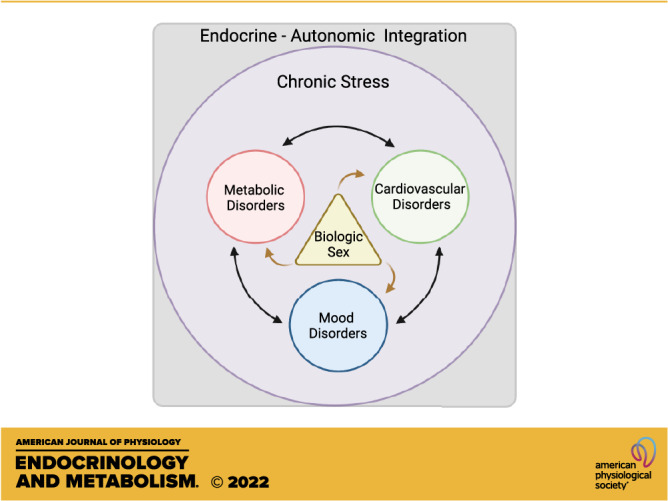
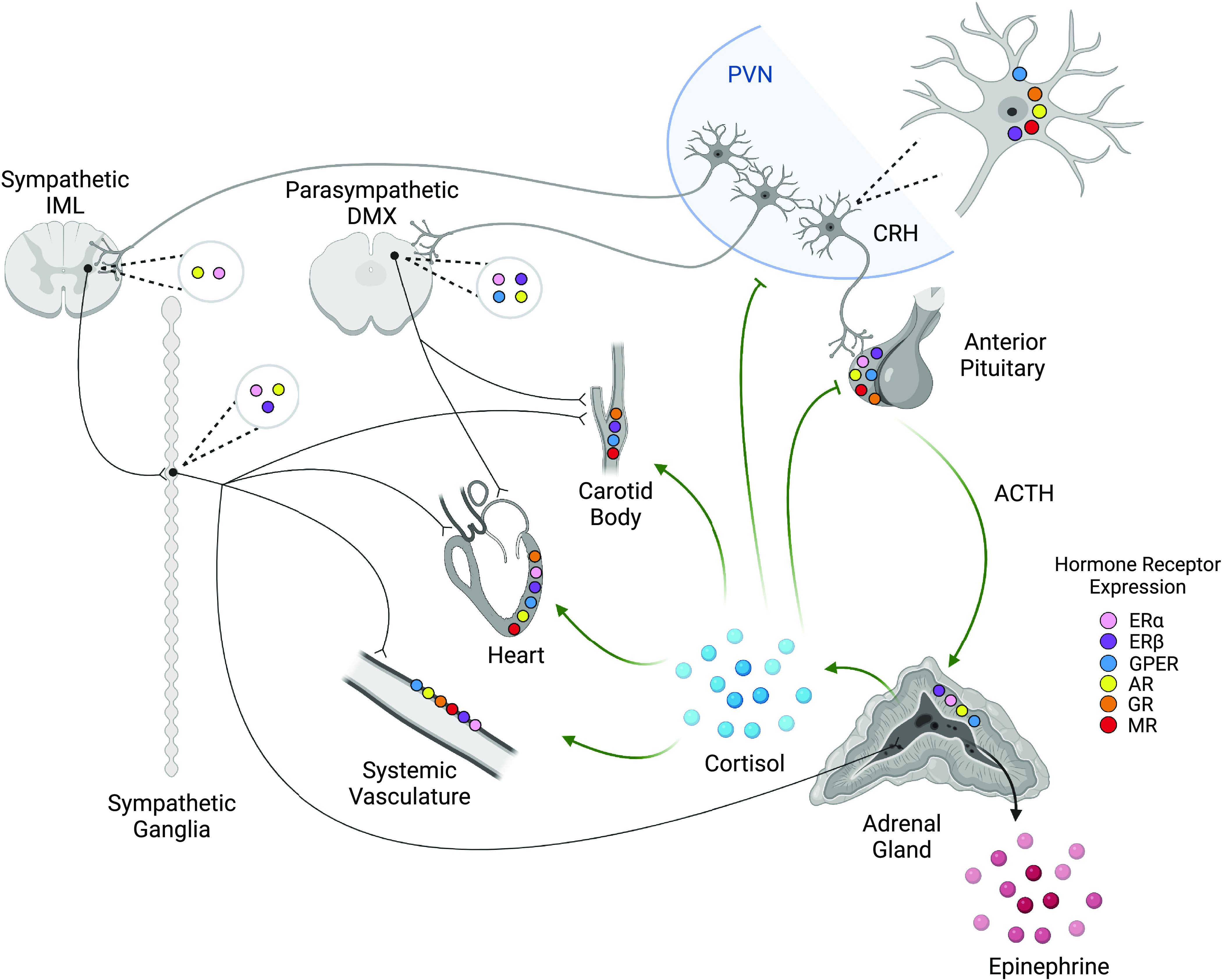
Chronic stress is a significant risk factor for negative health outcomes. Furthermore, imbalance of autonomic nervous system control leads to dysregulation of physiological responses to stress and contributes to the pathogenesis of cardiometabolic and psychiatric disorders. However, research on autonomic stress responses has historically focused on males, despite evidence that females are disproportionality affected by stress-related disorders. Accordingly, this mini-review focuses on the influence of biological sex on autonomic responses to stress in humans and rodent models. The reviewed literature points to sex differences in the consequences of chronic stress, including cardiovascular and metabolic disease. We also explore basic rodent studies of sex-specific autonomic responses to stress with a focus on sex hormones and hypothalamic-pituitary-adrenal axis regulation of cardiovascular and metabolic physiology. Ultimately, emerging evidence of sex differences in autonomic-endocrine integration highlights the importance of sex-specific studies to understand and treat cardiometabolic dysfunction.

Keywords: androgen; autonomic balance; chronic stress; estrogen; glucocorticoid.


Conflict of interest statement

No conflicts of interest, financial or otherwise, are declared by the authors.

Figures

None
Graphical abstract
Figure 1.
Figure 1.
Stress and gonadal hormone receptor expression across neuroendocrine and cardiovascular organs. Neurons in the paraventricular nucleus (PVN) of the hypothalamus initiate both autonomic and hypothalamic-pituitary-adrenal (HPA) axis responses to stress. Corticotropin-releasing hormone (CRH) acts on anterior pituitary corticotropes to cause the release of ACTH and the subsequent synthesis and release of glucocorticoids (cortisol in humans, corticosterone in rats and mice) from the adrenal cortex. Glucocorticoids have systemic action on cardiovascular responses and provide negative feedback on the PVN and anterior pituitary. PVN neurons also synapse in the sympathetic intermediolateral nucleus (IML) and the parasympathetic dorsal motor nucleus of the vagus (DMX). Efferents of the DMX innervate both the carotid body and heart to influence cardiac and baroreflex activity but are generally not found in the systemic vasculature. Efferents of the IML act through sympathetic ganglia to stimulate cardiovascular activity and the release of epinephrine from the adrenal medulla, which acts systemically. In addition, stress and gonadal hormone receptors regulate activity in a tissue-dependent manner. It is important to note that receptor distribution has not been completely characterized and further study is warranted. Thus, the absence of reports on expression does not indicate that receptors are not present. CRH neurons express estrogen receptors β (ERβ), G protein-coupled estrogen receptor (GPER) (52), and androgen receptors (AR) (53), as well as glucocorticoid receptors (GR) (54) and mineralocorticoid receptors (MR) (55). Similarly, the anterior pituitary expresses ERα, ERβ (56), GPER (57), AR (58), GR, and MR (59). The adrenal cortex expresses ERα, ERβ, GPER, and AR (60). The IML is known to express ERα (61) and AR (62). The DMX shows ERα, ERβ (63), GPER (64), and AR (62) expression. Sympathetic ganglia express ERα, ERβ (65), and AR (66). Cardiac tissue (–69) and systemic vasculature (–74) express ERα, ERβ, GPER, and AR, as well as GR and MR. The carotid body shows ERβ (75), GPER (76), GR (77), and MR expression (78). While many of the mechanistic interactions are yet to be elucidated, the integration of these systems modulates stress responses in sex-, age-, and tissue-dependent manners that impact the physiological outcomes of chronic stress. Created with BioRender.com.

References

    1. Anand SS, Yi Q, Gerstein H, Lonn E, Jacobs R, Vuksan V, Teo K, Davis B, Montague P, Yusuf S; Study of Health Assessment and Risk Evaluation in Aboriginal Peoples Investigators. Relationship of metabolic syndrome and fibrinolytic dysfunction to cardiovascular disease. Circulation 108: 420–425, 2003. doi:10.1161/01.CIR.0000080884.27358.49. - DOI - PubMed
    1. Isfort M, Stevens SCW, Schaffer S, Jong CJ, Wold LE. Metabolic dysfunction in diabetic cardiomyopathy. Heart Fail Rev 19: 35–48, 2014. doi:10.1007/s10741-013-9377-8. - DOI - PMC - PubMed
    1. McIntyre RS, Rasgon NL, Kemp DE, Nguyen HT, Law CWY, Taylor VH, Woldeyohannes HO, Alsuwaidan MT, Soczynska JK, Kim B, Lourenco MT, Kahn LS, Goldstein BI. Metabolic syndrome and major depressive disorder: co-occurrence and pathophysiologic overlap. Curr Diab Rep 9: 51–59, 2009. doi:10.1007/s11892-009-0010-0. - DOI - PubMed
    1. Dearing C, Morano R, Ptaskiewicz E, Mahbod P, Scheimann JR, Franco-Villanueva A, Wulsin L, Myers B. Glucoregulation and coping behavior after chronic stress in rats: sex differences across the lifespan. Horm Behav 136: 105060, 2021. doi:10.1016/j.yhbeh.2021.105060. - DOI - PMC - PubMed
    1. Myers B, McKlveen JM, Herman JP. Glucocorticoid actions on synapses, circuits, and behavior: implications for the energetics of stress. Front Neuroendocrinol 35: 180–196, 2014. doi:10.1016/j.yfrne.2013.12.003. - DOI - PMC - PubMed

Publication types

LinkOut - more resources