. 2022 Jul;10(7):e004698.
doi: 10.1136/jitc-2022-004698.

Spatial meta-transcriptomics reveal associations of intratumor bacteria burden with lung cancer cells showing a distinct oncogenic signature

Affiliations

Spatial meta-transcriptomics reveal associations of intratumor bacteria burden with lung cancer cells showing a distinct oncogenic signature

Abigail Wong-Rolle et al. J Immunother Cancer. 2022 Jul.

Abstract

Background: The lung intratumor microbiome influences lung cancer tumorigenesis and treatment responses, but detailed data on the extent, location, and effects of microbes within lung tumors are missing, information needed for improved prognosis and treatment.

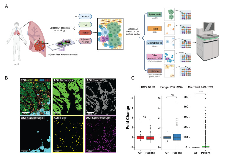
Methods: To address this gap, we developed a novel spatial meta-transcriptomic method simultaneously detecting the expression level of 1,811 host genes and 3 microbe targets (bacteria, fungi, and cytomegalovirus). After rigorous validation, we analyzed the spatial meta-transcriptomic profiles of tumor cells, T cells, macrophages, other immune cells, and stroma in surgically resected tumor samples from 12 patients with early-stage lung cancer.

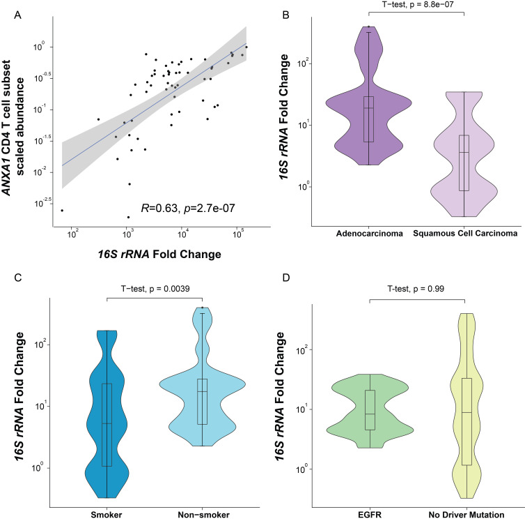
Results: Bacterial burden was significantly higher in tumor cells compared with T cells, macrophages, other immune cells, and stroma. This burden increased from tumor-adjacent normal lung and tertiary lymphoid structures to tumor cells to the airways, suggesting that lung intratumor bacteria derive from the latter route of entry. Expression of oncogenic β-catenin was strongly correlated with bacterial burden, as were tumor histological subtypes and environmental factors.

Conclusions: Intratumor bacteria were enriched with tumor cells and associated with multiple oncogenic pathways, supporting a rationale for reducing the local intratumor microbiome in lung cancer for patient benefit.

Trial registration number: NCT00242723, NCT02146170.

Keywords: lung neoplasms; translational medical research; tumor microenvironment.


Conflict of interest statement

Competing interests: PD is an employee and stockholder at NanoString Technologies. All other authors declare no competing interests. YZ is currently an employee at GlaxoSmithKline. All other authors declare no competing interests.

Figures

Figure 1
Figure 1
Spatial meta-transcriptomic analysis of lung cancers. (A) Study scheme: formalin-fixed, paraffin-embedded (FFPE) samples from 12 patients with lung cancers were sectioned onto 8 slides along with a consecutively sectioned germ-free mouse lung tumor sample on each slide as interslide negative control. Regions of interest (ROIs) (adjacent normal tissue n=56, tumor regions n=384, tertiary lymphoid structures (TLS) n=20, airway n=4) were selected based on morphology. Further area of interest (AOI) selection was done within these defined ROIs to investigate different cell types in the tumor microenvironment (TME), based on cell surface markers. RNAs from AOIs within each ROI were collected, separated, and sequenced as previously described. (B) Representative ROI and AOIs (tumor cells, macrophages, T cells, other immune cells, and stroma) collected from the ROI. Colored segments (AOIs) show areas collected for sequencing. (C) Cytomegalovirus (CMV), fungal, and microbial signals in germ-free KP mice tumors (GF) and patients with lung cancers (Patient) (***p<0.001, ns, not significant, t-test, error bars, SD).
Figure 2
Figure 2
Spatial distribution of lung intratumor bacteria. (A) 16S rRNA expression in tumor cells and adjacent normal tissue (paired two-sided Wilcoxon test). (B) 16S rRNA expression level across different cell types in the tumor microenvironment (TME) (**p<0.01, ns, not significant, two-sided Wilcoxon test against base mean; error bars, SD). (C) Representative RNAscope image showing the spatial distribution of bacterial 16S rRNA signal (rendered magenta squares) within tumor cells and in TME. (D) 16S rRNA expression level in adjacent normal tissues, tertiary lymphoid structures (TLS), and tumor cells (***p<0.001, ns, not significant, one-way analysis of variance (ANOVA) test; error bars, SD). (E) 16S rRNA expression level in adjacent normal tissues, TLS, tumor cells, and airway from one patient (*p<0.05, ***p<0.001, ns, not significant, one-way ANOVA test; error bars, SD).
Figure 3
Figure 3
Correlations of lung intratumor bacterial burden with tumor cells’ function. (A) Correlation of host genes with bacterial burden in tumor cells (Spearman’s rank correlation, R=0.7 cut-off shown in dotted red line). (B) Correlation between host CTNNB1 expression and bacterial burden in tumor cells (Spearman’s rank correlation, 95% CI shown in gray). (C) Representative images of immunohistochemistry staining of β-catenin in tumor regions with known low or high bacterial burden. (D) Ingenuity pathway analysis of genes correlated with bacterial burden in tumor cells.
Figure 4
Figure 4
Association of bacterial burden with immune cell abundance, histology type, patient history, and mutation profile. (A) Correlation between bacterial burden and the scaled abundance of ANXA1 CD4 T cell subset (Spearman’s rank correlation, 95% CI shown in gray). (B) Bacterial burden in tumor cells with different lung cancer histology subtypes (two-sided t-test). (C) Bacterial burden in tumor cells from patients with different smoking histories (two-sided t-test). (D) Bacterial burden in tumor cells with different driver mutation status (Spearman’s rank correlation, 95% CI shown in gray).

References

    1. Siegel RL, Miller KD, Jemal A. Cancer statistics, 2020. CA Cancer J Clin 2020;70:7–30. 10.3322/caac.21590 - DOI - PubMed
    1. Thai AA, Solomon BJ, Sequist LV, et al. Lung cancer. Lancet 2021;398:535–54. 10.1016/S0140-6736(21)00312-3 - DOI - PubMed
    1. Liu N-N, Ma Q, Ge Y, et al. Microbiome dysbiosis in lung cancer: from composition to therapy. NPJ Precis Oncol 2020;4:33. 10.1038/s41698-020-00138-z - DOI - PMC - PubMed
    1. Man WH, de Steenhuijsen Piters WAA, Bogaert D. The microbiota of the respiratory tract: gatekeeper to respiratory health. Nat Rev Microbiol 2017;15:259–70. 10.1038/nrmicro.2017.14 - DOI - PMC - PubMed
    1. Wong-Rolle A, Wei HK, Zhao C, et al. Unexpected guests in the tumor microenvironment: microbiome in cancer. Protein Cell 2021;12:426–35. 10.1007/s13238-020-00813-8 - DOI - PMC - PubMed

Publication types

Associated data