. 2022 Jul 6;20(1):304.
doi: 10.1186/s12967-022-03500-w.

SEMA4D/PlexinB1 promotes AML progression via activation of PI3K/Akt signaling

Affiliations

SEMA4D/PlexinB1 promotes AML progression via activation of PI3K/Akt signaling

Lu Liu et al. J Transl Med. .

Abstract

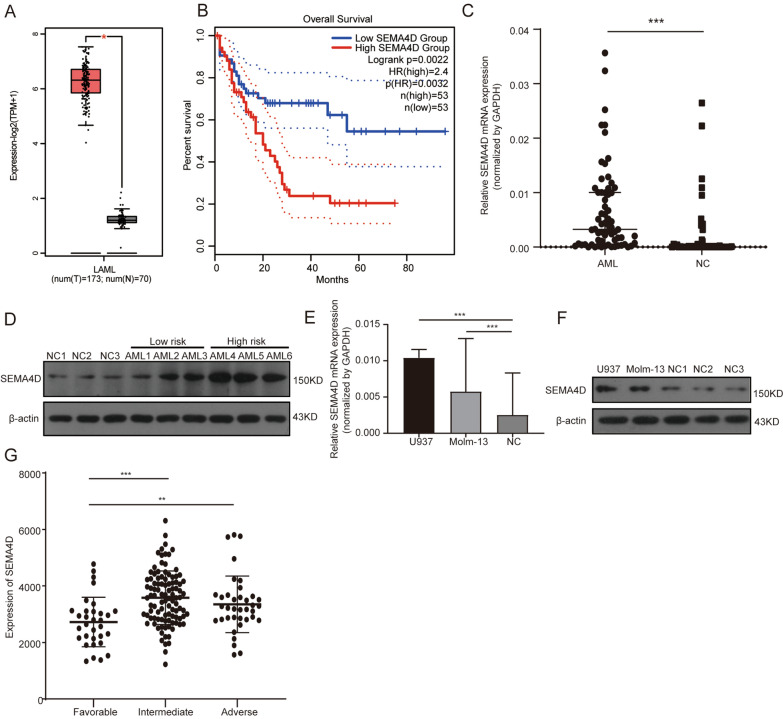
Background: Acute myeloid leukemia (AML) is the most common type of acute leukemia in adults. SEMA4D is a 150 kDa transmembrane protein that belongs to the IV class of the subfamily of semaphorin family. Previous studies have reported that SEMA4D is a multifunctional target in many solid tumors, involving multiple physiological systems, and there are emerging therapies to target these pathways. The role of SEMA4D in AML has not yet been explored.

Methods: The SEMA4D expression prolile, clinical data and potential prognostic analysis were acquired via the cBioPortal and GEPIA databases. SEMA4D expression was measured using real-time quantitative PCR and western blot. Cell counting kit-8 (CCK8) and flow cytometry were used to evaluate the malignant biological characteristics.

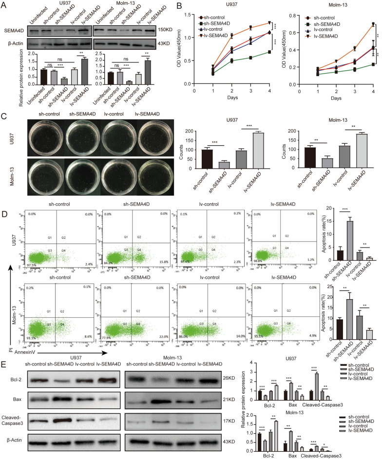
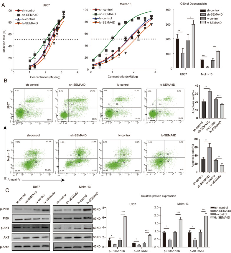
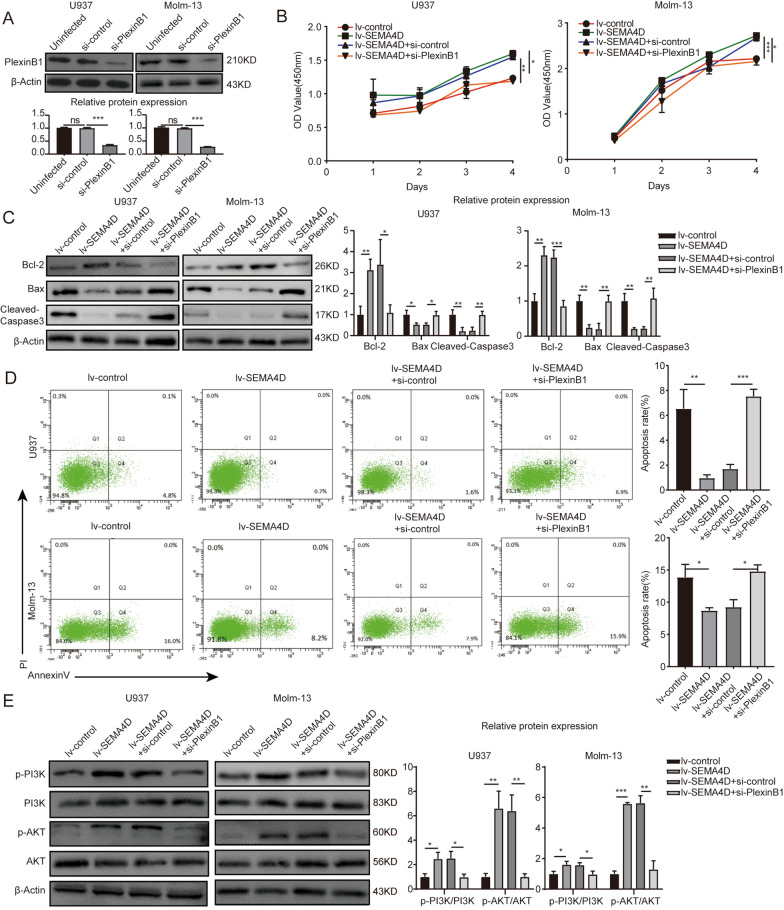
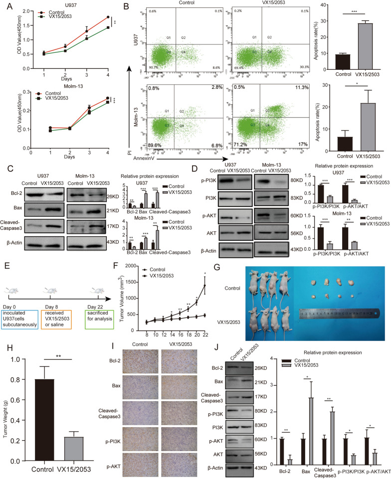
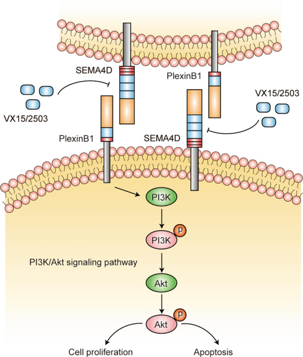
Results: We observed that SEMA4D was increased in AML patients and correlated with risk stratification and prognosis. Moreover, SEMA4D promotes the proliferation and inhibits apoptosis of AML cells by binding to its receptor, PlexinB1, and reduces the sensitivity of AML cells to daunorubicin. In addition, SEMA4D/PlexinB1 promotes the proliferation and survival of AML cells by activating the PI3K/Akt signaling pathway. VX15/2503, an anti-SEMA4D antibody, can inhibit the proliferation of AML cells in xenograft mouse models, thereby inhibiting the development of AML.

Conclusion: SEMA4D will serve as a unique predictive biomarker and a possible therapeutic target in AML.

Keywords: Acute myeloid leukemia; PI3K/Akt; PlexinB1; Prognostic; SEMA4D.


Conflict of interest statement

The authors declare that they have no competing interests.

Figures

Fig. 1
Fig. 1
Expression level of SEMA4D in AML. A Expression level of SEMA4D in LAML compared with healthy control in GEPIA Database. B The OS of patients with LAML in GEPIA Database. C qRT-PCR was used to detect SEMA4D mRNA level in BM-MNCs of AML patients and healthy donors. D Western blot was used to detect SEMA4D protein levels in BM-MNCs of AML patients and healthy donors. E qRT-PCR was used to detect SEMA4D mRNA level in U937 and Molm-13. F Western blot was used to detect SEMA4D protein levels in U937 and Molm-13. Results of densitometry analysis of relative expression levels after normalization to loading control β-actin are presented. G Expression of SEMA4D in LAML based on risk status in TCGA database. Data with statistical significance are as indicated, *P < 0.05, **P < 0.01, ***P < 0.001, ns not significant
Fig. 2
Fig. 2
SEMA4D promotes proliferation and inhibits apoptosis of AML cells. A Western blot was used to detect SEMA4D protein level when U937 and Molm-13 cells were transfected with stably knocking down or overexpressing SEMA4D lentivirus. B CCK-8 analysis of U937 and Molm-13 cells transfected with lentivirus targeting SEMA4D or control. C Colony formation assay of U937 and Molm-13 cells transfected with lentivirus targeting SEMA4D or control. D Cell apoptosis rate of U937 and Molm-13 cells transfected with lentivirus targeting SEMA4D or control was detected by flow cytometry using Annexin V-APC/PI staining. E Western blotting analysis was used to determine the expression of apoptosis-related proteins (Bcl-2, Bax, and cleaved-caspase3) in U937 and Molm-13 cells transfected with lentivirus targeting SEMA4D or control. Results of densitometry analysis of relative expression levels after normalization to loading control β-actin are presented. Data with statistical significance are as indicated, *P < 0.05, **P < 0.01, ***P < 0.001, ns not significant
Fig. 3
Fig. 3
SEMA4D affects chemotherapy sensitivity of daunorubicin and mediates PI3K/Akt phosphorylation in AML cells. A IC50 curves of Daunorubicin in U937 and Molm-13 cells transfected with lentivirus targeting SEMA4D or control. Transduced AML cells were treated with daunorubicin for 48 h and then measured by CCK-8 analysis. B Cell apoptosis rate of U937 and Molm-13 cells transfected with lentivirus targeting SEMA4D or control after Daunorubicin drug treatment was detected by flow cytometry using Annexin V-APC /PI staining. C Western blotting analysis was used to determine the expression of p-PI3K, PI3K, p-Akt, Akt in U937 and Molm-13 cells transfected with lentivirus targeting SEMA4D or control. Results of densitometry analysis of relative expression levels after normalization to loading control β-actin are presented. Data with statistical significance are as indicated, *P < 0.05, **P < 0.01, ***P < 0.001, ns not significant
Fig. 4
Fig. 4
SEMA4D functions through its receptor PlexinB1. A Western blot was used to detect PlexinB1 protein level when U937 and Molm-13 cells were transfected with siRNA-PlexinB1 or siRNA-control. B CCK-8 analysis of U937 and Molm-13 cells transfected with lentivirus targeting SEMA4D when PlexinB1 was knocked down or not. C Western blotting analysis was used to determine the expression of apoptosis-related proteins (Bcl-2, Bax, and cleaved-caspase3) in U937 and Molm-13 cells transfected with lentivirus targeting SEMA4D when PlexinB1 was knocked down or not. Results of densitometry analysis of relative expression levels after normalization to loading control β-actin are presented. D Cell apoptosis rate of U937 and Molm-13 cells transfected with lentivirus targeting SEMA4D when PlexinB1 was knocked down or not was detected by flow cytometry using Annexin V-APC/PI staining. E Western blotting analysis was used to determine the expression of p-PI3K, PI3K, p-Akt, Akt in U937 and Molm-13 cells transfected with lentivirus targeting SEMA4D when PlexinB1 was knocked down or not. Results of densitometry analysis of relative expression levels after normalization to loading control β-actin are presented. Data with statistical significance are as indicated, *P < 0.05, **P < 0.01, ***P < 0.001, ns not significant
Fig. 5
Fig. 5
Anti-SEMA4D antibody can inhibit the survival of AML cell lines in vivo and in vitro. A CCK-8 analysis of U937 and Molm-13 cells treated with VX15/2503 or not. B Cell apoptosis rate of U937 and Molm-13 cells treated with VX15/2503 or not was detected by flow cytometry using Annexin V-APC/PI staining. C Western blotting analysis was used to determine the expression of apoptosis-related proteins (Bcl-2, Bax, and cleaved-caspase3) in U937 and Molm-13 cells treated with VX15/2503 or not. Results of densitometry analysis of relative expression levels after normalization to loading control β-actin are presented. D Western blotting analysis was used to determine the expression of p-PI3K, PI3K, p-Akt, Akt in U937 and Molm-13 cells treated with VX15/2503 or not. Results of densitometry analysis of relative expression levels after normalization to loading control β-actin are presented. E Schematic outline of the mouse model delineating this experiment. F Nude mice were subcutaneously inoculated with U937 cells to establish AML xenograft tumors and treated with VX15/2503. Volumes of tumors were monitored by direct measurement. G Tumor size of xenograft mice in two groups. H Weights of tumors of xenograft mice in two groups. I Immunohistochemistry stain was used to measure the expression of Bcl-2, Bax, cleaved-caspase3, p-PI3K, p-Akt in xenograft tumors. J Western blotting analysis was used to determine the expression of Bcl-2, Bax, cleaved-caspase3, p-PI3K, PI3K, p-Akt, Akt in xenograft tumors. Results of densitometry analysis of relative expression levels after normalization to loading control β-actin are presented. Data with statistical significance are as indicated, *P < 0.05, **P < 0.01, ***P < 0.001, ns not significant
Fig. 6
Fig. 6
Schematic of proposed pathway depicting the role of SEMA4D/PlexinB1 in AML

References

    1. Dinmohamed AG, Visser O, van Norden Y, et al. Treatment, trial participation and survival in adult acute myeloid leukemia: a population-based study in the Netherlands, 1989–2012. Leukemia. 2016;30(1):24–31. doi: 10.1038/leu.2015.188. - DOI - PubMed
    1. Kolodkin AL, Matthes DJ, Goodman CS. The semaphorin genes encode a family of transmembrane and secreted growth cone guidance molecules. Cell. 1993;75(7):1389–1399. doi: 10.1016/0092-8674(93)90625-Z. - DOI - PubMed
    1. Janssen BJ, Robinson RA, Pérez-Brangulí F, et al. Structural basis of semaphorin-plexin signalling. Nature. 2010;467(7319):1118–1122. doi: 10.1038/nature09468. - DOI - PMC - PubMed
    1. Wu M, Li J, Gao Q, Ye F. The role of Sema4D/CD100 as a therapeutic target for tumor microenvironments and for autoimmune, neuroimmune and bone diseases. Expert Opin Ther Targets. 2016;20(7):885–901. doi: 10.1517/14728222.2016.1139083. - DOI - PubMed
    1. Franzolin G, Tamagnone L. Semaphorin signaling in cancer-associated inflammation. Int J Mol Sci. 2019;20(2):377. doi: 10.3390/ijms20020377. - DOI - PMC - PubMed

MeSH terms