. 2022 Jul 6;10(1):98.
doi: 10.1186/s40478-022-01403-x.

Nuclear alpha-synuclein is present in the human brain and is modified in dementia with Lewy bodies

Affiliations

Nuclear alpha-synuclein is present in the human brain and is modified in dementia with Lewy bodies

David J Koss et al. Acta Neuropathol Commun. .

Abstract

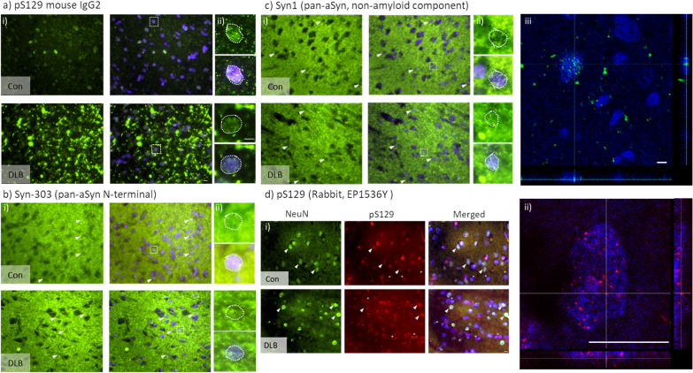
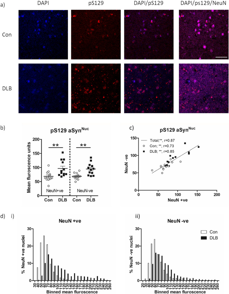
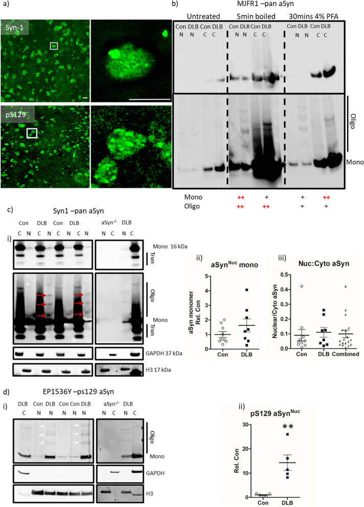
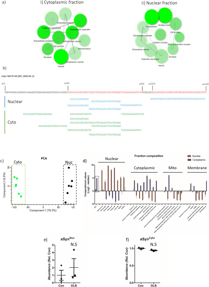
Dementia with Lewy bodies (DLB) is pathologically defined by the cytoplasmic accumulation of alpha-synuclein (aSyn) within neurons in the brain. Predominately pre-synaptic, aSyn has been reported in various subcellular compartments in experimental models. Indeed, nuclear alpha-synuclein (aSynNuc) is evident in many models, the dysregulation of which is associated with altered DNA integrity, transcription and nuclear homeostasis. However, the presence of aSynNuc in human brain cells remains controversial, yet the determination of human brain aSynNuc and its pathological modification is essential for understanding synucleinopathies. Here, using a multi-disciplinary approach employing immunohistochemistry, immunoblot, and mass-spectrometry (MS), we confirm aSynNuc in post-mortem brain tissue obtained from DLB and control cases. Highly dependent on antigen retrieval methods, in optimal conditions, intra-nuclear pan and phospho-S129 positive aSyn puncta were observed in cortical neurons and non-neuronal cells in fixed brain sections and in isolated nuclear preparations in all cases examined. Furthermore, an increase in nuclear phospho-S129 positive aSyn immunoreactivity was apparent in DLB cases compared to controls, in both neuronal and non-neuronal cell types. Our initial histological investigations identified that aSynNuc is affected by epitope unmasking methods but present under optimal conditions, and this presence was confirmed by isolation of nuclei and a combined approach of immunoblotting and mass spectrometry, where aSynNuc was approximately tenfold less abundant in the nucleus than cytoplasm. Notably, direct comparison of DLB cases to aged controls identified increased pS129 and higher molecular weight species in the nuclei of DLB cases, suggesting putative pathogenic modifications to aSynNuc in DLB. In summary, using multiple approaches we provide several lines of evidence supporting the presence of aSynNuc in autoptic human brain tissue and, notably, that it is subject to putative pathogenic modifications in DLB that may contribute to the disease phenotype.

Keywords: Alpha-synuclein; Dementia with Lewy bodies; Lewy body disease; Nuclear pathology; Synucleinopathies.


Conflict of interest statement

The authors declare that they have no competing interests.

Figures

Fig. 1
Fig. 1
In-situ immunohistochemical detection of nuclear aSyn. Example micrograph images from the cingulate cortex of control (con) and dementia with Lewy body (DLB) cases of phospho-serine 129 positive aSyn (pS129) mouse IgG2 (a), pan-aSyn N-terminal directed Syn-303 (b), pan-aSyn non-amyloid component directed Syn1 (c) and NeuN and pS129 rabbit EP1536Y (d) immunoreactivity. All sections were pre-treated with EDTA + formic acid antigen retrieval, nuclei in sections were counterstained with DAPI. Images captured via 40× (a–c, i), 60 × objective lens (d, i) with wide-field fluorescence microscope and 20 × objective lens with 6 × digital zoom (c, iii) or 63× objective lens with 3 × digital zoom (d, ii) with confocal microscope. Expanded area inserts (a–c, ii, dotted line boxes in i are shown for aSyn immunoreactive alone and in combination with DAPI nuclear stain, where the nuclear outline is highlighted (dotted outline). Note frequent detection of intranuclear aSyn immunoreactivity, examples highlighted by arrow heads, in d NeuN-negative nuclei positive for aSyn are further denotes with an asterisk. Scale bar in i = 10 µm and ii and iii = 5 µm
Fig. 2
Fig. 2
Quantification of nuclear phospho-S129 aSyn in temporal cortex of DLB and Control cases. Example of 60 × objective lens confocal images of lateral temporal cortex stained with phospho-serine 129 aSyn (pS129; EP1536Y) and NeuN antibodies with a DAPI nuclear co-stain (a). Images from a control (Con) and dementia with Lewy body (DLB) case are shown. Note prominent pS129 nuclear stain in both cases, with additional cytoplasmic pathology in the form of Lewy neurites in the DLB case. Quantification of nuclear pS129 immunoreactivity in control and DLB cases as segregated according to NeuN reactivity is shown (b) alongside correlative analysis of nuclear pS129 levels between NeuN positive (NeuN +ve) and negative (NeuN −ve) nuclei (c), with spearman’s correlation (r), given when analysed as a combined total single cohort or when divided into Con and DLB. Frequency plots of individual NeuN +ve (i) and NeuN −ve (ii) nuclei are additionally shown (d), reporting the % nuclei of con and DLB cases within a of 20 arbitrary units. Data in c) reported as scatter plots of mean fluorescence arbitrary units per case with group mean ± SEM indicated and in d) also as mean fluorescence units, dotted line demonstrates best-fit line when all cases (con + DLB) are considered combined. ** = p < 0.01. Scale bar in a = 50 µm
Fig. 3
Fig. 3
Quantification of nuclear aSyn pathology in cases of dementia with lewy bodies in isolated nuclei. a Representative confocal micrograph images of nuclei isolated from lateral temporal cortex (× 40 objective, inserts ii + iv taken with 10 × digital zoom, scale = 5 µm) stained for pan-aSyn (Syn-1, i + ii) and phospho-serine 129 aSyn (pS129, iii + iv). b Optimisation of immunoblot detection of aSyn from cytoplasmic (c) and nuclear (n) fractionates generated from control (Con) and dementia with Lewy bodies (DLB) cases. Comparisons of post-transfer membrane phosphate buffered saline heating (5 min boiling) or 4% paraformaldehyde chemical (30 min 4% paraformaldehyde) fixation with non-fixed (untreated) controls is shown, captured under optimal and overexposed settings. Monomeric (mono) and oligomeric (oligo) immunoreactivity is highlighted alongside a ranking of fixation methods-based retention of the aSyn species (+ = enhanced and++ = very enhanced). Quantification of c pan-aSyn via Syn-1, and d pS129 via EP1536Y, (i) example immunoblots following boiling fixation are shown at optimised and overexposed capture settings and mono, oligo and truncated (trunc) species are highlighted (i). Loading controls of GAPDH and Histone H3 demonstrate fractionation purity and resulting immunoreactivity from similarly fractionated aSyn knockout mouse brain tissue (aSyn−/−) demonstrated antibody specificity. Quantification of nuclear aSyn monomers between DLB and controls cases is also shown (ii), demonstrating increased pS129 Syn in DLB cases compared to controls (n = 5 per group), despite no change in total aSyn levels (n = 9 and 8 for con and DLB respectively) and no difference in the nuclear: cytoplasmic ratio of total aSyn (c iii). Data shown as scatter plots, expressed relative to mean control values (Rel. Con) with mean ± SEM also indicated, ** = p < 0.01
Fig. 4
Fig. 4
Mass spectrometry fractional analysis and aSyn peptide sequence alignment. a Gene ontology cell component enrichment analysis of mass-spectrometry identified proteosome from nuclear (i) and cytoplasmic fraction (ii) isolated from the lateral temporal cortex. Cell component networks are shown as produced from Shiny GO (V 0.66). For each node, darker shading represents significantly greater gene enrichment, node size indicates the number of genes aligning with the specific cellular component node and the thickness of connecting lines demonstrates the magnitude of overlapping genes between nodes. b Peptide mapping to the aSyn amino acid (aa) sequence, unique peptides recovered from mass spectrometry of nuclear fraction are shown in blue and those recovered from the cytoplasmic fraction shown in green. Full length aSyn sequence (NP_000336.1) is shown above with the sequence covered by peptides present in nuclear fraction shown in red. c Principal component analysis (PCA) of the relative abundance of proteins across pooled protein library, demonstrated clear distinction between cytoplasmic (Cyto) and Nuclear (Nuc) fractions. d Fractional composition of nuclear and cytoplasmic preparations, abundance of selective nuclear, cytoplasmic, mitochondrial (mito) and membrane proteins are show, alongside aSyn for comparison. Abundance values are also shown from nuclear (e; aSynNuc) and cytoplasmic (f; aSynCyto). Data expressed as compositional values (log2-median) + SEM. N.S = not significant

References

    1. Alafuzoff I, Ince PG, Arzberger T, Al-Sarraj S, Bell J, Bodi I, Bogdanovic N, Bugiani O, Ferrer I, Gelpi E, et al. Staging/typing of Lewy body related α-synuclein pathology: a study of the BrainNet Europe Consortium. Acta Neuropathol. 2009;117:635–652. doi: 10.1007/s00401-009-0523-2. - DOI - PubMed
    1. Anderson JP, Walker DE, Goldstein JM, de Laat R, Banducci K, Caccavello RJ, Barbour R, Huang J, Kling K, Lee M, et al. Phosphorylation of Ser-129 is the dominant pathological modification of alpha-synuclein in familial and sporadic Lewy body disease. J Biol Chem. 2006;281:29739–29752. doi: 10.1074/jbc.M600933200. - DOI - PubMed
    1. Bandopadhyay R. Sequential extraction of soluble and insoluble alpha-synuclein from parkinsonian brains. JoVE. 2016 doi: 10.3791/53415. - DOI - PMC - PubMed
    1. Beach TG, Adler CH, Lue L, Sue LI, Bachalakuri J, Henry-Watson J, Sasse J, Boyer S, Shirohi S, Brooks R, et al. Unified staging system for Lewy body disorders: correlation with nigrostriatal degeneration, cognitive impairment and motor dysfunction. Acta Neuropathol. 2009;117:613–634. doi: 10.1007/s00401-009-0538-8. - DOI - PMC - PubMed
    1. Beach TG, White CL, Hamilton RL, Duda JE, Iwatsubo T, Dickson DW, Leverenz JB, Roncaroli F, Buttini M, Hladik CL, et al. Evaluation of alpha-synuclein immunohistochemical methods used by invited experts. Acta Neuropathol. 2008;116:277–288. doi: 10.1007/s00401-008-0409-8. - DOI - PMC - PubMed

Publication types

Substances