. 2022 Jun 20:10:867608.
doi: 10.3389/fcell.2022.867608. eCollection 2022.

Alterations in Inflammatory Cytokines and Redox Homeostasis in LPS-Induced Pancreatic Beta-Cell Toxicity and Mitochondrial Stress: Protection by Azadirachtin

Affiliations

Alterations in Inflammatory Cytokines and Redox Homeostasis in LPS-Induced Pancreatic Beta-Cell Toxicity and Mitochondrial Stress: Protection by Azadirachtin

Annie John et al. Front Cell Dev Biol. .

Abstract

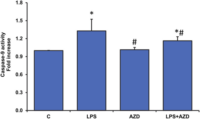
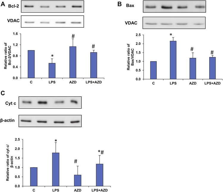
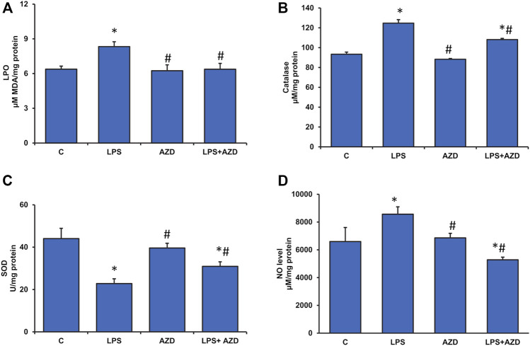
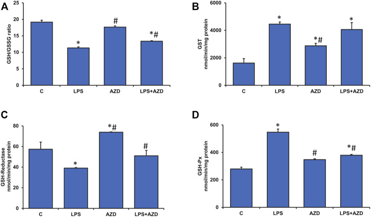
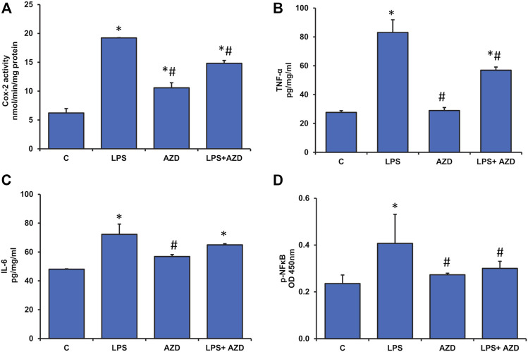
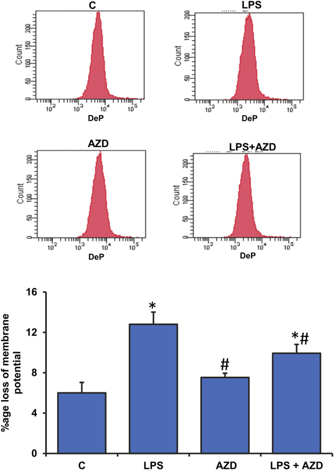
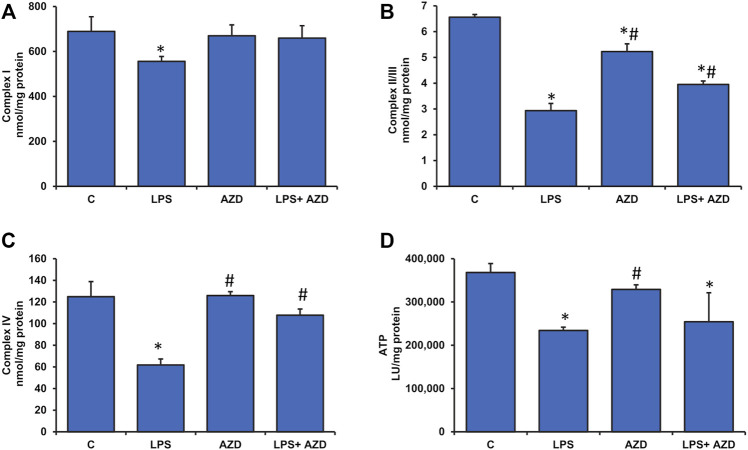
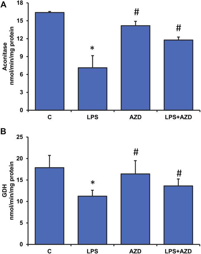
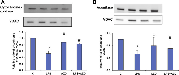
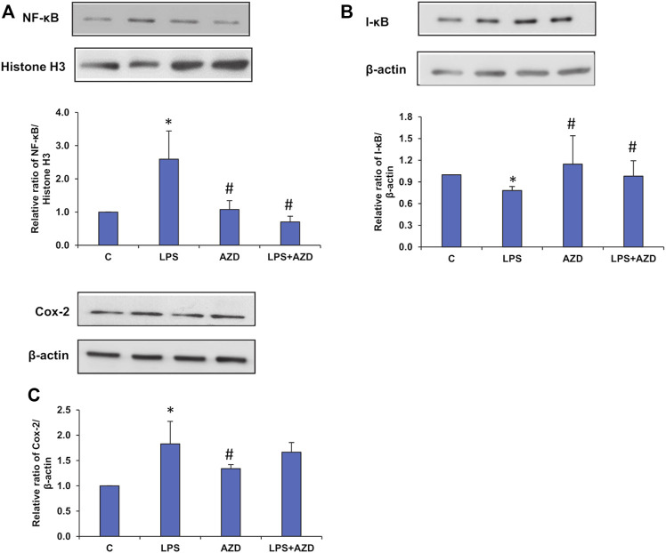
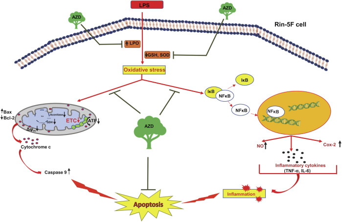
Inflammation and redox imbalance are hallmarks of cancer, diabetes, and other degenerative disorders. Pathophysiological response to these disorders leads to oxidative stress and mitochondrial dysfunction by alterations and reprogramming in cellular signaling and metabolism. Pancreatic beta cells are very sensitive to the inflammatory and altered nutrient signals and hence play a crucial role in diabetes and cancer. In this study, we treated insulin-secreting pancreatic beta cells, Rin-5F, with the bacterial endotoxin, LPS (1 μg/ml) to induce an inflammatory response in vitro and then treated the cells with a known anti-inflammatory, anticancer and antioxidant phytochemical, azadirachtin (AZD, 25 µM for 24 h). Our results demonstrated lipid peroxidation and nitric oxide production causing increased nitro/oxidative stress and alterations in the activities of anti-oxidant enzymes, superoxide dismutase and catalase after LPS treatment. Pro-inflammatory responses caused by translocation of nuclear factor kappa B and release of inflammatory cytokines were also observed. These changes were accompanied by GSH-dependent redox imbalance and alterations in mitochondrial membrane potential and respiratory complexes enzyme activities leading to mitochondrial respiratory dysfunction, reduced ATP synthesis, and intrinsic caspase-9 mediated apoptosis. Caspase-9 was activated due to alterations in Bcl-2 and Bax proteins and release of cytochrome c into the cytosol. The activities of oxidative stress-sensitive mitochondrial matrix enzymes, aconitase, and glutamate dehydrogenase were also inhibited. Treatment with AZD showed beneficial effects on the recovery of antioxidant enzymes, inflammatory responses, and mitochondrial functions. GSH-dependent redox homeostasis also recovered after the treatment with AZD. This study may help in better understanding the etiology and pathogenesis of inflammation-induced disorders in pancreatic beta cells to better manage therapeutic strategies.

Keywords: GSH redox metabolism; LPS; azadirachtin; inflammation signaling; mitochondria; pancreatic cell.


Conflict of interest statement

The authors declare that the research was conducted in the absence of any commercial or financial relationships that could be construed as a potential conflict of interest.

Figures

FIGURE 1
FIGURE 1
LPS-induced activation of caspase-9 activity and attenuation by AZD. Caspase-9 activity was measured in Rin-5F cells treated with LPS with/without AZD for 24 h, using the caspase-9 activity assay kit. Data are expressed as mean ± SD of three independent experiments. Statistical significances are shown as asterisks (*indicates significant difference (p ≤ 0.05) relative to control untreated cells whereas # indicates significant difference (p ≤ 0.05) relative to LPS-treated cells).
FIGURE 2
FIGURE 2
LPS-induced expression of mitochondrial oxidative stress markers. Mitochondrial and post-mitochondrial fractions (5–10 µg protein) from LPS and/or AZD-treated Rin-5F cells were separated electrophoretically and transferred onto nitrocellulose membranes by Western blotting. Immunoreactive proteins were probed using the respective antibodies against Bcl-2 (A), Bax (B), and post-mitochondrial (PMS) cytochrome c (C). Bands were detected by enhanced chemiluminescence using the Sapphire Biomolecular Imager (Azure biosystems, Dublin, United States) or developed using X-ray films. VDAC and β-actin were used as loading controls for mitochondrial and post-mitochondrial fractions, respectively. Proteins were quantitated and normalized against their respective loading controls and represented as histograms. A representative of three independent experiments is given. Statistical significances are shown as asterisks (* indicates significant difference (p ≤ 0.05) relative to control untreated cells whereas # indicates significant difference (p ≤ 0.05) relative to LPS-treated cells).
FIGURE 3
FIGURE 3
Assessment of oxidative stress in LPS-treated Rin-5F cells. Oxidative stress was assessed in LPS and/or AZD-treated Rin-5F cells using various oxidative stress parameters. LPO (lipid peroxidation) was measured as the total malondialdehyde produced (A) as per the manufacturer’s protocol. Catalase activity (B) was measured colorimetrically as the formaldehyde produced by oxidation of methanol by hydrogen peroxide. Percent inhibition of nitro blue tetrazolium (NBT)-diformazan was used as a measure of SOD activity (C) as per the vendor’s protocol. Total nitrite concentration was used as a measure of NO production (D) as per the manufacturer’s protocol. Data are expressed as mean ± SD of three independent experiments. Statistical significances are shown as asterisks (*indicates significant difference (p ≤ 0.05) relative to control untreated cells whereas # indicates significant difference (p ≤ 0.05) relative to LPS-treated cells).
FIGURE 4
FIGURE 4
Alterations in redox homeostasis by LPS in Rin-5F cells. GSH/GSSG ratio (A), GST (B), GSH-reductase (C), and GSH-Px (D) were measured in LPS and/or AZD-treated Rin-5F cells using the appropriate substrates. Data are expressed as mean ± SD of three independent experiments. Statistical significances are shown as asterisks (*indicates significant difference (p ≤ 0.05) relative to control untreated cells whereas # indicates significant difference (p ≤ 0.05) relative to LPS-treated cells).
FIGURE 5
FIGURE 5
Production of inflammatory markers in Rin-5F cells after treatment with LPS. Cox-2 activity (A) was measured at 590 nm by examining the appearance of oxidized TMPD as per the vendor’s protocol. Other inflammatory markers like TNF-α (B), IL-6 (C), and p-NF-κB (D) were measured using standard ELISA kits as per the manufacturer’s protocol. Data are expressed as mean ± SD of three independent experiments. Statistical significances are shown as asterisks (*indicates significant difference (p ≤ 0.05) relative to control untreated cells whereas # indicates significant difference (p ≤ 0.05) relative to LPS-treated cells).
FIGURE 6
FIGURE 6
Alterations in mitochondrial membrane potential by LPS. Mitochondrial membrane potential was measured in Rin-5F cells treated with LPS and/or AZD by flow cytometry using a fluorescent dye, as per the vendor’s protocol. The histogram represents the percentage loss of mitochondrial membrane potential and represents the mean ± SD of three independent experiments. Statistical significances are shown as asterisks (*indicates significant difference (p ≤ 0.05) relative to control untreated cells whereas # indicates significant difference (p ≤ 0.05) relative to LPS-treated cells).
FIGURE 7
FIGURE 7
Alterations in the activities of mitochondrial respiratory complexes and bioenergetics caused by LPS in Rin-5F cells. Mitochondrial respiratory complexes, Complex I (A), Complex II/III (B), and Complex IV (C) were measured in Rin-5F cells treated with LPS and/or AZD, using their specific substrates. ATP levels (D) were measured using the ATP Bioluminescent cell assay kit as per the manufacturer’s protocol. Data are expressed as mean ± SD of three independent experiments. Statistical significances are shown as asterisks (*indicates significant difference (p ≤ 0.05) relative to control untreated cells whereas # indicates significant difference (p ≤ 0.05) relative to LPS-treated cells).
FIGURE 8
FIGURE 8
Effects of LPS on Krebs’ cycle enzymes in Rin-5F cells. Activities of Krebs’ cycle enzymes, aconitase (A), and glutamate dehydrogenase (B), were measured in LPS and/or AZD-treated Rin-5F cells using the aconitase assay kit (Oxis Int, Inc. Portland, OR, United States) and the glutamate dehydrogenase kit (Abcam, Cambridge, England, United Kingdom) respectively as per the vendors’ protocols. Data are expressed as mean ± SD of three independent experiments. Statistical significances are shown as asterisks (*indicates significant difference (p ≤ 0.05) relative to control untreated cells whereas # indicates significant difference (p ≤ 0.05) relative to compared with LPS-treated cells).
FIGURE 9
FIGURE 9
Expression of mitochondrial matrix markers after LPS treatment. Mitochondrial fractions (5–10 µg protein) isolated from Rin-5F cells treated with LPS and/or AZD were separated electrophoretically and transferred on to nitrocellulose membrane by Western blotting. Immunoreactive proteins were detected using the appropriate probes against cytochrome c oxidase (A), and Aconitase (B). Bands were detected by enhanced chemiluminescence using the Sapphire Biomolecular Imager (Azure biosystems, Dublin United States) or developed using X-ray films. VDAC was used as the loading control. Proteins were quantitated and normalized against their respective loading controls and represented as histograms. A representative of three independent experiments is shown. Statistical significances are shown as asterisks (* indicates significant difference (p ≤ 0.05) relative to control untreated cells whereas # indicates significant difference (p ≤ 0.05) relative to LPS-treated cells).
FIGURE 10
FIGURE 10
Expression of inflammatory markers induced by LPS. Nuclear, post-mitochondrial, or total cell extracts (25–30 µg protein) from LPS and/or AZD treated Rin-5F cells were separated electrophoretically and transferred onto nitrocellulose membranes by Western blotting. Immunoreactive bands were probed with the appropriate primary antibodies against NF-κB (A), I-κB (B), and Cox-2 (C). Bands were detected by enhanced chemiluminescence using the Sapphire Biomolecular Imager (Azure biosystems, Dublin United States) or developed using X-ray films. Histone H3 and β-actin were used as loading controls for nuclear and post-mitochondrial/total extracts respectively. Proteins were quantitated and normalized against their respective loading controls and represented as histograms. A representative of three independent experiments is shown. Statistical significances are shown as asterisks (* indicates significant difference (p ≤ 0.05) relative to control untreated cells whereas # indicates significant difference (p ≤ 0.05) relative to LPS-treated cells).
FIGURE 11
FIGURE 11
Schematic representation showing the protective mechanism of azadirachtin (AZD) on LPS-induced oxidative stress and inflammatory response in Rin-5F pancreatic cells. The bacterial endotoxin, lipopolysaccharide (LPS) has been shown to increase lipid peroxidation and decrease the physiological antioxidants, GSH (reduced glutathione) and SOD (superoxide dismutase), causing increased oxidative stress in the Rin-5F pancreatic cells. This, in turn, resulted in translocation of NF-κB into the nucleus, thus activating the inflammatory responses. LPS also caused decrease in the activities of mitochondrial respiratory complexes in the electron transport chain (ETC), causing alterations in the mitochondrial bioenergetics resulting in mitochondrial dysfunction, release of cytochrome c and caspase 9 activation leading to apoptosis. As illustrated in the model, AZD protected the cells from oxidative and mitochondrial stress by increasing the levels of GSH and SOD, increasing the activities of mitochondrial complexes and decreasing the inflammatory response, thus suppressing apoptosis.

Similar articles

Cited by

References

    1. Al-Nahdi A. M. T., John A., Raza H. (2018). Cytoprotective Effects of N-Acetylcysteine on Streptozotocin- Induced Oxidative Stress and Apoptosis in RIN-5F Pancreatic β-Cells. Cell. Physiol. biochem. 51, 201–216. 10.1159/000495200 PubMed Abstract | 10.1159/000495200 | Google Scholar - DOI - DOI - PubMed
    1. Alnahdi A., John A., Raza H. (2019a). Augmentation of Glucotoxicity, Oxidative Stress, Apoptosis and Mitochondrial Dysfunction in HepG2 Cells by Palmitic Acid. Nutrients 11, 1979. 10.3390/nu11091979 PubMed Abstract | 10.3390/nu11091979 | Google Scholar - DOI - DOI - PMC - PubMed
    1. Alnahdi A., John A., Raza H. (2020). Mitigation of Glucolipotoxicity-Induced Apoptosis, Mitochondrial Dysfunction, and Metabolic Stress by N-Acetyl Cysteine in Pancreatic β-Cells. Biomolecules 10, 239. 10.3390/biom10020239 PubMed Abstract | 10.3390/biom10020239 | Google Scholar - DOI - DOI - PMC - PubMed
    1. Alnahdi A., John A., Raza H. (2019b). N-acetyl Cysteine Attenuates Oxidative Stress and Glutathione-dependent Redox Imbalance Caused by High Glucose/high Palmitic Acid Treatment in Pancreatic Rin-5F Cells. PLoS ONE 14, e0226696. 10.1371/journal.pone.0226696 PubMed Abstract | 10.1371/journal.pone.0226696 | Google Scholar - DOI - DOI - PMC - PubMed
    1. Amyot J., Semache M., Ferdaoussi M., Fontés G., Poitout V. (2012). Lipopolysaccharides Impair Insulin Gene Expression in Isolated Islets of Langerhans via Toll-like Receptor-4 and NF-Κb Signalling. PLOS ONE 7, e36200. 10.1371/journal.pone.0036200 PubMed Abstract | 10.1371/journal.pone.0036200 | Google Scholar - DOI - DOI - PMC - PubMed