[Preprint]. 2022 Jul 28:2022.06.27.497816.
doi: 10.1101/2022.06.27.497816.

Iterative computational design and crystallographic screening identifies potent inhibitors targeting the Nsp3 Macrodomain of SARS-CoV-2

Affiliations

Iterative computational design and crystallographic screening identifies potent inhibitors targeting the Nsp3 Macrodomain of SARS-CoV-2

Stefan Gahbauer et al. bioRxiv. .

Update in

Abstract

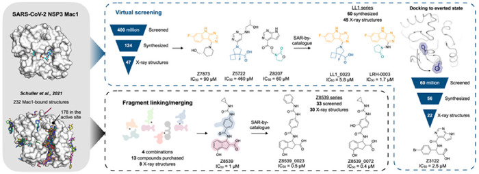
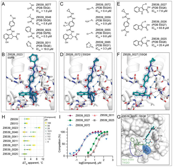
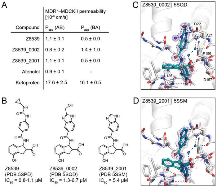
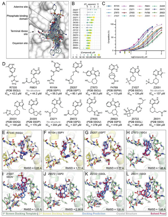
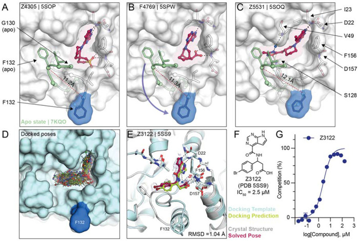
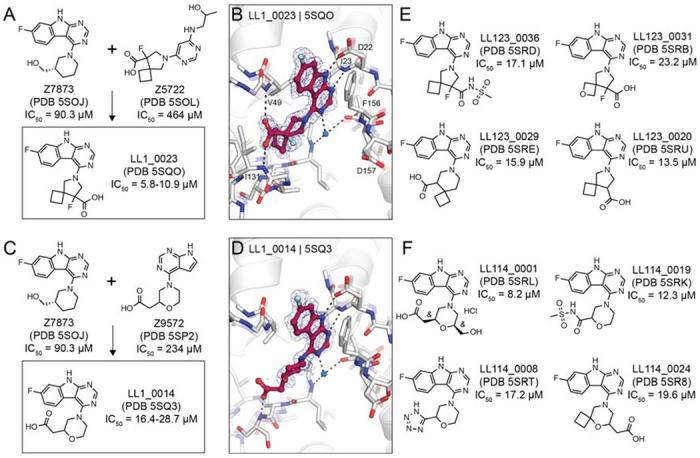
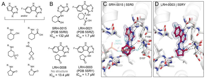
The nonstructural protein 3 (NSP3) of the severe acute respiratory syndrome coronavirus 2 (SARS-CoV-2) contains a conserved macrodomain enzyme (Mac1) that is critical for pathogenesis and lethality. While small molecule inhibitors of Mac1 have great therapeutic potential, at the outset of the COVID-19 pandemic there were no well-validated inhibitors for this protein nor, indeed, the macrodomain enzyme family, making this target a pharmacological orphan. Here, we report the structure-based discovery and development of several different chemical scaffolds exhibiting low- to sub-micromolar affinity for Mac1 through iterations of computer-aided design, structural characterization by ultra-high resolution protein crystallography, and binding evaluation. Potent scaffolds were designed with in silico fragment linkage and by ultra-large library docking of over 450 million molecules. Both techniques leverage the computational exploration of tangible chemical space and are applicable to other pharmacological orphans. Overall, 160 ligands in 119 different scaffolds were discovered, and 152 Mac1-ligand complex crystal structures were determined, typically to 1 Å resolution or better. Our analyses discovered selective and cell-permeable molecules, unexpected ligand-mediated protein dynamics within the active site, and key inhibitor motifs that will template future drug development against Mac1.

Keywords: Nsp3 macrodomain; coronavirus; fragment-based drug discovery; virtual screening.


Figures

Figure 1.
Figure 1.
Overview of the structure-based strategies used to discover ligands that bind to the NSP3 macrodomain of SARS-CoV-2 (Mac1).
Figure 2.
Figure 2.. In silico fragment-linking targeting the adenosine site of Mac1.
A) Binding pose of two fragments identified in the previously reported fragment screen (12). Fragment-protein hydrogen bonds are shown with dashed black lines. B) Theoretical linked scaffold of ZINC922 and ZINC337835 generated using Fragmenstein (14). C) Availability of corresponding chemical building blocks and reactions in the Enamine REAL database. D) Readily accessible analogs of the theoretical scaffold shown in (B). E) X-ray crystal structure of Mac1 bound to Z8539. Three conformations of Z8539 [(R,R) and two (S,S)] could be resolved in the PanDDA event map (blue mesh contoured at 2 σ). Water molecules that form bridging hydrogen bonds between Z8539 and the protein are shown as blue spheres. The apo state of Mac1 is shown with transparent white sticks. F) 2D structure of the most potent (R,R)-stereoisomer of Z8539 (Z8601). G) ADPr-peptide competition (%) of Z8539, Z8515 and Z8601 on Mac1 determined by an HTRF-based displacement assay. ADPr was used as reference. Data are presented as the mean ± SEM of at least two technical replicates.
Figure 3.
Figure 3.. Structure-based optimization of Z8539.
A) Modifications of the cyclopropyl-phenylurea group. B) X-ray crystal structure of Mac1 bound to Z8539_0023. The PanDDA event map is shown around the ligand (blue mesh contoured at 2 σ). C) Modifications of the central benzene. D) X-ray crystal structure of Mac1 bound to Z8539_0072. E) Modifications of the indane group. F) X-ray crystal structure of Mac1 bound to Z8539_0027. G) X-ray crystal structure of Mac1 bound to Z8539_0025. The Gly130-Phe132 loop is aligned to the apo-state conformation in green (PDB 7KQO). The Z8539_0025-Mac1 structure is shown with a transparent white surface. H) DSF-derived temperature upshifts. Data are presented for three technical replicates. I) HTRF-based peptide displacement dose-response curves. Data are presented as the mean ± SEM of at least two technical replicates.
Figure 4.
Figure 4.. Z8539 analog with enhanced cell membrane permeability.
A) Apparent permeability (Papp) assayed with MDR1-MDCKII cells. Permeability was measured in apical (A)-to-basolateral (B) direction and vice versa. Atenolol and Ketoprofen were included as control compounds. B) 2D structures of Z8539, Z8539_0002 and Z8539_2001. C) X-ray crystal structure of Mac1 bound to Z8539_0002. Hydrogen bonding interactions between ligand and the Lys11 backbone nitrogen of a symmetry mate are shown with purple dashes/spheres. PanDDA event maps are shown around the ligand (blue mesh contoured at 2 σ). D) Crystal structure of Mac1 bound to Z8539_2001.
Figure 5.
Figure 5.. Large scale docking targeting the adenosine site of Mac1.
A) Binding poses of 47 docking hits confirmed by X-ray crystallography. The ADPr-bound structure of Mac1 (PDB 6W02) is shown with a white surface. B) Thermal upshifts measured ibyn DSF. Data are presented for three technical replicates. C) HTRF-based peptide displacement dose-response curves. Data are presented as the mean ± SEM of at least 2 repeat measurements. D) 2D structures of docking hits with activity in the HTRF assay. E-L) Crystal structures of Mac1 bound to R7335, R1104, Z8207, Z7873, Z1027, Z9572, Z5722, Z6511, respectively. The protein structure used in the first docking screen is shown in green, the structure from the second screen is colored yellow. The predicted binding poses are shown in blue. Protein crystal structures are shown in gray and the solved binding poses are shown in red, with alternative ligand conformations colored salmon. Hydrogen bonding interactions between ligands and the Lys11 backbone nitrogen of a symmetry mate are shown with purple dashes/spheres. Hungarian Root Mean Square deviations (RMSD) between the docked and solved ligand poses were calculated with DOCK6. PanDDA event maps are shown for each ligand (contoured at 2 σ).
Figure 6.
Figure 6.. Stabilization of everted phosphate binding region by docking hits.
A,B,C) The ligand-bound Mac1 crystal structures are shown in gray with Phe132 highlighted in blue. The Gly130-Phe132 loop of the Mac1 apo structure is depicted in green. Experimentally determined ligand-binding poses are shown in red. D) Predicted binding poses of molecules docked against the Z4305-bound Mac1 structure (PDB 5SOP). E) Crystal structure of Z3122 (red) bound to Mac1 (gray) compared to the predicted complex (Mac1 in blue, Z3122 in green). The PanDDA event map is shown around the ligand (blue mesh contoured at 2 σ). The Hungarian RMSD between solved and docked binding poses was calculated with DOCK6. F) Chemical structure of Z3122. G) HTRF-derived ADPr-peptide competition curve of Z3122. Data are presented as the mean ± SEM of three technical repeats.
Figure 7.
Figure 7.. Structure-based optimization of docking hits.
A) Design of LL1_0023. B) X-ray crystal structure of LL1_0023. The PanDDA event map is shown around the ligand (contoured at 2 σ). Hydrogen bonds are shown with dashed black lines. C, D) Design and X-ray crystal structure of LL1_0014, respectively. E, F) Selected analogs of LL1_0023 and LL1_0014, respectively.
Figure 8.
Figure 8.. Probing neutral functional groups in the Mac1 oxyanion subsite.
A) Design strategy of analog set. B) Chemical structures of most potent hits. C) Crystal structure of Mac1 bound to SRH-0015. ADPr and the water-mediated hydrogen bond to the oxyanion subsite are shown for reference (PDB 7KQP, transparent cyan sticks/spheres). Both of the trans stereoisomers were modeled: the (S,R) is colored dark red and the (R,S) isomer is colored salmon. PanDDA event maps are shown around the ligand (blue mesh contoured at 2 σ). D) Crystal structure of the Mac1-LRH-0003 complex.

References

    1. Yan W., Zheng Y., Zeng X., He B., Cheng W., Structural biology of SARS-CoV-2: open the door for novel therapies. Signal Transduct Target Ther 7, 26 (2022). - PMC - PubMed
    1. Leung A. K. L., Griffin D. E., Bosch J., Fehr A. R., The Conserved Macrodomain Is a Potential Therapeutic Target for Coronaviruses and Alphaviruses. Pathogens 11, 94 (2022). - PMC - PubMed
    1. Yang H., Rao Z., Structural biology of SARS-CoV-2 and implications for therapeutic development. Nat. Rev. Microbiol. 19, 685–700 (2021). - PMC - PubMed
    1. Fu W., et al. , The search for inhibitors of macrodomains for targeting the readers and erasers of mono-ADP-ribosylation. Drug Discov. Today 26, 2547–2558 (2021). - PubMed
    1. Rack J. G. M., et al. , Viral macrodomains: a structural and evolutionary assessment of the pharmacological potential. Open Biol. 10, 200237 (2020). - PMC - PubMed

Publication types