ST-Segment Elevation Myocardial Infarction and Right Atrial Myxoma
- PMID: 35795298
- PMCID: PMC9252612
- DOI: 10.1055/s-0042-1749211
ST-Segment Elevation Myocardial Infarction and Right Atrial Myxoma
Abstract
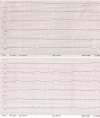
Background Cardiac myxoma is the most common primary cardiac tumor. Although benign, it can cause life-threatening complications due to embolization. Case Presentation We describe an ST-elevation myocardial infarction (STEMI) involving a giant right atrial myxoma and persisting foramen ovale (PFO) in a 64-year-old male patient and report on emergency percutaneous interventional therapy and subsequent cardiac surgery to remove the right atrial myxoma. Conclusion A right atrial myxoma, combined with a PFO, can cause a STEMI. Therefore, every acute coronary syndrome patient should undergo ultrafast exploratory emergency echocardiography to protect the physician from unpleasant surprises.
Keywords: cardiac catheterization/interventionpercutaneous coronary intervention; cardiovascular surgery; echocardiography; myocardial infarction; myxoma; tumor.
The Author(s). This is an open access article published by Thieme under the terms of the Creative Commons Attribution-NonDerivative-NonCommercial License, permitting copying and reproduction so long as the original work is given appropriate credit. Contents may not be used for commercial purposes, or adapted, remixed, transformed or built upon. ( https://creativecommons.org/licenses/by-nc-nd/4.0/ ).
Conflict of interest statement
Conflict of Interest None declared.
Figures


References
-
- Poterucha T J, Kochav J, O'Connor D S, Rosner G F. Cardiac tumors: clinical presentation, diagnosis, and management. Curr Treat Options Oncol. 2019;20(08):66. - PubMed
-
- Braun S, Schrötter H, Reynen K, Schwencke C, Strasser R H. Myocardial infarction as complication of left atrial myxoma. Int J Cardiol. 2005;101(01):115–121. - PubMed
-
- ESC Scientific Document Group . Ibanez B, James S, Agewall S. 2017 ESC Guidelines for the management of acute myocardial infarction in patients presenting with ST-segment elevation: The Task Force for the management of acute myocardial infarction in patients presenting with ST-segment elevation of the European Society of Cardiology (ESC) Eur Heart J. 2018;39(02):119–177. - PubMed
-
- Revankar S G, Clark R A. Infected cardiac myxoma. Case report and literature review. Medicine (Baltimore) 1998;77(05):337–344. - PubMed
-
- Balk A H, Wagenaar S S, Bruschke A V. Bilateral cardiac myxomas and peripheral myxomas in a patient with recent myocardial infarction. Am J Cardiol. 1979;44(04):767–770. - PubMed
Publication types
LinkOut - more resources
Full Text Sources

