. 2022 Jun 13:29:116-132.
doi: 10.1016/j.omtn.2022.06.005. eCollection 2022 Sep 13.

PK-modifying anchors significantly alter clearance kinetics, tissue distribution, and efficacy of therapeutics siRNAs

Affiliations

PK-modifying anchors significantly alter clearance kinetics, tissue distribution, and efficacy of therapeutics siRNAs

Bruno M D C Godinho et al. Mol Ther Nucleic Acids. .

Abstract

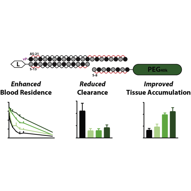
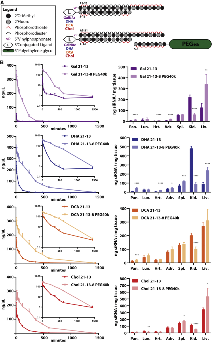
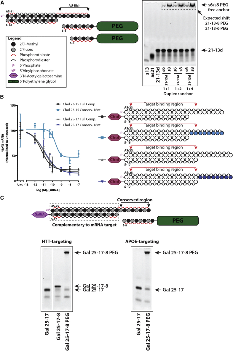
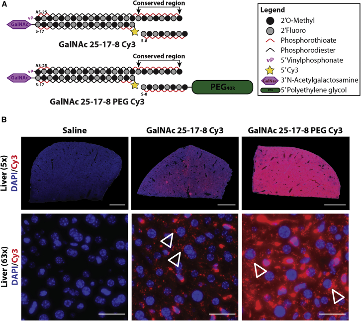
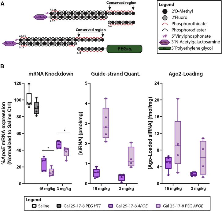
Effective systemic delivery of small interfering RNAs (siRNAs) to tissues other than liver remains a challenge. siRNAs are small (∼15 kDa) and therefore rapidly cleared by the kidneys, resulting in limited blood residence times and tissue exposure. Current strategies to improve the unfavorable pharmacokinetic (PK) properties of siRNAs rely on enhancing binding to serum proteins through extensive phosphorothioate modifications or by conjugation of targeting ligands. Here, we describe an alternative strategy for enhancing blood and tissue PK based on dynamic modulation of the overall size of the siRNA. We engineered a high-affinity universal oligonucleotide anchor conjugated to a high-molecular-weight moiety, which binds to the 3' end of the guide strand of an asymmetric siRNA. Data showed a strong correlation between the size of the PK-modifying anchor and clearance kinetics. Large 40-kDa PK-modifying anchors reduced renal clearance by ∼23-fold and improved tissue exposure area under the curve (AUC) by ∼26-fold, resulting in increased extrahepatic tissue retention (∼3- to 5-fold). Furthermore, PK-modifying oligonucleotide anchors allowed for straightforward and versatile modulation of blood residence times and biodistribution of a panel of chemically distinct ligands. The effects were more pronounced for conjugates with low lipophilicity (e.g., N-Acetylgalactosamine [GalNAc]), where significant improvement in uptake by hepatocytes and dose-dependent silencing in the liver was observed.

Keywords: GalNAc; Gene silencing; PEGylation; RNA interference; extrahepatic; oligonucleotide; siRNA conjugates.


Conflict of interest statement

B.M.D.C.G., M.R.H., and A.K. have filed a patent application for dynamic PK-modifying oligonucleotide anchors.

Figures

None
Graphical abstract
Figure 1
Figure 1
A minimum of eight fully modified nt bases are necessary to support formation of a multi-strand siRNA duplex for in vivo delivery (A) (Left) Schematic of fully modified fluorescently labeled asymmetric siRNA and of oligonucleotide anchors of varying length (5–8 nt) covalently attached to a polyethylene glycol (PEG) moiety. (Right) Gel-shift assay depicting the ability of each oligonucleotide anchor to hybridize with the parent asymmetric siRNA duplex in a range of molar ratios. The oligonucleotide anchor must have at least 8 nt to enable the shift of the parent duplex to the top of the gel, indicating formation of a multi-strand duplex. (B) (Top) Wild-type FVB/N female mice were treated intravenously (23.5 nmol, ∼12 mg/kg) with cholesterol-conjugated asymmetric siRNA duplex hybridized to a Cy3-labeled 8-nt-long oligonucleotide anchor (cholesterol [Chol] 21-13-8 cy3) or with the anchor alone (8-mer Cy3). (Bottom) Tiled fluorescent images (10× objective) were obtained from tissue sections of the liver and the kidney 48 h post-injection. The shift in biodistribution indicates that the 8-mer anchor enables the formation of a multi-strand duplex that is stable in vivo. n = 2/group. Blue: nuclei (DAPI), red: Cy3-labeled oligonucleotide. Scale bar, 2 mm.
Figure 2
Figure 2
Enhancement in blood residence and tissue accumulation is strongly correlated to the molecular weight of PK-modifying anchors (A) (Left) Schematic of fully modified fluorescently labeled asymmetric siRNA and of 8-mer oligonucleotide anchors covalently attached to a variety of high-molecular-weight PEG moieties. (Right) Gel-shift assay depicting binding and hybridization of each variant of the 8-mer anchor to the parent siRNA duplex (21–13), highlighting the differences in migration according to size. (B and C) Wild-type FVB/N female mice treated intravenously (28.5 nmol, ∼13 mg/kg) with siRNA duplexes containing different PEG sizes hybridized to the parent asymmetric compound through an 8-mer anchor. Concentrations of the guide strand in the blood and tissues were assessed by PNA-based hybridization assay and values normalized to the MW of the parent unconjugated siRNA duplex for comparison. (B) (Left) Concentration-time profiles of the parent siRNA using different size PK-modifying anchors. Serial blood samples were collected from the saphenous vein. (Right) Pharmacokinetic parameters calculated based on non-compartmental analysis. (C) Tissue biodistribution profile relative to the parent siRNA when using different size PK-modifying anchors at 48 h post-injection. n = 4–5/group. Mean ± Standard deviation (SD).
Figure 3
Figure 3
High-molecular-weight PK-modifying anchors delay kidney clearance and enhance distribution of fully modified asymmetric siRNA duplexes to the liver after single intravenous administration (A) Schematic of fully modified Cy3-labeled asymmetric siRNA and of 8-mer oligonucleotide anchors covalently attached to a variety of high-molecular-weight (MW) PEG moieties. (B) Wild-type FVB/N female mice were treated intravenously (single dose, 28.5 nmol) with siRNA duplexes containing different MW PEGs hybridized to the parent asymmetric compound through an 8-mer anchor. Tissue biodistribution was assessed 48 h post-injection. (Top) Tiled fluorescent images of sections of the liver. (Bottom) Tiled fluorescent images of the kidney (5× objective, scale bar, 2 mm). n = 4–5/group.
Figure 4
Figure 4
PK-modifying anchors allow efficient modulation of blood circulating times and biodistribution of chemically distinct siRNA conjugates (A) (Top) Schematic of fully modified asymmetric siRNAs conjugated to sugar (N-Acetylgalactosamine [GalNAc]) or lipid moieties (docosahexaenoic acid [DHA], docosanoic acid [DCA], Chol) at the 3′ end of the sense strand. (Bottom) Schematic of an 8-mer oligonucleotide anchor conjugated to a 40-kDa PEG moiety binding to a parent asymmetric siRNA containing a ligand. (B) Wild-type FVB/N female mice treated intravenously (28.5 nmol, ∼13 mg/kg) with parent asymmetric siRNA duplex (21–13) containing a ligand (sugar or lipid) or a PEGylated variant (21-13-8 PEG40k) of the parent compound. Concentrations of the guide strand in the blood and tissues were assessed by PNA-based hybridization assay, and values were normalized to the MW of an unconjugated 21–13 asymmetric siRNA duplex for comparison. (B) (Left) Concentration-time profiles for each parent asymmetric siRNA and corresponding PEGylated version using PK-modifying anchors. Serial blood samples were collected from the saphenous vein. (Right) Tissue biodistribution profile for each parent asymmetric siRNA and corresponding PEGylated version at 48 h post-injection. n = 4–5/group. Mean ± Standard deviation (SD).
Figure 5
Figure 5
Optimization of a standardized GC-rich PK-modifying anchor (A) (Left) Schematics of siRNA duplex containing AU-rich region at the tail of the guide strand and corresponding AU-rich oligonucleotide anchors (6- and 8-nt long) covalently attached to a PEG moiety. Legend applies only to oligo schematics in (A) and (C). (Right) Gel-shift assay illustrating poor binding for both 6- and 8-mer oligonucleotide anchors to the parent asymmetric siRNA duplex in a range of molar ratios. Dashed line box indicates the expected shift on the 21–13 duplex if successful hybridization of the anchor occurred (B) (Right) Schematics of Chol-conjugated siRNA duplexes of different lengths (23–15 and 25–17). White circles represent nucleotides that have full complementarity to the mRNA target and passenger strand. Blue circles represent a GC-rich conserved region that is not complementary to the mRNA target. The target binding region (nt 2–17) of the guide strand is indicated by the red bracket. (Left) Concentration-response curves assessed in HeLa cells using RNAiMax. After 72 h, incubation samples were analyzed by Quantigene bDNA assay. Data were normalized to housekeeping gene (Hprt) and displayed as a percentage of untreated control cells. n = 3. Mean ± Standard deviation (SD). (C) (Top) Schematics of GalNAc-conjugated siRNAs with 25-mer antisense strands, containing a GC-rich conserved region (region between arrows) from nt 18 through 23-25. The first 17 nt are fully complementary to the respective mRNA target (dashed box), and the GC-rich tail allows anchoring of an 8-mer covalently attached to a PEG moiety. (Bottom) Gel-shift assay demonstrating successful hybridization of the standard GC-rich 8-mer anchor to HTT-targeting and to APOE-targeting 25–17 siRNA duplexes. Gels were stained with SYBR gold.
Figure 6
Figure 6
Standardized GC-rich PK-modifying anchors enhance delivery of GalNAc conjugates to the liver (A) Schematics depict Cy3-labeled GalNAc-conjugated siRNA duplexes containing a GC-rich conserved region hybridizing to an 8-mer oligonucleotide anchor (with or without a PEG moiety). (B) Wild-type FVB/N female mice treated intravenously (28.5 nmol, ∼13 mg/kg) with Cy3-labeled GalNAc-conjugated siRNA duplexes as depicted above. (Top) Tiled fluorescent images of sections of the liver (5× objective, scale bar, 2 mm) imaged at 48 h post-injection. (Bottom) High-magnification images (63× objective, scale bar, 25 μm) with unfilled arrow heads indicating perinuclear localization of GalNAc-conjugated siRNAs within hepatocytes. n = 3/group. Blue: nuclei (DAPI), red: Cy3-labeled oligonucleotide.
Figure 7
Figure 7
Standardized GC-rich PK-modifying anchors enhance gene knockdown by GalNAc conjugates in the liver and display superior Ago-2 loading after intravenous (i.v.) administrations (A) Schematics depict GalNAc-conjugated siRNA duplexes containing a GC-rich conserved region hybridizing to an 8-mer oligonucleotide anchor (with or without a PEG moiety). (B) Wild-type FVB/N female mice were treated with a single i.v. injection (23.7 nmol, ∼15 mg/kg, or 4.7 nmol, ∼3 mg/kg) of APOE-targeting GalNAc-conjugated siRNAs (Gal 25-17-8 PEG APOE) with or without PK-modifying anchor (Gal 25-17-8 APOE). Huntingtin (HTT)-targeting siRNA (Gal 25-17-8 PEG HTT) was used as negative control for Apoe silencing. Animals were necropsied 7 days post-injection. (Left) Gene expression was assessed from tissue punch biopsies by Quantigene bDNA assay. Data were normalized to housekeeping gene (Cyclophilin B) and presented as a percentage of saline-treated control. (Center) Guide strand of the siRNA was quantified by miqPCR. (left) Ago2 loading was ascertained by protein pulldown followed by miqPCR for quantification of guide strands associated with Ago2. n = 4–5/group. ∗p < 0.05 by two-tailed t test. Mean ± Standard deviation (SD).

References

    1. Godinho B.M., Khvorova A. The era of RNA interference medicines: the clinical landscape of synthetic gene silencing drugs. Saúde Tecnol. 2019;21:05–17.
    1. Adams D., Gonzalez-Duarte A., O’Riordan W.D., Yang C.-C., Ueda M., Kristen A.V., Tournev I., Schmidt H.H., Coelho T., Berk J.L., et al. Patisiran, an RNAi therapeutic, for hereditary transthyretin amyloidosis. N. Engl. J. Med. 2018;379:11–21. - PubMed
    1. Balwani M., Sardh E., Ventura P., Peiró P.A., Rees D.C., Stölzel U., Bissell D.M., Bonkovsky H.L., Windyga J., Anderson K.E., et al. Phase 3 trial of RNAi therapeutic givosiran for acute intermittent porphyria. N. Engl. J. Med. 2020;382:2289–2301. - PubMed
    1. Morrissey D.V., Lockridge J.A., Shaw L., Blanchard K., Jensen K., Breen W., Hartsough K., Machemer L., Radka S., Jadhav V., et al. Potent and persistent in vivo anti-HBV activity of chemically modified siRNAs. Nat. Biotechnol. 2005;23:1002–1007. - PubMed
    1. Christensen J., Litherland K., Faller T., Van de Kerkhof E., Natt F., Hunziker J., Krauser J., Swart P. Metabolism studies of unformulated internally [3H]-labeled short interfering RNAs in mice. Drug Metab. Dispos. 2013;41:1211–1219. - PubMed

LinkOut - more resources