Review
. 2022 Jun 20:9:926573.
doi: 10.3389/fmed.2022.926573. eCollection 2022.

The Role of Ultrasound in Temporomandibular Joint Disorders: An Update and Future Perspectives

Affiliations
Review

The Role of Ultrasound in Temporomandibular Joint Disorders: An Update and Future Perspectives

Beatrice Maranini et al. Front Med (Lausanne). .

Abstract

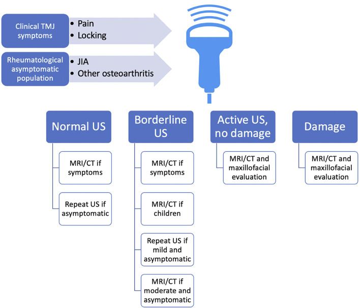
Temporomandibular joint (TMJ) disorder is the second most common chronic pain condition affecting the general population after back pain. It encompasses a complex set of conditions, manifesting with jaw pain and limitation in mouth opening, influencing chewing, eating, speaking, and facial expression. TMJ dysfunction could be related to mechanical abnormalities or underlying inflammatory arthropathies, such as rheumatoid arthritis (RA) or juvenile idiopathic arthritis (JIA). TMJ exhibits a complex anatomy, and thus a thorough investigation is required to detect the TMJ abnormalities. Importantly, TMJ involvement can be completely asymptomatic during the early stages of the disease, showing no clinically detectable signs, exposing patients to delayed diagnosis, and progressive irreversible condylar damage. For the prevention of JIA complications, early diagnosis is therefore essential. Currently, magnetic resonance imaging (MRI) is described in the literature as the gold standard method to evaluate TMJ. However, it is a high-cost procedure, not available in all centers, and requires a long time for image acquisition, which could represent a problem notably in the pediatric population. It also suffers restricted usage in patients with claustrophobia. Ultrasonography (US) has emerged in recent years as an alternative diagnostic method, as it is less expensive, not invasive, and does not demand special facilities. In this narrative review, we will investigate the power of US in TMJ disorders based on the most relevant literature data, from an early screening of TMJ changes to differential diagnosis and monitoring. We then propose a potential algorithm to optimize the management of TMJ pathology, questioning what would be the role of ultrasonographic study.

Keywords: articular disc; capsular width; diagnostic imaging; joint pain; temporomandibular joint; temporomandibular joint disorders; ultrasonography.


Conflict of interest statement

The authors declare that the research was conducted in the absence of any commercial or financial relationships that could be construed as a potential conflict of interest.

Figures

Figure 1
Figure 1
Flow diagram describing the inclusion decision of papers under the scope of this review.
Figure 2
Figure 2
Transverse image of the right (A) and left (B) TMJs showing the condyle and capsular width (distance between markers). White arrows show the condylar process and red arrows show the articular capsule (Personal archive).
Figure 3
Figure 3
Conventional US transducer positions are parallel to the Frankfort horizontal plane (a plane connecting the highest point of the opening of the external auditory canal with the lowest point on the lower margin of the orbit) in closed-mouth (A) position and open-mouth (B) position, as well as parallel to the ramus of mandible, both in closed-mouth (C) and open-mouth (D) positions. Normal ultrasound image of TMJ in transverse sections in closed- (E) and open-mouth positions (F). The normal ultrasound appearance of the articular disk in the sagittal plane is an inverted hypoechoic C-shaped structure, outlined by the red circle. During the mouth opening, the mandibular condyle translates anteriorly as defined by the distance between the center of the condylar oval at the two positions (yellow dotted line). Notably, the disk maintains a constant central appearance with respect to the center of the mandibular condyle in normal anatomy, while it may be displaced anteriorly or posteriorly in the pathological findings. Normal ultrasound image of TMJ in longitudinal closed-mouth (G) and open-mouth position (H). Red arrows show the articular capsule. JD, joint disk; MC, mandibular condyle (Personal archive).
Figure 4
Figure 4
Overview of possible proposed employment of US in TMJ pathology screening as a first-level examination, guiding choice on the follow-up methods. As no validated protocol suggests any early imaging methodic screening in the rheumatological population at-risk for TMD, and also TMD may present rather asymptomatic during the early stages of the disease, we propose US as an “entry-level” method, which is rapidly accessible and of relatively low cost. US could approach all the patients with rheumatic conditions and hopefully also RA-at-risk patients, even if asymptomatic, and of course those with TMJ symptoms. Because US was found to be specific, but not particularly sensitive, we advocate MRI execution even for borderline suspicious findings at US, as baseline MRI could improve anatomic US accuracy during the follow-up. CT, computed tomography; JIA, juvenile idiopathic arthritis; MRI, magnetic resonance imaging; RA, rheumatoid arthritis; TMJ, temporomandibular joint; US, ultrasonography.

References

    1. Alomar X, Medrano J, Cabratosa J, Clavero JA, Lorente M, Serra I, et al. . Anatomy of the temporomandibular joint. Semin Ultrasound CT MR. (2007) 28:170–83. 10.1053/j.sult.2007.02.002 - DOI - PubMed
    1. Covert L, Mater HV, Hechler BL. Comprehensive management of rheumatic diseases affecting the temporomandibular joint. Diagnostics (Basel). (2021) 11:409. 10.3390/diagnostics11030409 - DOI - PMC - PubMed
    1. Research, NIoDaC . Facial Pain. Available online at: https://www.nidcr.nih.gov/research/data-statistics/facial-pain (accessed March 22, 2022).
    1. Hunter DJ, Bierma-Zeinstra S. Osteoarthritis. Lancet. (2019) 393:1745–59. 10.1016/S0140-6736(19)30417-9 - DOI - PubMed
    1. Song H, Lee JY, Huh KH, Park JW. Long-Term changes of temporomandibular joint osteoarthritis on computed tomography. Sci Rep. (2020) 10:6731. 10.1038/s41598-020-63493-8 - DOI - PMC - PubMed