

Comparison of Two Surgical Techniques for the Treatment of Canine Disc Associated-Cervical Spondylomyelopathy
- PMID: 35795784
- PMCID: PMC9251543
- DOI: 10.3389/fvets.2022.880018
Comparison of Two Surgical Techniques for the Treatment of Canine Disc Associated-Cervical Spondylomyelopathy
Abstract
Objective: To compare prosthetic disc and vertebral distraction stabilization in dogs with disc-associated cervical spondylomyelopathy (DA-CSM).
Study design: A retrospective clinical study.
Animals: 25 dogs.
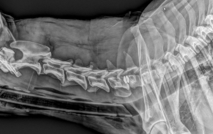
Methods: Dogs presenting with clinical signs and MRI findings compatible with DA-CSM underwent surgery. Implantation of the Adamo's prosthetic disc (PD) or vertebral distraction-stabilization (DS) with intervertebral cage, ventral locking plates, and dorsal transarticular screws was performed. All dogs were followed-up and evaluated clinically for a minimum of 1 year and radiographically for at least 3 months. In particular, we focused on the evaluation of subsidence (the degree of vertebral collapse).
Results: Twenty-five dogs were enrolled: 12 with PD implantation and 13 with DS implantation. Of these, 24 dogs were followed-up at 1 year. Overall, 12 dogs improved (4 PD and 8 DS), eight were stable (4 PD and 4 DS), and four deteriorated (3 PD and 1 DS). Deterioration was more common in PD cases, especially soon after surgery. In a few PD cases, a second surgery was necessary. The most common complication in dogs with DS was discospondylitis. Subsidence was detected in 11 PD and 7 DS dogs. Subsidence was more severe and occurred sooner after surgery in PD cases compared to DS cases. DS cases were more prone to clinical improvement and less prone to subsidence than PD cases in this study. However, the statistical evidence was weak owing to the small sample size.
Conclusion: The preliminary results suggest that prosthetic disc implantation is more prone to clinical and radiographic failures than distraction stabilization.
Clinical relevance: The DS technique is a valuable surgical option for treating dogs with DA-CSM, with favorable short- and long-term clinical and radiographic outcomes.
Keywords: Wobbler syndrome; cervical spondylomyelopathy (CSM); distraction stabilization; prosthetic disc; surgical procedures.
Copyright © 2022 Falzone, Tranquillo and Gasparinetti.
Conflict of interest statement
The authors declare that the research was conducted in the absence of any commercial or financial relationships that could be construed as a potential conflict of interest.
Figures


References
-
- Lewis DG. Cervical spondylomyelopathy (‘wobbler’ syndrome) in the dog: a study based on 224 cases. J Small Anim Pract. (1989) 30:657–65. 10.1111/j.1748-5827.1989.tb01909.x - DOI
-
- Seim H, Withrow S. Pathophysiology and diagnosis of caudal cervical spondylo-myelopathy with emphasis on the Doberman Pinscher. J Am Anim Hosp Assoc. (1982) 18:241–51.
-
- Seim H. Wobbler syndrome. In: Fossum T. editor, Small Animal Surgery. 2nd ed. St Louis, MO: Mosby Inc. (2002). p. 1237–49.
LinkOut - more resources
Full Text Sources