. 2022 Sep 21;43(36):3417-3431.
doi: 10.1093/eurheartj/ehac184.

Pulmonary vascular disease in pulmonary hypertension due to left heart disease: pathophysiologic implications

Affiliations

Pulmonary vascular disease in pulmonary hypertension due to left heart disease: pathophysiologic implications

Kazunori Omote et al. Eur Heart J. .

Abstract

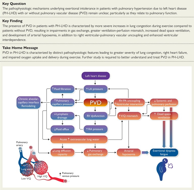
Aims: Pulmonary hypertension (PH) and pulmonary vascular disease (PVD) are common and associated with adverse outcomes in left heart disease (LHD). This study sought to characterize the pathophysiology of PVD across the spectrum of PH in LHD.

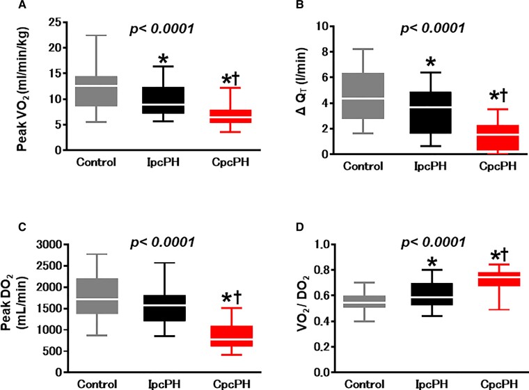
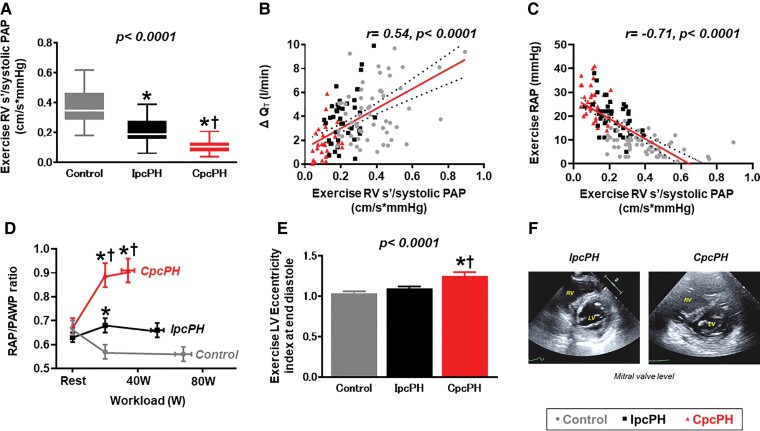
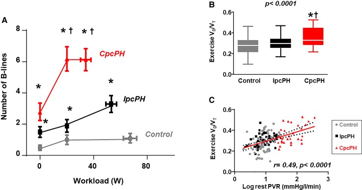
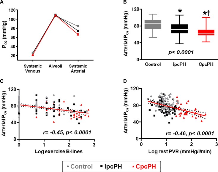
Methods and results: Patients with PH-LHD [mean pulmonary artery (PA) pressure >20 mmHg and PA wedge pressure (PAWP) ≥15 mmHg] and controls free of PH or LHD underwent invasive haemodynamic exercise testing with simultaneous echocardiography, expired air and blood gas analysis, and lung ultrasound in a prospective study. Patients with PH-LHD were divided into isolated post-capillary PH (IpcPH) and PVD [combined post- and pre-capillary PH (CpcPH)] based upon pulmonary vascular resistance (PVR <3.0 or ≥3.0 WU). As compared with controls (n = 69) and IpcPH-LHD (n = 55), participants with CpcPH-LHD (n = 40) displayed poorer left atrial function and more severe right ventricular (RV) dysfunction at rest. With exercise, patients with CpcPH-LHD displayed similar PAWP to IpcPH-LHD, but more severe RV-PA uncoupling, greater ventricular interaction, and more severe impairments in cardiac output, O2 delivery, and peak O2 consumption. Despite higher PVR, participants with CpcPH developed more severe lung congestion compared with both IpcPH-LHD and controls, which was associated lower arterial O2 tension, reduced alveolar ventilation, decreased pulmonary O2 diffusion, and greater ventilation-perfusion mismatch.

Conclusions: Pulmonary vascular disease in LHD is associated with a distinct pathophysiologic signature marked by greater exercise-induced lung congestion, arterial hypoxaemia, RV-PA uncoupling, ventricular interdependence, and impairment in O2 delivery, impairing aerobic capacity. Further study is required to identify novel treatments targeting the pulmonary vasculature in PH-LHD.

Keywords: Combined post- and pre-capillary pulmonary hypertension; Exercise haemodynamics; Heart failure; Left heart disease; Pulmonary hypertension; Pulmonary vascular resistance.


Conflict of interest statement

Conflict of interest: none declared.

Figures

Structured Graphical Abstract
Impact of pulmonary vascular disease on the heart and lungs in heart failure.
Figure 1
Figure 1
Compared with controls and participants with isolated post-capillary pulmonary hypertension, participants with combined post- and pre-capillary pulmonary hypertension  displayed lower peak VO2 (A) and less increase in the cardiac output response to exercise (B). Oxygen delivery during exercise was lowest in combined post- and pre-capillary pulmonary hypertension (C), with the highest O2 extraction (D). QT, cardiac output; CpcPH, combined post-and pre-capillary pulmonary hypertension; IpcPH, isolated post-capillary pulmonary hypertension; VO2, oxygen consumption; DO2, oxygen delivery. *P < 0.05 vs. controls. †P < 0.05 vs. IpcPH groups.
Figure 2
Figure 2
Right ventricular–pulmonary artery coupling during exercise was worse in the combined post- and pre-capillary pulmonary hypertension group compared with other groups (A) and was associated with impairments in CO reserve (B) and greater elevation in central venous pressure (C). Pericardial restraint and diastolic ventricular interdependence are enhanced in combined post- and pre-capillary pulmonary hypertension, evidenced by higher right atrial pressure/pulmonary artery wedge pressure ratio (D) and a greater exercise left ventricular eccentricity index compared with isolated post-capillary pulmonary hypertension and controls (E, F). PA, pulmonary artery; PAP, pulmonary artery pressure; RAP, right atrial pressure; PAWP, pulmonary artery wedge pressure; RV, right ventricular; s′, systolic tissue doppler velocity; QT, cardiac output; CpcPH, combined post-and pre-capillary pulmonary hypertension; IpcPH, isolated post-capillary pulmonary hypertension; VO2, oxygen consumption; DO2, oxygen delivery. ‘r’ determined by Pearson’s correlation analysis. *P < 0.05 vs. controls. †P < 0.05 vs. IpcPH groups.
Figure 3
Figure 3
Participants with combined post- and pre-capillary pulmonary hypertension-left heart disease display greater increases in extravascular lung water during exercise, indicated by increased B-line artefacts on lung ultrasound (A). This was coupled with increases in dead space ventilation (VD/VT ratio), which was directly correlated with resting pulmonary vascular resistance (B, C). EVLW, extravascular lung water; PVR; pulmonary vascular resistance, VD, pulmonary dead space; VT, tidal volume; QT, cardiac output; CpcPH, combined post-and pre-capillary pulmonary hypertension; IpcPH, isolated post-capillary pulmonary hypertension; VO2, oxygen consumption; DO2, oxygen delivery. ‘r’ determined by Pearson’s correlation analysis. *P < 0.05 vs. controls. †P < 0.05 vs. IpcPH groups.
Figure 4
Figure 4
As compared with individuals with isolated post-capillary pulmonary hypertension and controls, individuals with combined post- and pre-capillary pulmonary hypertension displayed lower arterial pO2 with exercise despite similar alveolar O2 tension (A, B). Arterial pO2 during exercise decreased with greater increases in extravascular lung water during exercise (C) and higher resting pulmonary vascular resistance (D). PA, pulmonary artery; PAP, pulmonary artery pressure; RAP, right atrial pressure; PAWP, pulmonary artery wedge pressure; RV, right ventricular; s′, systolic tissue doppler velocity; QT, cardiac output; CpcPH, combined post-and pre-capillary pulmonary hypertension; IpcPH, isolated post-capillary pulmonary hypertension; VO2, oxygen consumption; DO2, oxygen delivery. ‘r’ determined by Pearson’s correlation analysis. *P < 0.05 vs. controls. †P < 0.05 vs. IpcPH groups.

Comment in

References

    1. Vachiery JL, Tedford RJ, Rosenkranz S, Palazzini M, Lang I, Guazzi M, et al. . Pulmonary hypertension due to left heart disease. Eur Respir J 2019;53:1801897. - PMC - PubMed
    1. Guazzi M, Naeije R. Pulmonary hypertension in heart failure: pathophysiology, pathobiology, and emerging clinical perspectives. J Am Coll Cardiol 2017;69:1718–1734. - PubMed
    1. Verbrugge FH, Guazzi M, Testani JM, Borlaug BA. Altered hemodynamics and end-organ damage in heart failure: impact on the lung and kidney. Circulation 2020;142:998–1012. - PMC - PubMed
    1. Reddy YNV, Borlaug BA. Pulmonary hypertension in left heart disease. Clin Chest Med 2021;42:39–58. - PubMed
    1. Wijeratne DT, Lajkosz K, Brogly SB, Lougheed MD, Jiang L, Housin A, et al. . Increasing incidence and prevalence of world health organization groups 1 to 4 pulmonary hypertension: a population-based cohort study in Ontario, Canada. Circ Cardiovasc Qual Outcomes 2018;11:e003973. - PMC - PubMed

Publication types