The acute effect of exercise intensity on peripheral and cerebral vascular function in healthy adults
- PMID: 35796612
- PMCID: PMC9377787
- DOI: 10.1152/japplphysiol.00772.2021
The acute effect of exercise intensity on peripheral and cerebral vascular function in healthy adults
Abstract
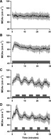
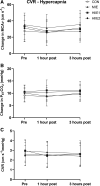
The acute effect of exercise intensity on cerebrovascular reactivity and whether this mirrors changes in peripheral vascular function have not been investigated. The aim of this study was to explore the acute effect of exercise intensity on cerebrovascular reactivity (CVR) and peripheral vascular function in healthy young adults (n = 10, 6 females, 22.7 ± 3.5 yr). Participants completed four experimental conditions on separate days: high-intensity interval exercise (HIIE) with intervals performed at 75% maximal oxygen uptake (V̇o2max; HIIE1), HIIE with intervals performed at 90% V̇o2max (HIIE2), continuous moderate-intensity exercise (MIE) at 60% V̇o2max and a sedentary control condition (CON). All exercise conditions were completed on a cycle ergometer and matched for time (30 min) and average intensity (60% V̇o2max). Brachial artery flow-mediated dilation (FMD) and CVR of the middle cerebral artery were measured before exercise, and 1- and 3-h after exercise. CVR was assessed using transcranial Doppler ultrasonography to both hypercapnia (6% carbon dioxide breathing) and hypocapnia (hyperventilation). FMD was significantly elevated above baseline 1 and 3 h following both HIIE conditions (P < 0.05), but FMD was unchanged following the MIE and CON trials (P > 0.33). CVR to both hypercapnia and hypocapnia, and when expressed across the end-tidal CO2 range, was unchanged in all conditions, at all time points (all P > 0.14). In conclusion, these novel findings show that the acute increases in peripheral vascular function following HIIE, compared with MIE, were not mirrored by changes in cerebrovascular reactivity, which was unaltered following all exercise conditions in healthy young adults.NEW & NOTEWORTHY This is the first study to identify that acute improvements in peripheral vascular function following high-intensity interval exercise are not mirrored by improvements in cerebrovascular reactivity in healthy young adults. High-intensity interval exercise completed at both 75% and 90% V̇o2max increased brachial artery flow-mediated dilation 1 and 3 h following exercise, compared with continuous moderate-intensity exercise and a sedentary control condition. By contrast, cerebrovascular reactivity was unchanged following all four conditions.
Keywords: HIIE; cerebrovascular reactivity; endothelial function; flow-mediated dilation.
Conflict of interest statement
No conflicts of interest, financial or otherwise, are declared by the authors.
Figures


Similar articles
-
A high salt meal does not impair cerebrovascular reactivity in healthy young adults.Physiol Rep. 2020 Oct;8(19):e14585. doi: 10.14814/phy2.14585. Physiol Rep. 2020. PMID: 33038066 Free PMC article.
-
Comparison of cerebrovascular reactivity recovery following high-intensity interval training and moderate-intensity continuous training.Physiol Rep. 2020 Jun;8(11):e14467. doi: 10.14814/phy2.14467. Physiol Rep. 2020. PMID: 32506845 Free PMC article. Clinical Trial.
-
High-intensity interval exercise attenuates but does not eliminate endothelial dysfunction after a fast food meal.Am J Physiol Heart Circ Physiol. 2018 Feb 1;314(2):H188-H194. doi: 10.1152/ajpheart.00384.2017. Epub 2017 Nov 3. Am J Physiol Heart Circ Physiol. 2018. PMID: 29101171 Clinical Trial.
-
Effects of high intensity interval exercise on cerebrovascular function: A systematic review.PLoS One. 2020 Oct 29;15(10):e0241248. doi: 10.1371/journal.pone.0241248. eCollection 2020. PLoS One. 2020. PMID: 33119691 Free PMC article.
-
A framework of transient hypercapnia to achieve an increased cerebral blood flow induced by nasal breathing during aerobic exercise.Cereb Circ Cogn Behav. 2023 Sep 13;5:100183. doi: 10.1016/j.cccb.2023.100183. eCollection 2023. Cereb Circ Cogn Behav. 2023. PMID: 37745894 Free PMC article. Review.
Cited by
-
Vascular endothelial function and its response to moderate-intensity aerobic exercise in trained and untrained healthy young men.Sci Rep. 2024 Sep 3;14(1):20450. doi: 10.1038/s41598-024-71471-7. Sci Rep. 2024. PMID: 39242762 Free PMC article.
-
Effect of Exercise Prior to Sedentary Behavior on Vascular Health Parameters: A Systematic Review and Meta-Analysis of Crossover Trials.Sports Med Open. 2024 Jun 9;10(1):69. doi: 10.1186/s40798-024-00734-4. Sports Med Open. 2024. PMID: 38853205 Free PMC article.
-
Hippocampal blood flow rapidly and preferentially increases after a bout of moderate-intensity exercise in older adults with poor cerebrovascular health.Cereb Cortex. 2023 Apr 25;33(9):5297-5306. doi: 10.1093/cercor/bhac418. Cereb Cortex. 2023. PMID: 36255379 Free PMC article.
-
The interrelationship among exercise intensity, endothelial function, and ghrelin in healthy humans.Physiol Rep. 2025 Apr;13(7):e70213. doi: 10.14814/phy2.70213. Physiol Rep. 2025. PMID: 40214273 Free PMC article.
-
The independent and combined effects of aerobic exercise intensity and dose differentially increase post-exercise cerebral shear stress and blood flow.Exp Physiol. 2024 Oct;109(10):1796-1805. doi: 10.1113/EP091856. Epub 2024 Aug 14. Exp Physiol. 2024. PMID: 39141846 Free PMC article. Clinical Trial.
References
Publication types
MeSH terms
Grants and funding
LinkOut - more resources
Full Text Sources

