

Top-Down Detection of Oxidative Protein Footprinting by Collision-Induced Dissociation, Electron-Transfer Dissociation, and Electron-Capture Dissociation
- PMID: 35797180
- PMCID: PMC9311227
- DOI: 10.1021/acs.analchem.1c05476
Top-Down Detection of Oxidative Protein Footprinting by Collision-Induced Dissociation, Electron-Transfer Dissociation, and Electron-Capture Dissociation
Abstract
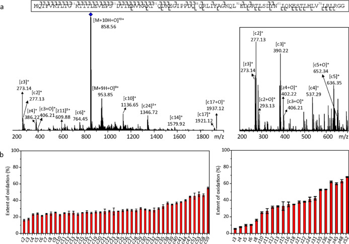
Fast photochemical oxidation of proteins (FPOP) footprinting is a structural mass spectrometry method that maps proteins by fast and irreversible chemical reactions. The position of oxidative modification reflects solvent accessibility and site reactivity and thus provides information about protein conformation, structural dynamics, and interactions. Bottom-up mass spectrometry is an established standard method to analyze FPOP samples. In the bottom-up approach, all forms of the protein are digested together by a protease of choice, which results in a mixture of peptides from various subpopulations of proteins with varying degrees of photochemical oxidation. Here, we investigate the possibility to analyze a specifically selected population of only singly oxidized proteins. This requires utilization of more specific top-down mass spectrometry approaches. The key element of any top-down experiment is the selection of a suitable method of ion isolation, excitation, and fragmentation. Here, we employ and compare collision-induced dissociation, electron-transfer dissociation, and electron-capture dissociation combined with multi-continuous accumulation of selected ions. A singly oxidized subpopulation of FPOP-labeled ubiquitin was used to optimize the method. The top-down approach in FPOP is limited to smaller proteins, but its usefulness was demonstrated by using it to visualize structural changes induced by co-factor removal from the holo/apo myoglobin system. The top-down data were compared with the literature and with the bottom-up data set obtained on the same samples. The top-down results were found to be in good agreement, which indicates that monitoring a singly oxidized FPOP ion population by the top-down approach is a functional workflow for oxidative protein footprinting.
Conflict of interest statement
The authors declare no competing financial interest.
Figures


Similar articles
-
Toward a MALDI in-source decay (ISD) method for top-down analysis of protein footprinting.Eur J Mass Spectrom (Chichester). 2023 Oct;29(5-6):292-302. doi: 10.1177/14690667231202695. Epub 2023 Sep 26. Eur J Mass Spectrom (Chichester). 2023. PMID: 37750197 Free PMC article.
-
New protein footprinting: fast photochemical iodination combined with top-down and bottom-up mass spectrometry.J Am Soc Mass Spectrom. 2012 Aug;23(8):1306-18. doi: 10.1007/s13361-012-0403-1. Epub 2012 Jun 6. J Am Soc Mass Spectrom. 2012. PMID: 22669760 Free PMC article.
-
Protein Footprinting by Carbenes on a Fast Photochemical Oxidation of Proteins (FPOP) Platform.J Am Soc Mass Spectrom. 2016 Mar;27(3):552-5. doi: 10.1007/s13361-015-1313-9. Epub 2015 Dec 17. J Am Soc Mass Spectrom. 2016. PMID: 26679355 Free PMC article.
-
Mass Spectrometry-Based Fast Photochemical Oxidation of Proteins (FPOP) for Higher Order Structure Characterization.Acc Chem Res. 2018 Mar 20;51(3):736-744. doi: 10.1021/acs.accounts.7b00593. Epub 2018 Feb 16. Acc Chem Res. 2018. PMID: 29450991 Free PMC article. Review.
-
Fast Photochemical Oxidation of Proteins Coupled with Mass Spectrometry.Protein Pept Lett. 2019;26(1):27-34. doi: 10.2174/0929866526666181128124554. Protein Pept Lett. 2019. PMID: 30484399 Free PMC article. Review.
Cited by
-
Isotopic Depletion Increases the Spatial Resolution of FPOP Top-Down Mass Spectrometry Analysis.Anal Chem. 2024 Jan 30;96(4):1478-1487. doi: 10.1021/acs.analchem.3c03759. Epub 2024 Jan 16. Anal Chem. 2024. PMID: 38226459 Free PMC article.
-
Top-Down Proteoform Analysis by 2D MS with Quadrupolar Detection.Anal Chem. 2023 Nov 7;95(44):16123-16130. doi: 10.1021/acs.analchem.3c02225. Epub 2023 Oct 25. Anal Chem. 2023. PMID: 37877738 Free PMC article.
-
Quantifying the Impact of the Peptide Identification Framework on the Results of Fast Photochemical Oxidation of Protein Analysis.J Proteome Res. 2024 Feb 2;23(2):609-617. doi: 10.1021/acs.jproteome.3c00390. Epub 2023 Dec 29. J Proteome Res. 2024. PMID: 38158558 Free PMC article.
-
MALDI Peptide Mapping for Fast Analysis in Protein Footprinting.Int J Mass Spectrom. 2023 Aug;490:117080. doi: 10.1016/j.ijms.2023.117080. Epub 2023 May 11. Int J Mass Spectrom. 2023. PMID: 38465269 Free PMC article.
-
Quantification of pharmaceutical compounds in tissue and plasma samples using selective ion accumulation with multiple mass isolation windows.J Mass Spectrom. 2023 Jul;58(7):e4958. doi: 10.1002/jms.4958. J Mass Spectrom. 2023. PMID: 37431164 Free PMC article.
References
Publication types
MeSH terms
Substances
LinkOut - more resources
Full Text Sources
Molecular Biology Databases
