Review
. 2022 Jul 7;18(7):e1010602.
doi: 10.1371/journal.ppat.1010602. eCollection 2022 Jul.

Mycobacteriophages: From Petri dish to patient

Affiliations
Review

Mycobacteriophages: From Petri dish to patient

Graham F Hatfull. PLoS Pathog. .

Abstract

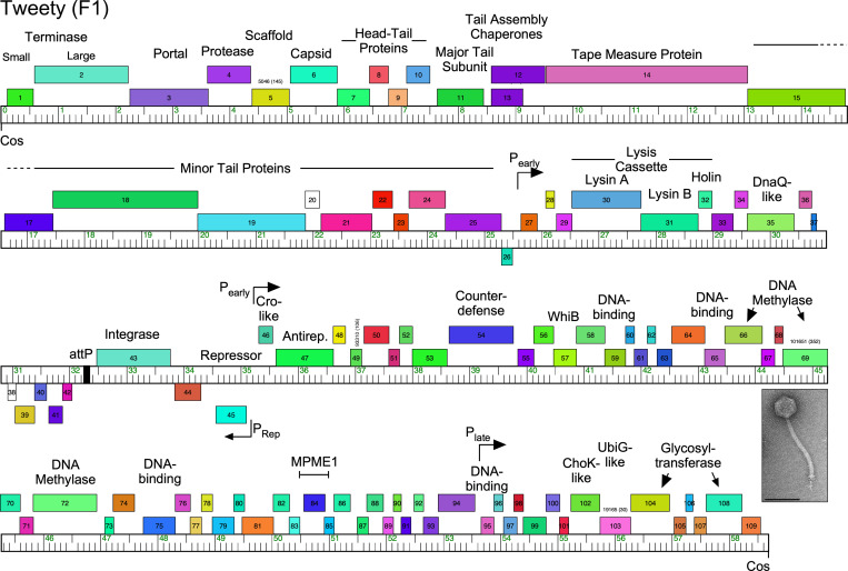
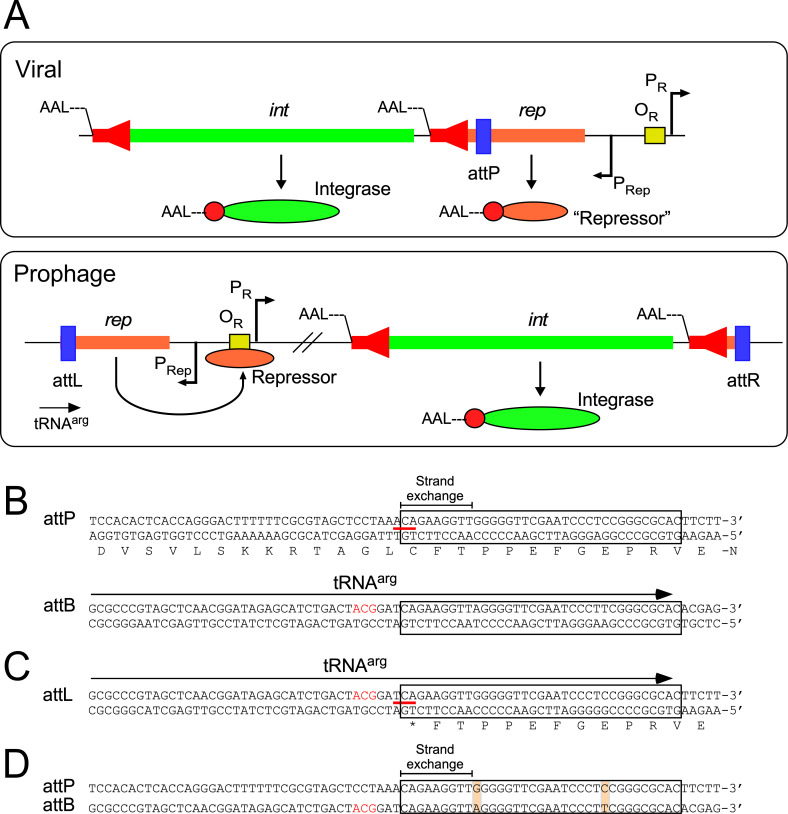
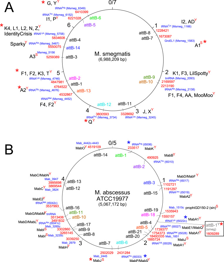
Mycobacteriophages-bacteriophages infecting Mycobacterium hosts-contribute substantially to our understanding of viral diversity and evolution, provide resources for advancing Mycobacterium genetics, are the basis of high-impact science education programs, and show considerable therapeutic potential. Over 10,000 individual mycobacteriophages have been isolated by high school and undergraduate students using the model organism Mycobacterium smegmatis mc2155 and 2,100 have been completely sequenced, giving a high-resolution view of the phages that infect a single common host strain. The phage genomes are revealed to be highly diverse and architecturally mosaic and are replete with genes of unknown function. Mycobacteriophages have provided many widely used tools for Mycobacterium genetics including integration-proficient vectors and recombineering systems, as well as systems for efficient delivery of reporter genes, transposons, and allelic exchange substrates. The genomic insights and engineering tools have facilitated exploration of phages for treatment of Mycobacterium infections, although their full therapeutic potential has yet to be realized.


Conflict of interest statement

I have read the journal’s policy and have the following conflicts: I receive research support from Janssen Pharmaceuticals, and am a consultant for Tessera Inc. and Janssen.

Figures

Fig 1
Fig 1. Genome organization of Mycobacteriophage Tweety.
Phage Tweety is a member of Subcluster F1, is temperate, has a siphoviral morphology (inset, electron micropraph; scale marker, 100 nm), and its genome organization illustrates several common features. The genome is represented as a bar with markers every 1 kbp, and the predicted genes are shown as colored boxes above or below the genome, indicating rightwards- and leftwards-transcription, respectively. Gene numbers are shown within the boxes and predicted functions are shown above. The virion structure and assembly genes are arranged in a rightwards-transcribed operon at the left end of the genome (genes 1 to 25), followed by the lysis cassette. The repressor (45) and putative Cro-like (46) genes are divergently transcribed, and the integration cassette (attP and 43) are located nearby, with attP defining a “left arm” from cosattP and a “right arm” from attP–cos. Note that the left arm genes—predominantly the virion structure and lysis genes—are relatively large and have known functions, whereas the right arm genes are relatively small, and most have unknown functions. Shown are the positions of the MPME mobile element and the counterdefense gene, 54. The positions of putative early and later lytic promoters as determined by RNAseq are indicated. Note that genes are arranged in long operons such that there are relatively few transcriptional changes (tdc), such as at the 25/26 and 45/46 junctions.
Fig 2
Fig 2. Diversity of mycobacteriophage genomes displayed as network phylogenies based on shared gene content.
(A) Relationships among representative members of clusters, subclusters, and singleton genomes. One member of each mycobacteriophage cluster and subcluster together with the 7 singletons were compared using Splitstree [39] with a nexus file recording the numbers of shared genes. Clusters are illustrated with colored shading; note that some clusters (e.g., Cluster A) contain several subclusters indicated as nodes, whereas other clusters are not subdivided. Singletons are shown as unlabeled black circles. (B) Diversity of Cluster F mycobacteriophages. All currently sequenced Cluster F mycobacteriophages (n = 188) are displayed as nodes in a network phylogeny using Splitstree. Colored circles show the positions of the Subclusters F2 to F5 genomes; all of the others (n = 177) are grouped in Subcluster F1. This illustrates the substantial intracluster diversity, and pairwise comparisons of Subcluster F1 phages show they may share as few as 40% of their genes.
Fig 3
Fig 3. Integration-dependent superinfection immunity systems.
(A) Typical organization of the immunity regions of phages encoding integration-dependent immunity systems. In the viral genome, the repressor (rep) and integrase (int) genes are cotranscribed from the PRep promoter, and the phage attachment site (attP, blue box) is located within the repressor gene. The virally encoded Integrase and Repressor proteins both carry C-terminal ssrA-like tags (——LAA, or similar) targeting the proteins for proteolytic degradation (red arrows in the int and rep genes; red circles in the proteins; the N-termini are indicated). The virally encoded form of the Repressor is not active in conferring superinfection immunity. The establishment of lysogeny requires integrase-mediated site-specific recombination between the phage attP site and a chromosomal attB site (which overlaps a host tRNA gene) to form a prophage. Integrative site-specific recombination removes the ssrA-like tag from the repressor and the stable, active form of the repressor binds to the operator (OR, yellow box) to shut down the early lytic promoter (PR) and confer superinfection immunity. (B) Organization of the attP and attB sites of phage BPs and M. smegmatis. Both DNA strands are shown, and the amino acid sequence of part of the leftwards-transcribed repressor is shown; DNA and protein polarities are indicated. The 35-bp common core sequence (conserved in attP, attB, attL, and attR) is boxed, and the region within which strand exchange for recombination must occur is indicated. The codon spanning the left side of the common core is indicated by a red line, and the third position base is shown in bold pink type. The location of the tRNAarg gene at attB is shown by an arrow; the anticodon is shown in red type. (C) Organization of the BPs attL site. Conservation of the common sequence between attP and attB results in construction of an active tRNAarg gene when the prophage is established. However, at attL the repressor gene encounters a termination codon (TGA) formed at the junction of the bacterial and phage sequences, changing the third base of a cysteine codon (red line) to a nonsense codon; the third position base is shown in bold pink type. (D) Alignment of attP and attB showing the common core (boxed), and the positions of 2 mismatches in the common core, neither of which introduced mispairing in the tRNAarg product. Only the top strand of each site is shown.
Fig 4
Fig 4. Chromosomal attB attachment sites used by temperate phages in M. smegmatis and M. abscessus.
(A) attB sites in the M. smegmatis mc2155 genome. The 7-Mbp M. smegmatis genome is represented as a circle with markers at each kbp indicated. The attB sites (e.g., attB-1, attB-2, etc.) are shown, together with the genome coordinates in red type; the genes are shown in blue type. The font color of the M. smegmatis attB sites (i.e., attB-xsmeg) label is coordinated with homologous sites in M. abscessus (i.e., attB-xMab) shown in panel B. The names of clusters or subclusters (or the phage name if it is a singleton) within which one or more phages use that site for integration are shown in large black type. “Y” or “S” superscripts on the cluster names denote whether the site is used by tyrosine- (Y) or serine (S) -integrases. attB sites for which integration-proficient vectors have been developed are indicated with a red asterisk. (B) attB sites in the M. abscessus ATCC19977 genome mapped by identification of integrated prophages. The font color of the attB sites (i.e., attB-xMab) is coordinated with their homologues in M. smegmatis (i.e., attB-xsmeg) shown in panel A. Genome coordinates are shown in red type; genes are shown in blue type. The clusters/subclusters of prophages for which members are found integrated in those sites are shown in black type. “Y” or “S” superscripts on the cluster names denote whether the site is used by tyrosine- (Y) or serine (S) -integrases. The integration vectors based on phage L5 (attB-1smeg) integrate at a homologous site in M. abscessus (shown in box) although no prophages have been identified there. This and the attB-7Mab site are only ones for which integration vectors have been shown to work (marked with red asterisks), but several vectors developed for M. smegmatis are predicted to also work in M. abscessus (blue asterisks).

References

    1. Rohwer F, Youle M, Maughan H, Hisakawa N. Life in our phage world: A centenial field guide to the earth’s most diverse inhiabitants. San Diego, CA: Wholon; 2014.
    1. Mushegian AR. Are There 10(31) Virus Particles on Earth, or More, or Fewer? J Bacteriol. 2020;202(9). Epub 2020/02/20. doi: 10.1128/JB.00052-20 ; PubMed Central PMCID: PMC7148134. - DOI - PMC - PubMed
    1. Hendrix RW, Smith MC, Burns RN, Ford ME, Hatfull GF. Evolutionary relationships among diverse bacteriophages and prophages: all the world’s a phage. Proc Natl Acad Sci U S A. 1999;96(5):2192–7. Epub 1999/03/03. doi: 10.1073/pnas.96.5.2192 ; PubMed Central PMCID: PMC26759. - DOI - PMC - PubMed
    1. Hendrix RW. Bacteriophages: evolution of the majority. Theor Popul Biol. 2002;61 (4):471–80. doi: 10.1006/tpbi.2002.1590 . - DOI - PubMed
    1. Hatfull GF, Hendrix RW. Bacteriophages and their Genomes. Curr Opin Virol. 2011;1:298–303. doi: 10.1016/j.coviro.2011.06.009 - DOI - PMC - PubMed

Publication types