

Glomus Tumor of the Lower Extremity Previously Misdiagnosed as Complex Regional Pain Syndrome in Close Proximity to a Myxofibrosarcoma: A Case Report
- PMID: 35797605
- PMCID: PMC9263485
- DOI: 10.5435/JAAOSGlobal-D-21-00311
Glomus Tumor of the Lower Extremity Previously Misdiagnosed as Complex Regional Pain Syndrome in Close Proximity to a Myxofibrosarcoma: A Case Report
Abstract
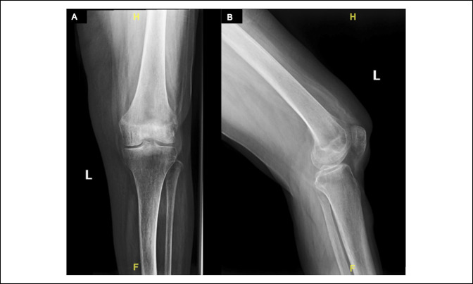
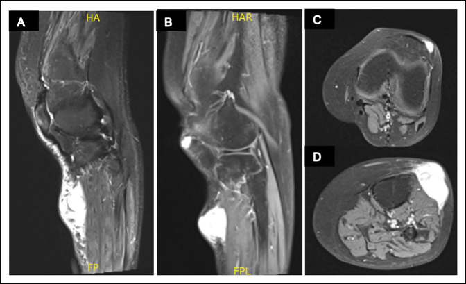
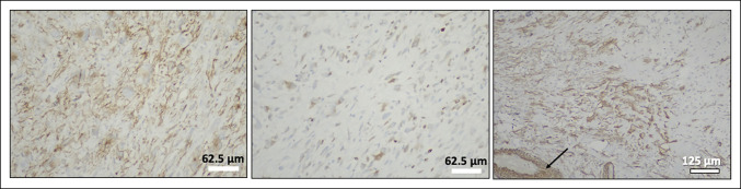
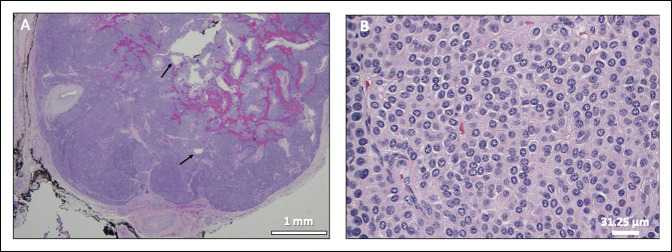
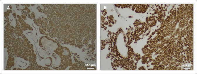
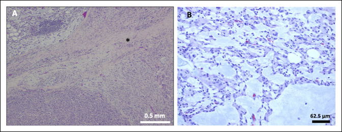
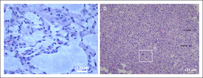
Complex regional pain syndrome (CRPS) is a potentially devastating condition that can result in severe psychological and social morbidity. It is a diagnosis of exclusion, and other pathologic entities must be ruled out first. Glomus tumors are exquisitely painful benign vascular tumors that are most common in the hand and are rarely found in the lower extremity. Here, we present a case of a patient who developed a focus of severe anterior knee pain and tenderness a few months after a car accident that had been misdiagnosed as CRPS for 15 years. She coincidentally developed a sarcoma of her ipsilateral leg distal to this site. Magnetic resonance imaging of the sarcoma included the area of knee pain where, interestingly, it identified a separate small soft-tissue mass. A glomus tumor was diagnosed histologically in a needle biopsy specimen from this mass, which was resected along with the sarcoma. For the first time in 15 years, despite the additional sarcoma surgery, she reported relief of her pain and complete resolution of her "CRPS."
Copyright © 2022 The Authors. Published by Wolters Kluwer Health, Inc. on behalf of the American Academy of Orthopaedic Surgeons.
Figures
Similar articles
-
Glomus tumor presenting as complex regional pain syndrome of the left upper limb: a case report.J Med Case Rep. 2015 Dec 29;9:293. doi: 10.1186/s13256-015-0793-3. J Med Case Rep. 2015. PMID: 26715068 Free PMC article.
-
Glomus tumors around or in the knee: a case report and literature review.BMC Surg. 2022 Mar 16;22(1):97. doi: 10.1186/s12893-022-01545-8. BMC Surg. 2022. PMID: 35296290 Free PMC article. Review.
-
Glomus Tumor in the Tarsal Tunnel: A Case Report.J Foot Ankle Surg. 2017 Jul-Aug;56(4):865-867. doi: 10.1053/j.jfas.2017.03.005. J Foot Ankle Surg. 2017. PMID: 28633794
-
Glomus tumors.J Hand Surg Am. 2006 Oct;31(8):1397-400. doi: 10.1016/j.jhsa.2006.05.018. J Hand Surg Am. 2006. PMID: 17027805 Review.
-
Glomus tumor masquerading for 22 years as osteoarthritis of the hip.Cutis. 2008 Apr;81(4):339-42. Cutis. 2008. PMID: 18491482
Cited by
-
Complex Regional Pain Syndrome in Cancer Cases: Current Knowledge and Perspectives.Int Med Case Rep J. 2024 May 18;17:497-506. doi: 10.2147/IMCRJ.S451291. eCollection 2024. Int Med Case Rep J. 2024. PMID: 38778887 Free PMC article.
-
Acrometastasis as a mimic of complex regional pain syndrome.Interv Pain Med. 2023 Jun 6;2(2):100250. doi: 10.1016/j.inpm.2023.100250. eCollection 2023 Jun. Interv Pain Med. 2023. PMID: 39238669 Free PMC article.
References
Publication types
MeSH terms
LinkOut - more resources
Full Text Sources
Medical
